 ムービー
ムービー コントローラー


+ データを開く

- 基本情報
基本情報
| 登録情報 |  | |||||||||
|---|---|---|---|---|---|---|---|---|---|---|
| タイトル | Sub-tomogram average of the RSV F pairs (with over-dosed particles removed) from the surface of native virions released from RSV-infected BEAS-2B cells cultured on EM grids collected via montage parallel array cryo- tomography (MPACT) | |||||||||
 マップデータ マップデータ | STA of RSV F pair from native RSV virion collected via parallel array montage cryo-ET | |||||||||
 試料 試料 |
| |||||||||
 キーワード キーワード | natively folded / class I viral fusion protein / pre-fusion native state / whole virion / VIRUS | |||||||||
| 生物種 |  Respiratory syncytial virus A2 (ウイルス) Respiratory syncytial virus A2 (ウイルス) | |||||||||
| 手法 | サブトモグラム平均法 / クライオ電子顕微鏡法 / 解像度: 18.5 Å | |||||||||
 データ登録者 データ登録者 | Yang J / Wright ER | |||||||||
| 資金援助 |  米国, 2件 米国, 2件
| |||||||||
 引用 引用 | ジャーナル: Nat Methods / 年: 2023 タイトル: Correlative montage parallel array cryo-tomography for in situ structural cell biology. 著者: Jie E Yang / Matthew R Larson / Bryan S Sibert / Joseph Y Kim / Daniel Parrell / Juan C Sanchez / Victoria Pappas / Anil Kumar / Kai Cai / Keith Thompson / Elizabeth R Wright / 要旨: Imaging large fields of view while preserving high-resolution structural information remains a challenge in low-dose cryo-electron tomography. Here we present robust tools for montage parallel array ...Imaging large fields of view while preserving high-resolution structural information remains a challenge in low-dose cryo-electron tomography. Here we present robust tools for montage parallel array cryo-tomography (MPACT) tailored for vitrified specimens. The combination of correlative cryo-fluorescence microscopy, focused-ion-beam milling, substrate micropatterning, and MPACT supports studies that contextually define the three-dimensional architecture of cells. To further extend the flexibility of MPACT, tilt series may be processed in their entirety or as individual tiles suitable for sub-tomogram averaging, enabling efficient data processing and analysis. | |||||||||
| 履歴 |
|
- 構造の表示
構造の表示
| 添付画像 |
|---|

- ダウンロードとリンク
-EMDBアーカイブ
| マップデータ | emd_40307.map.gz | 7.3 MB |  EMDBマップデータ形式 EMDBマップデータ形式 | |
|---|---|---|---|---|
| ヘッダ (付随情報) | emd-40307-v30.xmlemd-40307.xml | 17.9 KB 17.9 KB | 表示 表示 | EMDBヘッダ |
| FSC (解像度算出) | emd_40307_fsc.xml | 5.2 KB | 表示 | FSCデータファイル |
| 画像 |  emd_40307.png emd_40307.png | 19.2 KB | ||
| Filedesc metadata | emd-40307.cif.gz | 5.2 KB | ||
| その他 | emd_40307_half_map_1.map.gzemd_40307_half_map_2.map.gz | 7.3 MB 7.3 MB | ||
| アーカイブディレクトリ |  http://ftp.pdbj.org/pub/emdb/structures/EMD-40307ftp://ftp.pdbj.org/pub/emdb/structures/EMD-40307 http://ftp.pdbj.org/pub/emdb/structures/EMD-40307ftp://ftp.pdbj.org/pub/emdb/structures/EMD-40307 | HTTPS FTP |
-検証レポート
| 文書・要旨 | emd_40307_validation.pdf.gz | 602.5 KB | 表示 | EMDB検証レポート |
|---|---|---|---|---|
| 文書・詳細版 | emd_40307_full_validation.pdf.gz | 602.1 KB | 表示 | |
| XML形式データ | emd_40307_validation.xml.gz | 10.8 KB | 表示 | |
| CIF形式データ | emd_40307_validation.cif.gz | 13.8 KB | 表示 | |
| アーカイブディレクトリ | https://ftp.pdbj.org/pub/emdb/validation_reports/EMD-40307ftp://ftp.pdbj.org/pub/emdb/validation_reports/EMD-40307 | HTTPS FTP |
-関連構造データ


-リンク
| EMDBのページ | EMDB (EBI/PDBe) / EMDataResource |
|---|
-マップ
| ファイル | ダウンロード / ファイル: emd_40307.map.gz / 形式: CCP4 / 大きさ: 8 MB / タイプ: IMAGE STORED AS FLOATING POINT NUMBER (4 BYTES) | ||||||||||||||||||||||||||||||||||||
|---|---|---|---|---|---|---|---|---|---|---|---|---|---|---|---|---|---|---|---|---|---|---|---|---|---|---|---|---|---|---|---|---|---|---|---|---|---|
| 注釈 | STA of RSV F pair from native RSV virion collected via parallel array montage cryo-ET | ||||||||||||||||||||||||||||||||||||
| 投影像・断面図 | 画像のコントロール
画像は Spider により作成 | ||||||||||||||||||||||||||||||||||||
| ボクセルのサイズ | X=Y=Z: 2.503 Å | ||||||||||||||||||||||||||||||||||||
| 密度 |
| ||||||||||||||||||||||||||||||||||||
| 対称性 | 空間群: 1 | ||||||||||||||||||||||||||||||||||||
| 詳細 | EMDB XML:
|
 Z (Sec.)
 Y (Row.)
 X (Col.)


-添付データ
-ハーフマップ: STA of RSV F pair from native RSV...
| ファイル | emd_40307_half_map_1.map | ||||||||||||
|---|---|---|---|---|---|---|---|---|---|---|---|---|---|
| 注釈 | STA of RSV F pair from native RSV virion collected via parallel array montage cryo-ET | ||||||||||||
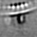
| 投影像・断面図 |
| ||||||||||||
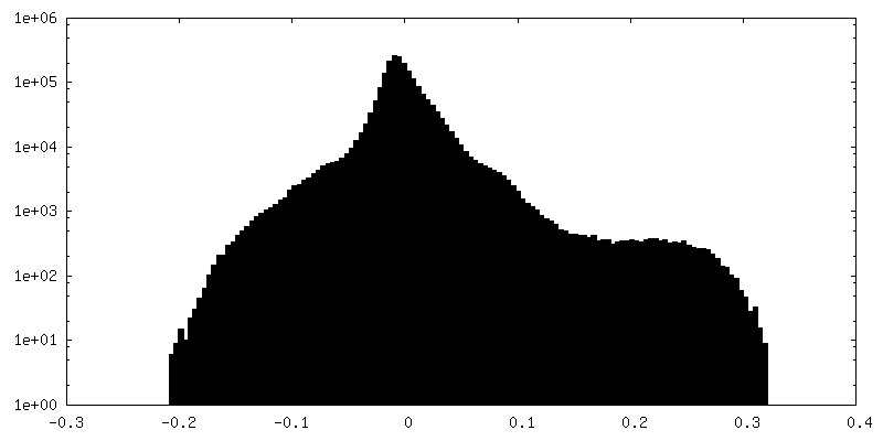
| 密度ヒストグラム |


-ハーフマップ: STA of RSV F pair from native RSV...
| ファイル | emd_40307_half_map_2.map | ||||||||||||
|---|---|---|---|---|---|---|---|---|---|---|---|---|---|
| 注釈 | STA of RSV F pair from native RSV virion collected via parallel array montage cryo-ET | ||||||||||||
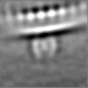
| 投影像・断面図 |
| ||||||||||||
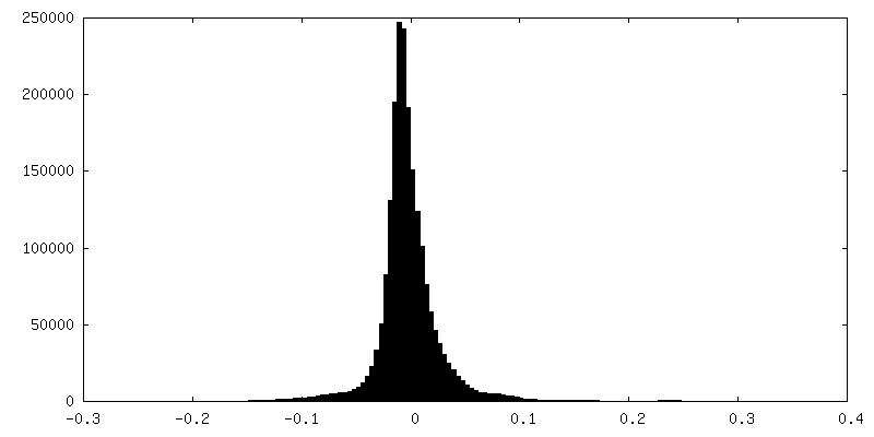
| 密度ヒストグラム |


- 試料の構成要素
-全体 : Respiratory syncytial virus A2
| 全体 | 名称: Respiratory syncytial virus A2 (ウイルス) |
|---|---|
| 要素 |
|
-超分子 #1: Respiratory syncytial virus A2
| 超分子 | 名称: Respiratory syncytial virus A2 / タイプ: virus / ID: 1 / 親要素: 0 詳細: The strain was kindly provided by Dr. Martin L. Moore. The reference is PMCID: PMC3492879/PMID:23062737 NCBI-ID: 1972429 / 生物種: Respiratory syncytial virus A2 / Sci species strain: rA2-mK+ / ウイルスタイプ: VIRION / ウイルス・単離状態: STRAIN / ウイルス・エンベロープ: Yes / ウイルス・中空状態: No |
|---|---|
| 宿主 | 生物種:  Homo sapiens (ヒト) Homo sapiens (ヒト) |
-実験情報
-構造解析
| 手法 | クライオ電子顕微鏡法 |
|---|---|
 解析 解析 | サブトモグラム平均法 |
| 試料の集合状態 | cell |
-試料調製
| 緩衝液 | pH: 7 |
|---|---|
| グリッド | モデル: Quantifoil R2/1 / 材質: GOLD / メッシュ: 200 / 支持フィルム - 材質: CARBON / 支持フィルム - トポロジー: HOLEY / 支持フィルム - Film thickness: 17 |
| 凍結 | 凍結剤: ETHANE / チャンバー内湿度: 80 % / チャンバー内温度: 303 K / 装置: GATAN CRYOPLUNGE 3 |
| 詳細 | Respiratory syncytial virus released from infected whole BEAS-2B mammalian cells (human non-tumorigenic lung epithelial cell) |
- 電子顕微鏡法
| 顕微鏡 | TFS KRIOS |
|---|---|
| 特殊光学系 | エネルギーフィルター - 名称: GIF Bioquantum / エネルギーフィルター - スリット幅: 20 eV |
| 撮影 | フィルム・検出器のモデル: GATAN K3 BIOQUANTUM (6k x 4k) デジタル化 - サイズ - 横: 5760 pixel / デジタル化 - サイズ - 縦: 4092 pixel / 平均露光時間: 0.5 sec. / 平均電子線量: 1.8 e/Å2 詳細: Tilt series of defocus collection ranging from -3 to -6 micrometer, with a step of -0.5 micrometer |
| 電子線 | 加速電圧: 300 kV / 電子線源:  FIELD EMISSION GUN FIELD EMISSION GUN |
| 電子光学系 | C2レンズ絞り径: 100.0 µm 最大 デフォーカス(補正後): 6.8500000000000005 µm 最小 デフォーカス(補正後): 3.3200000000000003 µm 照射モード: FLOOD BEAM / 撮影モード: BRIGHT FIELD / Cs: 2.7 mm / 最大 デフォーカス(公称値): 6.0 µm / 最小 デフォーカス(公称値): 3.0 µm |
| 試料ステージ | 試料ホルダーモデル: FEI TITAN KRIOS AUTOGRID HOLDER ホルダー冷却材: NITROGEN |
| 実験機器 |  モデル: Titan Krios / 画像提供: FEI Company |

