 Movie
Movie Controller

[English] 日本語
 Yorodumi
- EMDB-40007: Local map of SARS-Cov2 S protein structure in complex with neutra... -
+ Open data

- Basic information
| Entry |  | |||||||||
|---|---|---|---|---|---|---|---|---|---|---|
| Title | Local map of SARS-Cov2 S protein structure in complex with neutralizing monoclonal antibody 002-S21B10 | |||||||||
 Map data Map data | Local refinement map of SARS-CoV-2 spike glycoprotein bound to antibody 002-S21B10. Used in construction of composite map EMD-29950. | |||||||||
 Sample Sample |
| |||||||||
 Keywords Keywords | antibody / spike / SARS-CoV-2 / viral protein-immune system complex / viral protein | |||||||||
| Biological species |  Homo sapiens (human) / Homo sapiens (human) /   Severe acute respiratory syndrome coronavirus 2 Severe acute respiratory syndrome coronavirus 2 | |||||||||
| Method | single particle reconstruction / cryo EM / Resolution: 3.85 Å | |||||||||
 Authors Authors | Patel A / Ortlund EA | |||||||||
| Funding support |  United States, 1 items United States, 1 items
| |||||||||
 Citation Citation | Journal: To Be Published Title: Elucidating the mechanism of SARS-CoV-2 Omicron variant escape from a RBD class-3 human antibody Authors: Patel A / Kumar S / Lai L / Chakravarthy C / Valanparambil R / Keen M / Laughlin ZT / Frank F / Cheedarla N / Verkerke HP / Neish AS / Roback JD / Davis CW / Wrammert J / Ahmed R / Suthar MS ...Authors: Patel A / Kumar S / Lai L / Chakravarthy C / Valanparambil R / Keen M / Laughlin ZT / Frank F / Cheedarla N / Verkerke HP / Neish AS / Roback JD / Davis CW / Wrammert J / Ahmed R / Suthar MS / Murali-Krishna K / Chandele A / Ortlund EA | |||||||||
| History |
|
- Structure visualization
| Supplemental images |
|---|

- Downloads & links
-EMDB archive
| Map data | emd_40007.map.gz | 259.1 MB |  EMDB map data format EMDB map data format | |
|---|---|---|---|---|
| Header (meta data) | emd-40007-v30.xmlemd-40007.xml | 18.4 KB 18.4 KB | Display Display | EMDB header |
| FSC (resolution estimation) | emd_40007_fsc.xml | 13.8 KB | Display | FSC data file |
| Images | 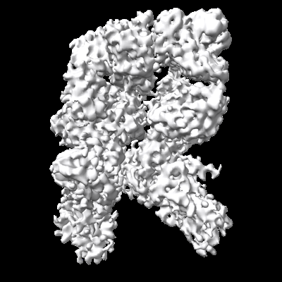 emd_40007.png emd_40007.png | 133 KB | ||
| Filedesc metadata | emd-40007.cif.gz | 4.7 KB | ||
| Others | emd_40007_half_map_1.map.gzemd_40007_half_map_2.map.gz | 255 MB 255 MB | ||
| Archive directory |  http://ftp.pdbj.org/pub/emdb/structures/EMD-40007ftp://ftp.pdbj.org/pub/emdb/structures/EMD-40007 http://ftp.pdbj.org/pub/emdb/structures/EMD-40007ftp://ftp.pdbj.org/pub/emdb/structures/EMD-40007 | HTTPS FTP |
-Related structure data


-Links
| EMDB pages | EMDB (EBI/PDBe) / EMDataResource |
|---|
-Map
| File | Download / File: emd_40007.map.gz / Format: CCP4 / Size: 274.6 MB / Type: IMAGE STORED AS FLOATING POINT NUMBER (4 BYTES) | ||||||||||||||||||||||||||||||||||||
|---|---|---|---|---|---|---|---|---|---|---|---|---|---|---|---|---|---|---|---|---|---|---|---|---|---|---|---|---|---|---|---|---|---|---|---|---|---|
| Annotation | Local refinement map of SARS-CoV-2 spike glycoprotein bound to antibody 002-S21B10. Used in construction of composite map EMD-29950. | ||||||||||||||||||||||||||||||||||||
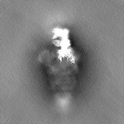
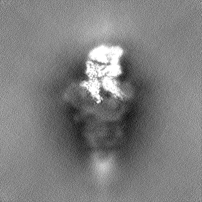
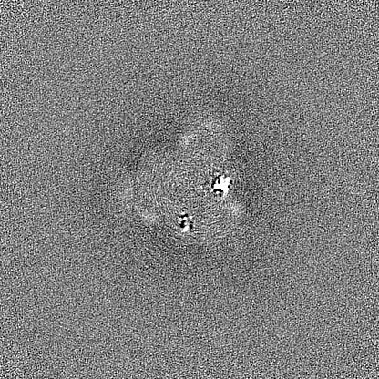
| Projections & slices | Image control
Images are generated by Spider. | ||||||||||||||||||||||||||||||||||||
| Voxel size | X=Y=Z: 1.0582 Å | ||||||||||||||||||||||||||||||||||||
| Density |
| ||||||||||||||||||||||||||||||||||||
| Symmetry | Space group: 1 | ||||||||||||||||||||||||||||||||||||
| Details | EMDB XML:
|
 Z (Sec.)
 Y (Row.)
 X (Col.)


-Supplemental data
-Half map: Half map of local refinement map of SARS-CoV-2...
| File | emd_40007_half_map_1.map | ||||||||||||
|---|---|---|---|---|---|---|---|---|---|---|---|---|---|
| Annotation | Half map of local refinement map of SARS-CoV-2 spike glycoprotein bound to antibody 002-S21B10. | ||||||||||||
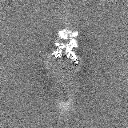
| Projections & Slices |
| ||||||||||||
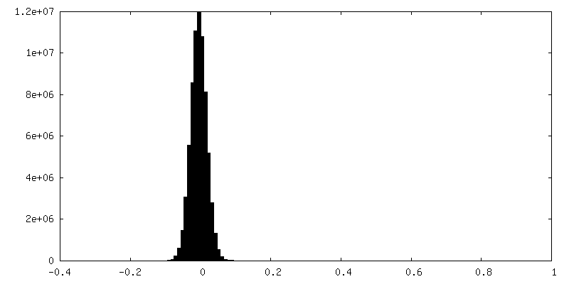
| Density Histograms |


-Half map: Half map of local refinement map of SARS-CoV-2...
| File | emd_40007_half_map_2.map | ||||||||||||
|---|---|---|---|---|---|---|---|---|---|---|---|---|---|
| Annotation | Half map of local refinement map of SARS-CoV-2 spike glycoprotein bound to antibody 002-S21B10. | ||||||||||||
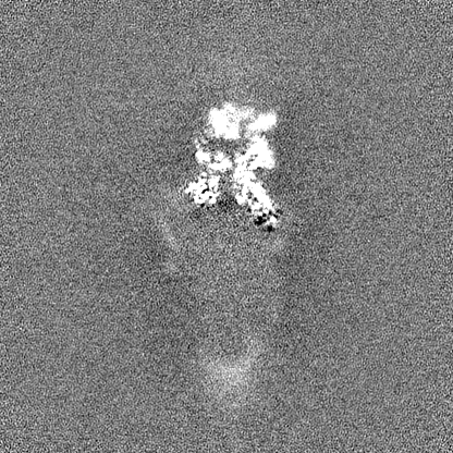
| Projections & Slices |
| ||||||||||||
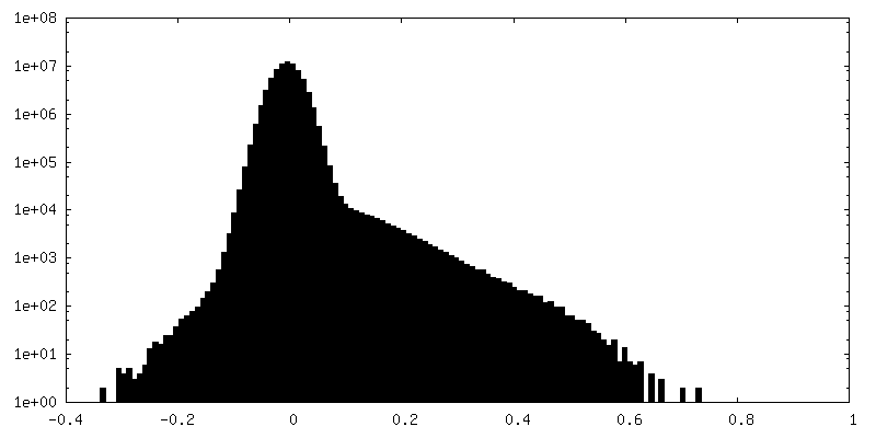
| Density Histograms |


- Sample components
-Entire : Antibody 002-S21B10 bound to spike glycoprotein trimer
| Entire | Name: Antibody 002-S21B10 bound to spike glycoprotein trimer |
|---|---|
| Components |
|
-Supramolecule #1: Antibody 002-S21B10 bound to spike glycoprotein trimer
| Supramolecule | Name: Antibody 002-S21B10 bound to spike glycoprotein trimer type: complex / ID: 1 / Parent: 0 / Macromolecule list: #1-#3 |
|---|
-Supramolecule #2: Antibody 002-S21B10
| Supramolecule | Name: Antibody 002-S21B10 / type: complex / ID: 2 / Parent: 1 / Macromolecule list: #2-#3 |
|---|---|
| Source (natural) | Organism: Homo sapiens (human) |
-Supramolecule #3: Spike glycoprotein trimer
| Supramolecule | Name: Spike glycoprotein trimer / type: complex / ID: 3 / Parent: 1 / Macromolecule list: #1 |
|---|---|
| Source (natural) | Organism: Severe acute respiratory syndrome coronavirus 2 |
-Experimental details
-Structure determination
| Method | cryo EM |
|---|---|
 Processing Processing | single particle reconstruction |
| Aggregation state | particle |
-Sample preparation
| Concentration | 0.7 mg/mL | |||||||||||||||
|---|---|---|---|---|---|---|---|---|---|---|---|---|---|---|---|---|
| Buffer | pH: 7.4 Component:
| |||||||||||||||
| Grid | Model: C-flat-1.2/1.3 / Material: COPPER / Mesh: 400 / Support film - Material: CARBON / Support film - topology: HOLEY | |||||||||||||||
| Vitrification | Cryogen name: ETHANE / Chamber humidity: 100 % / Chamber temperature: 295 K / Instrument: FEI VITROBOT MARK IV |
- Electron microscopy
| Microscope | TFS KRIOS |
|---|---|
| Specialist optics | Energy filter - Slit width: 20 eV |
| Image recording | Film or detector model: GATAN K3 (6k x 4k) / Number grids imaged: 1 / Number real images: 9054 / Average electron dose: 54.37 e/Å2 |
| Electron beam | Acceleration voltage: 300 kV / Electron source:  FIELD EMISSION GUN FIELD EMISSION GUN |
| Electron optics | Illumination mode: FLOOD BEAM / Imaging mode: BRIGHT FIELD / Cs: 2.7 mm / Nominal defocus max: 2.4 µm / Nominal defocus min: 1.0 µm / Nominal magnification: 81000 |
| Sample stage | Cooling holder cryogen: NITROGEN |
| Experimental equipment |  Model: Titan Krios / Image courtesy: FEI Company |
+Image processing

-Atomic model buiding 1
| Refinement | Space: REAL / Protocol: RIGID BODY FIT / Overall B value: 100.25 / Target criteria: Correlation coefficient |
|---|
