Movie
Movie Controller


+ Open data

- Basic information
| Entry |  | |||||||||
|---|---|---|---|---|---|---|---|---|---|---|
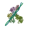
| Title | Human left ventricle ATM complex | |||||||||
 Map data Map data | EM map | |||||||||
 Sample Sample |
| |||||||||
 Keywords Keywords | protein fibril / protein complex | |||||||||
| Function / homology |  Function and homology information Function and homology informationregulation of slow-twitch skeletal muscle fiber contraction / regulation of the force of skeletal muscle contraction / positive regulation of heart rate by epinephrine / muscle thin filament tropomyosin / actin filament-based movement / bleb / muscle myosin complex / actin-myosin filament sliding / regulation of muscle contraction / cardiac myofibril assembly ...regulation of slow-twitch skeletal muscle fiber contraction / regulation of the force of skeletal muscle contraction / positive regulation of heart rate by epinephrine / muscle thin filament tropomyosin / actin filament-based movement / bleb / muscle myosin complex / actin-myosin filament sliding / regulation of muscle contraction / cardiac myofibril assembly / Formation of the dystrophin-glycoprotein complex (DGC) / regulation of the force of heart contraction / transition between fast and slow fiber / myosin filament / ruffle organization / adult heart development / cardiac muscle tissue morphogenesis / actomyosin structure organization / Striated Muscle Contraction / cardiac muscle hypertrophy in response to stress / muscle filament sliding / myosin complex / myosin II complex / I band / structural constituent of muscle / sarcomere organization / RHOB GTPase cycle / ventricular cardiac muscle tissue morphogenesis / microfilament motor activity / heart contraction / regulation of heart contraction / myosin binding / negative regulation of vascular associated smooth muscle cell migration / myofibril / mesenchyme migration / negative regulation of vascular associated smooth muscle cell proliferation / skeletal muscle thin filament assembly / RHOA GTPase cycle / Smooth Muscle Contraction / skeletal muscle contraction / striated muscle contraction / ATP metabolic process / cytoskeletal protein binding / stress fiber / cardiac muscle contraction / positive regulation of stress fiber assembly / cytoskeleton organization / positive regulation of cell adhesion / muscle contraction / regulation of heart rate / actin filament organization / negative regulation of cell migration / sarcomere / cellular response to reactive oxygen species / actin filament / filopodium / wound healing / structural constituent of cytoskeleton / Hydrolases; Acting on acid anhydrides; Acting on acid anhydrides to facilitate cellular and subcellular movement / ruffle membrane / Z disc / actin filament binding / actin cytoskeleton / lamellipodium / regulation of cell shape / actin binding / cell body / blood microparticle / response to ethanol / cytoskeleton / calmodulin binding / hydrolase activity / response to xenobiotic stimulus / protein heterodimerization activity / focal adhesion / positive regulation of gene expression / negative regulation of apoptotic process / glutamatergic synapse / protein homodimerization activity / extracellular space / extracellular exosome / ATP binding / identical protein binding / membrane / cytosol / cytoplasm Similarity search - Function | |||||||||
| Biological species |  Homo sapiens (human) Homo sapiens (human) | |||||||||
| Method | helical reconstruction / cryo EM / Resolution: 3.19 Å | |||||||||
 Authors Authors | Li DN / Zhao QY / Liu C | |||||||||
| Funding support | 1 items
| |||||||||
 Citation Citation | Journal: To Be Published Title: Cryo-EM structure of human left ventricle ATM complex Authors: Li DN / Zhao QY / Liu C | |||||||||
| History |
|
- Structure visualization
| Supplemental images |
|---|

- Downloads & links
-EMDB archive
| Map data | emd_39896.map.gz | 43.2 MB | EMDB map data format | |
|---|---|---|---|---|
| Header (meta data) | emd-39896-v30.xmlemd-39896.xml | 15.4 KB 15.4 KB | Display Display | EMDB header |
| FSC (resolution estimation) | emd_39896_fsc.xml | 17 KB | Display | FSC data file |
| Images |  emd_39896.png emd_39896.png | 116.4 KB | ||
| Filedesc metadata | emd-39896.cif.gz | 5.7 KB | ||
| Others | emd_39896_half_map_1.map.gzemd_39896_half_map_2.map.gz | 337.3 MB 337.3 MB | ||
| Archive directory |  http://ftp.pdbj.org/pub/emdb/structures/EMD-39896ftp://ftp.pdbj.org/pub/emdb/structures/EMD-39896 http://ftp.pdbj.org/pub/emdb/structures/EMD-39896ftp://ftp.pdbj.org/pub/emdb/structures/EMD-39896 | HTTPS FTP |
-Validation report
| Summary document | emd_39896_validation.pdf.gz | 932.8 KB | Display | EMDB validaton report |
|---|---|---|---|---|
| Full document | emd_39896_full_validation.pdf.gz | 932.3 KB | Display | |
| Data in XML | emd_39896_validation.xml.gz | 24.1 KB | Display | |
| Data in CIF | emd_39896_validation.cif.gz | 31.6 KB | Display | |
| Arichive directory | https://ftp.pdbj.org/pub/emdb/validation_reports/EMD-39896ftp://ftp.pdbj.org/pub/emdb/validation_reports/EMD-39896 | HTTPS FTP |
-Related structure data
| Related structure data |  8zb7MC M: atomic model generated by this map C: citing same article ( |
|---|---|
| Similar structure data |
-Links
| EMDB pages | EMDB (EBI/PDBe) / EMDataResource |
|---|---|
| Related items in Molecule of the Month |


-Map
| File | Download / File: emd_39896.map.gz / Format: CCP4 / Size: 421.9 MB / Type: IMAGE STORED AS FLOATING POINT NUMBER (4 BYTES) | ||||||||||||||||||||||||||||||||||||
|---|---|---|---|---|---|---|---|---|---|---|---|---|---|---|---|---|---|---|---|---|---|---|---|---|---|---|---|---|---|---|---|---|---|---|---|---|---|
| Annotation | EM map | ||||||||||||||||||||||||||||||||||||
| Projections & slices | Image control
Images are generated by Spider. | ||||||||||||||||||||||||||||||||||||
| Voxel size | X=Y=Z: 0.83 Å | ||||||||||||||||||||||||||||||||||||
| Density |
| ||||||||||||||||||||||||||||||||||||
| Symmetry | Space group: 1 | ||||||||||||||||||||||||||||||||||||
| Details | EMDB XML:
|
 Z (Sec.)
 Y (Row.)
 X (Col.)


-Supplemental data
-Half map: #2
| File | emd_39896_half_map_1.map | ||||||||||||
|---|---|---|---|---|---|---|---|---|---|---|---|---|---|
| Projections & Slices |
| ||||||||||||
| Density Histograms |


-Half map: #1
| File | emd_39896_half_map_2.map | ||||||||||||
|---|---|---|---|---|---|---|---|---|---|---|---|---|---|
| Projections & Slices |
| ||||||||||||
| Density Histograms |


- Sample components
-Entire : Human left ventricle ATM complex
| Entire | Name: Human left ventricle ATM complex |
|---|---|
| Components |
|
-Supramolecule #1: Human left ventricle ATM complex
| Supramolecule | Name: Human left ventricle ATM complex / type: complex / ID: 1 / Parent: 0 / Macromolecule list: #1-#3 |
|---|---|
| Source (natural) | Organism: Homo sapiens (human) |
-Macromolecule #1: Myosin-7
| Macromolecule | Name: Myosin-7 / type: protein_or_peptide / ID: 1 / Number of copies: 6 / Enantiomer: LEVO |
|---|---|
| Source (natural) | Organism: Homo sapiens (human) |
| Molecular weight | Theoretical: 88.41218 KDa |
| Sequence | String: MAVFGAAAPY LRKSEKERLE AQTRPFDLKK DVFVPDDKQE FVKAKIVSRE GGKVTAETEY GKTVTVKEDQ VMQQNPPKFD KIEDMAMLT FLHEPAVLYN LKDRYGSWMI YTYSGLFCVT VNPYKWLPVY TPEVVAAYRG KKRSEAPPHI FSISDNAYQY M LTDRENQS ...String: MAVFGAAAPY LRKSEKERLE AQTRPFDLKK DVFVPDDKQE FVKAKIVSRE GGKVTAETEY GKTVTVKEDQ VMQQNPPKFD KIEDMAMLT FLHEPAVLYN LKDRYGSWMI YTYSGLFCVT VNPYKWLPVY TPEVVAAYRG KKRSEAPPHI FSISDNAYQY M LTDRENQS ILITGESGAG KTVNTKRVIQ YFAVIAAIGD RSKKDQSPGK GTLEDQIIQA NPALEAFGNA KTVRNDNSSR FG KFIRIHF GATGKLASAD IETYLLEKSR VIFQLKAERD YHIFYQILSN KKPELLDMLL ITNNPYDYAF ISQGETTVAS IDD AEELMA TDNAFDVLGF TSEEKNSMYK LTGAIMHFGN MKFKLKQREE QAEPDGTEEA DKSAYLMGLN SADLLKGLCH PRVK VGNEY VTKGQNVQQV IYATGALAKA VYERMFNWMV TRINATLETK QPRQYFIGVL DIAGFEIFDF NSFEQLCINF TNEKL QQFF NHHMFVLEQE EYKKEGIEWT FIDFGMDLQA CIDLIEKPMG IMSILEEECM FPKATDMTFK AKLFDNHLGK SANFQK PRN IKGKPEAHFS LIHYAGIVDY NIIGWLQKNK DPLNETVVGL YQKSSLKLLS TLFANYAGAD APIEKGKGKA KKGSSFQ TV SALHRENLNK LMTNLRSTHP HFVRCIIPNE TKSPGVMDNP LVMHQLRCNG VLEGIRICRK GFPNRILYGD FRQRYRIL N PAAIPEGQFI DSRKGAEKLL SSLDIDHNQY KFGHTKVFFK AGLLGLLEEM RDERL UniProtKB: Myosin-7 |
-Macromolecule #2: Actin, alpha cardiac muscle 1
| Macromolecule | Name: Actin, alpha cardiac muscle 1 / type: protein_or_peptide / ID: 2 / Number of copies: 6 / Enantiomer: LEVO EC number: Hydrolases; Acting on acid anhydrides; Acting on acid anhydrides to facilitate cellular and subcellular movement |
|---|---|
| Source (natural) | Organism: Homo sapiens (human) |
| Molecular weight | Theoretical: 41.342145 KDa |
| Sequence | String: TTALVCDNGS GLVKAGFAGD DAPRAVFPSI VGRPRHQGVM VGMGQKDSYV GDEAQSKRGI LTLKYPIEHG IITNWDDMEK IWHHTFYNE LRVAPEEHPT LLTEAPLNPK ANREKMTQIM FETFNVPAMY VAIQAVLSLY ASGRTTGIVL DSGDGVTHNV P IYEGYALP ...String: TTALVCDNGS GLVKAGFAGD DAPRAVFPSI VGRPRHQGVM VGMGQKDSYV GDEAQSKRGI LTLKYPIEHG IITNWDDMEK IWHHTFYNE LRVAPEEHPT LLTEAPLNPK ANREKMTQIM FETFNVPAMY VAIQAVLSLY ASGRTTGIVL DSGDGVTHNV P IYEGYALP HAIMRLDLAG RDLTDYLMKI LTERGYSFVT TAEREIVRDI KEKLCYVALD FENEMATAAS SSSLEKSYEL PD GQVITIG NERFRCPETL FQPSFIGMES AGIHETTYNS IMKCDIDIRK DLYANNVLSG GTTMYPGIAD RMQKEITALA PST MKIKII APPERKYSVW IGGSILASLS TFQQMWISKQ EYDEAGPSIV HRKCF UniProtKB: Actin, alpha cardiac muscle 1 |
-Macromolecule #3: Tropomyosin alpha-1 chain
| Macromolecule | Name: Tropomyosin alpha-1 chain / type: protein_or_peptide / ID: 3 / Number of copies: 4 / Enantiomer: LEVO |
|---|---|
| Source (natural) | Organism: Homo sapiens (human) |
| Molecular weight | Theoretical: 19.000168 KDa |
| Sequence | String: SLQKKLKGTE DELDKYSEAL KDAQEKLELA EKKATDAEAD VASLNRRIQL VEEELDRAQE RLATALQKLE EAEKAADESE RGMKVIESR AQKDEEKMEI QEIQLKEAKH IAEDADRKYE EVARKLVIIE SDLERAEERA ELSEGKCAEL EEELKTVTNN L KSLEAQ UniProtKB: Tropomyosin alpha-1 chain |
-Macromolecule #4: ADENOSINE-5'-DIPHOSPHATE
| Macromolecule | Name: ADENOSINE-5'-DIPHOSPHATE / type: ligand / ID: 4 / Number of copies: 6 / Formula: ADP |
|---|---|
| Molecular weight | Theoretical: 427.201 Da |
| Chemical component information |  ChemComp-ADP: |
-Experimental details
-Structure determination
| Method | cryo EM |
|---|---|
 Processing Processing | helical reconstruction |
| Aggregation state | filament |
-Sample preparation
| Buffer | pH: 8 |
|---|---|
| Vitrification | Cryogen name: ETHANE |
- Electron microscopy
| Microscope | FEI TITAN KRIOS |
|---|---|
| Image recording | Film or detector model: GATAN K3 BIOCONTINUUM (6k x 4k) / Average electron dose: 55.0 e/Å2 |
| Electron beam | Acceleration voltage: 300 kV / Electron source:  FIELD EMISSION GUN FIELD EMISSION GUN |
| Electron optics | Illumination mode: FLOOD BEAM / Imaging mode: BRIGHT FIELD / Nominal defocus max: 2.0 µm / Nominal defocus min: 1.0 µm |
| Experimental equipment |  Model: Titan Krios / Image courtesy: FEI Company |
-Image processing
| Final reconstruction | Applied symmetry - Helical parameters - Δz: 27.3 Å Applied symmetry - Helical parameters - Δ&Phi: -166.66 ° Applied symmetry - Helical parameters - Axial symmetry: C1 (asymmetric) Resolution.type: BY AUTHOR / Resolution: 3.19 Å / Resolution method: FSC 0.143 CUT-OFF / Number images used: 69013 |
|---|---|
| Startup model | Type of model: PDB ENTRY PDB model - PDB ID: |
| Final angle assignment | Type: NOT APPLICABLE |
| FSC plot (resolution estimation) |  |