+ Open data
- Basic information
| Entry |  | |||||||||
|---|---|---|---|---|---|---|---|---|---|---|
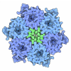
| Title | The structure of type III CRISPR-associated deaminase apo form | |||||||||
 Map data Map data | ||||||||||
 Sample Sample |
| |||||||||
 Keywords Keywords | defense system / deaminase / IMMUNE SYSTEM | |||||||||
| Function / homology |  Function and homology information Function and homology informationinosine biosynthetic process / adenosine deaminase / hypoxanthine salvage / adenosine deaminase activity / adenosine catabolic process / cytosol Similarity search - Function | |||||||||
| Biological species |  Limisphaera ngatamarikiensis (bacteria) Limisphaera ngatamarikiensis (bacteria) | |||||||||
| Method | single particle reconstruction / cryo EM / Resolution: 3.26 Å | |||||||||
 Authors Authors | Chen MR / Li ZX / Xiao YB | |||||||||
| Funding support |  China, 1 items China, 1 items
| |||||||||
 Citation Citation |  Journal: Science / Year: 2025 Journal: Science / Year: 2025Title: Antiviral signaling of a type III CRISPR-associated deaminase. Authors: Yutao Li / Zhaoxing Li / Purui Yan / Chenyang Hua / Jianping Kong / Wanqian Wu / Yurong Cui / Yan Duan / Shunxiang Li / Guanglei Li / Shunli Ji / Yijun Chen / Yucheng Zhao / Peng Yang / ...Authors: Yutao Li / Zhaoxing Li / Purui Yan / Chenyang Hua / Jianping Kong / Wanqian Wu / Yurong Cui / Yan Duan / Shunxiang Li / Guanglei Li / Shunli Ji / Yijun Chen / Yucheng Zhao / Peng Yang / Chunyi Hu / Meiling Lu / Meirong Chen / Yibei Xiao /   Abstract: Prokaryotes have evolved diverse defense strategies against viral infection, including foreign nucleic acid degradation by CRISPR-Cas systems and DNA and RNA synthesis inhibition through nucleotide ...Prokaryotes have evolved diverse defense strategies against viral infection, including foreign nucleic acid degradation by CRISPR-Cas systems and DNA and RNA synthesis inhibition through nucleotide pool depletion. Here, we report an antiviral mechanism of type III CRISPR-Cas-regulated adenosine triphosphate (ATP) depletion in which ATP is converted into inosine triphosphate (ITP) by CRISPR-Cas-associated adenosine deaminase (CAAD) upon activation by either cA or cA, followed by hydrolysis into inosine monophosphate (IMP) by Nudix hydrolase, ultimately resulting in cell growth arrest. The cryo-electron microscopy structures of CAAD in its apo and activated forms, together with biochemical evidence, revealed how cA or cA binds to the CRISPR-associated Rossmann fold (CARF) domain and abrogates CAAD autoinhibition, inducing substantial conformational changes that reshape the structure of CAAD and induce its deaminase activity. Our results reveal the mechanism of a CRISPR-Cas-regulated ATP depletion antiviral strategy. | |||||||||
| History |
|
- Structure visualization
| Supplemental images |
|---|
- Downloads & links
-EMDB archive
| Map data |  emd_39759.map.gz emd_39759.map.gz | 108 MB |  EMDB map data format EMDB map data format | |
|---|---|---|---|---|
| Header (meta data) |  emd-39759-v30.xml emd-39759-v30.xml emd-39759.xml emd-39759.xml | 18.3 KB 18.3 KB | Display Display |  EMDB header EMDB header |
| FSC (resolution estimation) |  emd_39759_fsc.xml emd_39759_fsc.xml | 12.8 KB | Display |  FSC data file FSC data file |
| Images |  emd_39759.png emd_39759.png | 37.8 KB | ||
| Filedesc metadata |  emd-39759.cif.gz emd-39759.cif.gz | 6.3 KB | ||
| Others |  emd_39759_half_map_1.map.gz emd_39759_half_map_1.map.gz emd_39759_half_map_2.map.gz emd_39759_half_map_2.map.gz | 200.3 MB 200.3 MB | ||
| Archive directory |  http://ftp.pdbj.org/pub/emdb/structures/EMD-39759 http://ftp.pdbj.org/pub/emdb/structures/EMD-39759 ftp://ftp.pdbj.org/pub/emdb/structures/EMD-39759 ftp://ftp.pdbj.org/pub/emdb/structures/EMD-39759 | HTTPS FTP |
-Related structure data
| Related structure data | 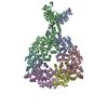 8z40MC 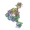 8z3kC 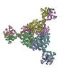 8z3pC 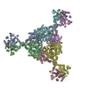 8z3rC M: atomic model generated by this map C: citing same article ( |
|---|---|
| Similar structure data | Similarity search - Function & homology  F&H Search F&H Search |
- Links
Links
| EMDB pages |  EMDB (EBI/PDBe) / EMDB (EBI/PDBe) /  EMDataResource EMDataResource |
|---|---|
| Related items in Molecule of the Month |
- Map
Map
| File |  Download / File: emd_39759.map.gz / Format: CCP4 / Size: 216 MB / Type: IMAGE STORED AS FLOATING POINT NUMBER (4 BYTES) Download / File: emd_39759.map.gz / Format: CCP4 / Size: 216 MB / Type: IMAGE STORED AS FLOATING POINT NUMBER (4 BYTES) | ||||||||||||||||||||||||||||||||||||
|---|---|---|---|---|---|---|---|---|---|---|---|---|---|---|---|---|---|---|---|---|---|---|---|---|---|---|---|---|---|---|---|---|---|---|---|---|---|
| Projections & slices | Image control
Images are generated by Spider. | ||||||||||||||||||||||||||||||||||||
| Voxel size | X=Y=Z: 0.92 Å | ||||||||||||||||||||||||||||||||||||
| Density |
| ||||||||||||||||||||||||||||||||||||
| Symmetry | Space group: 1 | ||||||||||||||||||||||||||||||||||||
| Details | EMDB XML:
|
-Supplemental data
-Half map: #1
| File | emd_39759_half_map_1.map | ||||||||||||
|---|---|---|---|---|---|---|---|---|---|---|---|---|---|
| Projections & Slices |
| ||||||||||||
| Density Histograms |
-Half map: #2
| File | emd_39759_half_map_2.map | ||||||||||||
|---|---|---|---|---|---|---|---|---|---|---|---|---|---|
| Projections & Slices |
| ||||||||||||
| Density Histograms |
- Sample components
-Entire : CRISPR-associated adenosine deaminase
| Entire | Name: CRISPR-associated adenosine deaminase |
|---|---|
| Components |
|
-Supramolecule #1: CRISPR-associated adenosine deaminase
| Supramolecule | Name: CRISPR-associated adenosine deaminase / type: complex / ID: 1 / Parent: 0 / Macromolecule list: all |
|---|---|
| Source (natural) | Organism:  Limisphaera ngatamarikiensis (bacteria) Limisphaera ngatamarikiensis (bacteria) |
-Macromolecule #1: Adenosine deaminase domain-containing protein
| Macromolecule | Name: Adenosine deaminase domain-containing protein / type: protein_or_peptide / ID: 1 / Number of copies: 6 / Enantiomer: LEVO |
|---|---|
| Source (natural) | Organism:  Limisphaera ngatamarikiensis (bacteria) Limisphaera ngatamarikiensis (bacteria) |
| Molecular weight | Theoretical: 70.859859 KDa |
| Recombinant expression | Organism:  |
| Sequence | String: MNVQAHLFVS LGTAPAIVPE AFLLPGARFV SVHVLTTERP DVTLIREFFR RHAPGVNLTI TRVAGFQDLK SEEDHFRFEE VMFRWFLAS RTGPEQRFVC LTGGFKTMSA AMQKAATVLG AAEVFHVLAD DCCVGPQGRL MPPSTLEEIL WARDQGHLHW I RLGPERGW ...String: MNVQAHLFVS LGTAPAIVPE AFLLPGARFV SVHVLTTERP DVTLIREFFR RHAPGVNLTI TRVAGFQDLK SEEDHFRFEE VMFRWFLAS RTGPEQRFVC LTGGFKTMSA AMQKAATVLG AAEVFHVLAD DCCVGPQGRL MPPSTLEEIL WARDQGHLHW I RLGPERGW PQLRRIAPEQ FPLQVVEEKG DERRVQAEDR AFGTFLQDLL QRASRIAGAW EMLPELPFAD LATWSEGELA WL REPLDPR APADQRWVAG LPKIELHCHL GGFATHGELL RRVRNAAENP GKLPPLEEPR LPEGWPLPAQ PIPLAEYMKL GNA NGTALL RDPGCLREQC RLLYRHLVDQ GVCYAEVRCS PANYAEVRSP WDVLADIRAA FQECMEGART APGGLPACHV NLIL IATRR ASGDYRAAIA RHLALAVTAA EHWRDENACR VVGVDLAGYE DEKTRAHYFR EEFTAVHRCG LAVTVHAGEN DDAEG IWRA VFDLNARRLG HALSLGQSRE LLRSVADRGI GVELCPYANL QIKGFRLDGS DRAGPADPRH EAHAPGPYPL LDYLRE GVR VTVNTDNIGI SAASLTDNLL LAARLCPGLT RLDLLHLQRH ALETAFCTAT QRLTLLRRIS SGIPRPHHHH HH UniProtKB: adenosine deaminase |
-Experimental details
-Structure determination
| Method | cryo EM |
|---|---|
 Processing Processing | single particle reconstruction |
| Aggregation state | particle |
- Sample preparation
| Buffer | pH: 7.5 |
|---|---|
| Vitrification | Cryogen name: ETHANE |
- Electron microscopy
| Microscope | FEI TITAN KRIOS |
|---|---|
| Image recording | Film or detector model: FEI FALCON IV (4k x 4k) / Average electron dose: 40.0 e/Å2 |
| Electron beam | Acceleration voltage: 300 kV / Electron source:  FIELD EMISSION GUN FIELD EMISSION GUN |
| Electron optics | Illumination mode: FLOOD BEAM / Imaging mode: BRIGHT FIELD / Nominal defocus max: 2.4 µm / Nominal defocus min: 0.8 µm |
| Experimental equipment |  Model: Titan Krios / Image courtesy: FEI Company |
 Movie
Movie Controller


 Z (Sec.)
 Y (Row.)
 X (Col.)


