Movie
Movie Controller


+ Open data

- Basic information
| Entry |  | |||||||||||||||
|---|---|---|---|---|---|---|---|---|---|---|---|---|---|---|---|---|
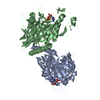
| Title | Kinesin-14 in nucleotide-free state bound to 13 PF Microtubule | |||||||||||||||
 Map data Map data | ||||||||||||||||
 Sample Sample |
| |||||||||||||||
 Keywords Keywords | Kinesin Motor Proteins / Force Production / Power Stroke Fluctuations / Motor Spring-like Element / Reversed Motility / Mechanochemical Coupling / Mechanical States / CELL CYCLE | |||||||||||||||
| Function / homology |  Function and homology information Function and homology informationminus-end directed microtubule sliding / distributive segregation / regulation of mitotic spindle elongation / meiotic spindle assembly / Regulation of PLK1 Activity at G2/M Transition / Loss of Nlp from mitotic centrosomes / Loss of proteins required for interphase microtubule organization from the centrosome / Anchoring of the basal body to the plasma membrane / AURKA Activation by TPX2 / Recruitment of mitotic centrosome proteins and complexes ...minus-end directed microtubule sliding / distributive segregation / regulation of mitotic spindle elongation / meiotic spindle assembly / Regulation of PLK1 Activity at G2/M Transition / Loss of Nlp from mitotic centrosomes / Loss of proteins required for interphase microtubule organization from the centrosome / Anchoring of the basal body to the plasma membrane / AURKA Activation by TPX2 / Recruitment of mitotic centrosome proteins and complexes / mitotic spindle elongation / odontoblast differentiation / mitotic spindle microtubule / meiotic spindle organization / mitotic centrosome separation / Neutrophil degranulation / microtubule bundle formation / regulation of mitotic spindle assembly / Microtubule-dependent trafficking of connexons from Golgi to the plasma membrane / Resolution of Sister Chromatid Cohesion / Hedgehog 'off' state / Cilium Assembly / Intraflagellar transport / COPI-dependent Golgi-to-ER retrograde traffic / Mitotic Prometaphase / Carboxyterminal post-translational modifications of tubulin / RHOH GTPase cycle / EML4 and NUDC in mitotic spindle formation / Sealing of the nuclear envelope (NE) by ESCRT-III / Kinesins / PKR-mediated signaling / Separation of Sister Chromatids / The role of GTSE1 in G2/M progression after G2 checkpoint / Aggrephagy / meiotic spindle / spindle assembly involved in female meiosis / RHO GTPases activate IQGAPs / RHO GTPases Activate Formins / HSP90 chaperone cycle for steroid hormone receptors (SHR) in the presence of ligand / minus-end-directed microtubule motor activity / spindle organization / MHC class II antigen presentation / Recruitment of NuMA to mitotic centrosomes / COPI-mediated anterograde transport / nuclear envelope lumen / regulation of synapse organization / MHC class I protein binding / mitotic spindle assembly / mRNA transport / intercellular bridge / spindle assembly / mitotic spindle organization / chromosome segregation / microtubule cytoskeleton organization / structural constituent of cytoskeleton / Hydrolases; Acting on acid anhydrides; Acting on acid anhydrides to facilitate cellular and subcellular movement / spindle / cytoplasmic ribonucleoprotein granule / mitotic spindle / mitotic cell cycle / microtubule cytoskeleton / cell body / microtubule binding / microtubule / Hydrolases; Acting on acid anhydrides; Acting on GTP to facilitate cellular and subcellular movement / cell division / hydrolase activity / GTPase activity / centrosome / ubiquitin protein ligase binding / GTP binding / protein homodimerization activity / ATP binding / metal ion binding / nucleus / cytosol / cytoplasm Similarity search - Function | |||||||||||||||
| Biological species |   | |||||||||||||||
| Method | helical reconstruction / cryo EM / Resolution: 3.6 Å | |||||||||||||||
 Authors Authors | Shibata S / Imasaki T / Shigematsu H / Endow SA / Nitta R | |||||||||||||||
| Funding support |  Japan, 4 items Japan, 4 items
| |||||||||||||||
 Citation Citation | Journal: bioRxiv / Year: 2024 Title: Structural transitions in kinesin minus-end directed microtubule motility. Authors: Satoki Shibata / Matthew Y Wang / Tsuyoshi Imasaki / Hideki Shigematsu / Yuanyuan Wei / Chacko Jobichen / Hajime Hagio / J Sivaraman / Sharyn A Endow / Ryo Nitta /   Abstract: Kinesin motor proteins hydrolyze ATP to produce force for spindle assembly and vesicle transport, performing essential functions in cell division and motility, but the structural changes required for ...Kinesin motor proteins hydrolyze ATP to produce force for spindle assembly and vesicle transport, performing essential functions in cell division and motility, but the structural changes required for force generation are uncertain. We now report high-resolution structures showing new transitions in the kinesin mechanochemical cycle, including power stroke fluctuations upon ATP binding and a post-hydrolysis state with bound ADP + free phosphate. We find that rate-limiting ADP release occurs upon microtubule binding, accompanied by central β-sheet twisting, which triggers the power stroke - stalk rotation and neck mimic docking - upon ATP binding. Microtubule release occurs with β-strand-to-loop transitions, implying that β-strand refolding induces Pi release and the recovery stroke. The strained β-sheet during the power stroke and strand-to-loop transitions identify the β-sheet as the long-sought motor spring. | |||||||||||||||
| History |
|
- Structure visualization
| Supplemental images |
|---|

- Downloads & links
-EMDB archive
| Map data | emd_39664.map.gz | 773.4 MB | EMDB map data format | |
|---|---|---|---|---|
| Header (meta data) | emd-39664-v30.xmlemd-39664.xml | 28.4 KB 28.4 KB | Display Display | EMDB header |
| FSC (resolution estimation) | emd_39664_fsc.xml | 21.3 KB | Display | FSC data file |
| Images |  emd_39664.png emd_39664.png | 107.6 KB | ||
| Filedesc metadata | emd-39664.cif.gz | 8.1 KB | ||
| Others | emd_39664_half_map_1.map.gzemd_39664_half_map_2.map.gz | 667.9 MB 668.4 MB | ||
| Archive directory |  http://ftp.pdbj.org/pub/emdb/structures/EMD-39664ftp://ftp.pdbj.org/pub/emdb/structures/EMD-39664 http://ftp.pdbj.org/pub/emdb/structures/EMD-39664ftp://ftp.pdbj.org/pub/emdb/structures/EMD-39664 | HTTPS FTP |
-Related structure data
| Related structure data |  8yy2MC  8yueC  8yy3C  8yy4C  8yy5C M: atomic model generated by this map C: citing same article ( |
|---|---|
| Similar structure data |


-Links
| EMDB pages | EMDB (EBI/PDBe) / EMDataResource |
|---|---|
| Related items in Molecule of the Month |


-Map
| File | Download / File: emd_39664.map.gz / Format: CCP4 / Size: 824 MB / Type: IMAGE STORED AS FLOATING POINT NUMBER (4 BYTES) | ||||||||||||||||||||||||||||||||||||
|---|---|---|---|---|---|---|---|---|---|---|---|---|---|---|---|---|---|---|---|---|---|---|---|---|---|---|---|---|---|---|---|---|---|---|---|---|---|
| Projections & slices | Image control
Images are generated by Spider. | ||||||||||||||||||||||||||||||||||||
| Voxel size | X=Y=Z: 0.752 Å | ||||||||||||||||||||||||||||||||||||
| Density |
| ||||||||||||||||||||||||||||||||||||
| Symmetry | Space group: 1 | ||||||||||||||||||||||||||||||||||||
| Details | EMDB XML:
|
 Z (Sec.)
 Y (Row.)
 X (Col.)


-Supplemental data
-Half map: #1
| File | emd_39664_half_map_1.map | ||||||||||||
|---|---|---|---|---|---|---|---|---|---|---|---|---|---|
| Projections & Slices |
| ||||||||||||
| Density Histograms |


-Half map: #2
| File | emd_39664_half_map_2.map | ||||||||||||
|---|---|---|---|---|---|---|---|---|---|---|---|---|---|
| Projections & Slices |
| ||||||||||||
| Density Histograms |


- Sample components
+Entire : Kinesin-microtubule complex
+Supramolecule #1: Kinesin-microtubule complex
+Supramolecule #2: Tubulin
+Supramolecule #3: NCD
+Macromolecule #1: Tubulin alpha-1B chain
+Macromolecule #2: Tubulin beta chain
+Macromolecule #3: Protein claret segregational

+Macromolecule #4: GUANOSINE-5'-TRIPHOSPHATE

+Macromolecule #5: GUANOSINE-5'-DIPHOSPHATE

+Macromolecule #6: MAGNESIUM ION
+Macromolecule #7: ADENOSINE-5'-DIPHOSPHATE

-Experimental details
-Structure determination
| Method | cryo EM |
|---|---|
 Processing Processing | helical reconstruction |
| Aggregation state | filament |
-Sample preparation
| Buffer | pH: 6.8 Component:
Details: 100 mM PIPES pH 6.8, 1 mM MgCl2, 1 mM EGTA, and 1 mM GTP | |||||||||||||||
|---|---|---|---|---|---|---|---|---|---|---|---|---|---|---|---|---|
| Vitrification | Cryogen name: ETHANE / Chamber humidity: 90 % / Chamber temperature: 310 K / Instrument: FEI VITROBOT MARK IV |
- Electron microscopy
| Microscope | JEOL CRYO ARM 300 |
|---|---|
| Image recording | Film or detector model: GATAN K3 BIOQUANTUM (6k x 4k) / Average electron dose: 50.0 e/Å2 |
| Electron beam | Acceleration voltage: 300 kV / Electron source:  FIELD EMISSION GUN FIELD EMISSION GUN |
| Electron optics | Illumination mode: FLOOD BEAM / Imaging mode: BRIGHT FIELD / Nominal defocus max: 1.5 µm / Nominal defocus min: 0.8 µm |