Movie
Movie Controller

[English] 日本語
 Yorodumi
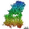
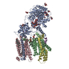
- EMDB-39574: Cryo-EM structure of human gamma-secretase in complex with Abeta43 -
+ Open data

- Basic information
| Entry |  | |||||||||
|---|---|---|---|---|---|---|---|---|---|---|
| Title | Cryo-EM structure of human gamma-secretase in complex with Abeta43 | |||||||||
 Map data Map data | ||||||||||
 Sample Sample |
| |||||||||
 Keywords Keywords | Intramembrane protease / gamma-secretase / MEMBRANE PROTEIN / MEMBRANE PROTEIN-HYDROLASE complex | |||||||||
| Biological species | synthetic construct (others) | |||||||||
| Method | single particle reconstruction / cryo EM / Resolution: 4.5 Å | |||||||||
 Authors Authors | Guo X / Zhou R / Shi Y | |||||||||
| Funding support |  China, 1 items China, 1 items
| |||||||||
 Citation Citation | Journal: Science / Year: 2024 Title: Molecular mechanism of substrate recognition and cleavage by human γ-secretase. Authors: Xuefei Guo / Haotian Li / Chuangye Yan / Jianlin Lei / Rui Zhou / Yigong Shi / Abstract: Successive cleavages of amyloid precursor protein C-terminal fragment with 99 residues (APP-C99) by γ-secretase result in amyloid-β (Aβ) peptides of varying lengths. Most cleavages have a step ...Successive cleavages of amyloid precursor protein C-terminal fragment with 99 residues (APP-C99) by γ-secretase result in amyloid-β (Aβ) peptides of varying lengths. Most cleavages have a step size of three residues. To elucidate the underlying mechanism, we determined the atomic structures of human γ-secretase bound individually to APP-C99, Aβ49, Aβ46, and Aβ43. In all cases, the substrate displays the same structural features: a transmembrane α-helix, a three-residue linker, and a β-strand that forms a hybrid β-sheet with presenilin 1 (PS1). Proteolytic cleavage occurs just ahead of the substrate β-strand. Each cleavage is followed by unwinding and translocation of the substrate α-helix by one turn and the formation of a new β-strand. This mechanism is consistent with existing biochemical data and may explain the cleavages of other substrates by γ-secretase. | |||||||||
| History |
|
- Structure visualization
| Supplemental images |
|---|

- Downloads & links
-EMDB archive
| Map data | emd_39574.map.gz | 25.4 MB |  EMDB map data format EMDB map data format | |
|---|---|---|---|---|
| Header (meta data) | emd-39574-v30.xmlemd-39574.xml | 12.5 KB 12.5 KB | Display Display | EMDB header |
| Images |  emd_39574.png emd_39574.png | 31.7 KB | ||
| Filedesc metadata | emd-39574.cif.gz | 4 KB | ||
| Others | emd_39574_half_map_1.map.gzemd_39574_half_map_2.map.gz | 24.8 MB 24.8 MB | ||
| Archive directory |  http://ftp.pdbj.org/pub/emdb/structures/EMD-39574ftp://ftp.pdbj.org/pub/emdb/structures/EMD-39574 http://ftp.pdbj.org/pub/emdb/structures/EMD-39574ftp://ftp.pdbj.org/pub/emdb/structures/EMD-39574 | HTTPS FTP |
-Validation report
| Summary document | emd_39574_validation.pdf.gz | 778.2 KB | Display | EMDB validaton report |
|---|---|---|---|---|
| Full document | emd_39574_full_validation.pdf.gz | 777.8 KB | Display | |
| Data in XML | emd_39574_validation.xml.gz | 10.5 KB | Display | |
| Data in CIF | emd_39574_validation.cif.gz | 12.2 KB | Display | |
| Arichive directory | https://ftp.pdbj.org/pub/emdb/validation_reports/EMD-39574ftp://ftp.pdbj.org/pub/emdb/validation_reports/EMD-39574 | HTTPS FTP |
-Related structure data


-Links
| EMDB pages | EMDB (EBI/PDBe) / EMDataResource |
|---|
-Map
| File | Download / File: emd_39574.map.gz / Format: CCP4 / Size: 27 MB / Type: IMAGE STORED AS FLOATING POINT NUMBER (4 BYTES) | ||||||||||||||||||||||||||||||||||||
|---|---|---|---|---|---|---|---|---|---|---|---|---|---|---|---|---|---|---|---|---|---|---|---|---|---|---|---|---|---|---|---|---|---|---|---|---|---|
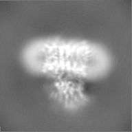
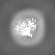
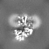
| Projections & slices | Image control
Images are generated by Spider. | ||||||||||||||||||||||||||||||||||||
| Voxel size | X=Y=Z: 1.0979 Å | ||||||||||||||||||||||||||||||||||||
| Density |
| ||||||||||||||||||||||||||||||||||||
| Symmetry | Space group: 1 | ||||||||||||||||||||||||||||||||||||
| Details | EMDB XML:
|
 Z (Sec.)
 Y (Row.)
 X (Col.)


-Supplemental data
-Half map: #1
| File | emd_39574_half_map_1.map | ||||||||||||
|---|---|---|---|---|---|---|---|---|---|---|---|---|---|
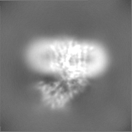
| Projections & Slices |
| ||||||||||||
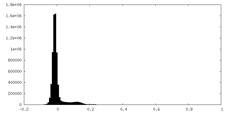
| Density Histograms |


-Half map: #2
| File | emd_39574_half_map_2.map | ||||||||||||
|---|---|---|---|---|---|---|---|---|---|---|---|---|---|
| Projections & Slices |
| ||||||||||||
| Density Histograms |


- Sample components
-Entire : Cryo-EM structure of human gamma-secretase in complex with Abeta43
| Entire | Name: Cryo-EM structure of human gamma-secretase in complex with Abeta43 |
|---|---|
| Components |
|
-Supramolecule #1: Cryo-EM structure of human gamma-secretase in complex with Abeta43
| Supramolecule | Name: Cryo-EM structure of human gamma-secretase in complex with Abeta43 type: complex / ID: 1 / Parent: 0 / Macromolecule list: all |
|---|---|
| Source (natural) | Organism: synthetic construct (others) |
-Macromolecule #1: Abeta43
| Macromolecule | Name: Abeta43 / type: other / ID: 1 / Classification: other |
|---|---|
| Source (natural) | Organism: synthetic construct (others) |
| Sequence | String: DAEFRHDSGY EVHHQKLVFF AEDVGSNKGA IIGLMVGGVV IAT |
-Experimental details
-Structure determination
| Method | cryo EM |
|---|---|
 Processing Processing | single particle reconstruction |
| Aggregation state | particle |
-Sample preparation
| Buffer | pH: 7.4 |
|---|---|
| Vitrification | Cryogen name: ETHANE |
- Electron microscopy
| Microscope | FEI TITAN KRIOS |
|---|---|
| Image recording | Film or detector model: GATAN K3 (6k x 4k) / Average electron dose: 50.0 e/Å2 |
| Electron beam | Acceleration voltage: 300 kV / Electron source:  FIELD EMISSION GUN FIELD EMISSION GUN |
| Electron optics | Illumination mode: FLOOD BEAM / Imaging mode: BRIGHT FIELD / Nominal defocus max: 1.8 µm / Nominal defocus min: 1.5 µm |
| Experimental equipment |  Model: Titan Krios / Image courtesy: FEI Company |
-Image processing
| Startup model | Type of model: EMDB MAP EMDB ID: |
|---|---|
| Final reconstruction | Resolution.type: BY AUTHOR / Resolution: 4.5 Å / Resolution method: FSC 0.5 CUT-OFF / Number images used: 578094 |
| Initial angle assignment | Type: MAXIMUM LIKELIHOOD |
| Final angle assignment | Type: MAXIMUM LIKELIHOOD |