 Movie
Movie Controller


+ Open data

- Basic information
| Entry |  | |||||||||
|---|---|---|---|---|---|---|---|---|---|---|
| Title | Iota toxin Ib pore serine-clamp mutant | |||||||||
 Map data Map data | ||||||||||
 Sample Sample |
| |||||||||
 Keywords Keywords | Bacterial binary toxin / Protein translocation channel / ADP-ribosylation / Toxin | |||||||||
| Function / homology |  Function and homology information Function and homology information | |||||||||
| Biological species |   Clostridium perfringens (bacteria) Clostridium perfringens (bacteria) | |||||||||
| Method | single particle reconstruction / cryo EM / Resolution: 2.36 Å | |||||||||
 Authors Authors | Ninomiya Y / Yoshida T / Yamada T / Kishikawa J / Tsuge H | |||||||||
| Funding support |  Japan, 1 items Japan, 1 items
| |||||||||
 Citation Citation | Journal: Commun Biol / Year: 2025 Title: Serine clamp of Clostridium perfringens binary toxin BECb (CPILEb)-pore confers cytotoxicity and enterotoxicity. Authors: Toru Yoshida / Chie Monma / Yuki Ninomiya / Sotaro Takiguchi / Shoko Fujita / Yuto Uchida / Noriaki Sakoda / Vladimir A Karginov / Jun-Ichi Kishikawa / Tomohito Yamada / Ryuji Kawano / Hideaki Tsuge /  Abstract: BEC (CPILE) is a virulence factor of the pathogen, Clostridium perfringens, which has caused foodborne outbreaks in Japan. BEC is a binary toxin that comprises the enzymatic A-component (BECa) and ...BEC (CPILE) is a virulence factor of the pathogen, Clostridium perfringens, which has caused foodborne outbreaks in Japan. BEC is a binary toxin that comprises the enzymatic A-component (BECa) and the B-component (BECb); the latter forms a membrane pore to translocate the A-component into target cells. Although BEC differs from other binary toxins in that the B-component alone shows enterotoxic activity, the reason for this remains unclear. We focus on the narrowest region of BECb-pore formed by not phenylalanine residues conserved in other binary toxins including iota toxin B-component (Ib) but serine residues. Comparisons between BECb and BECb (S405F) where the serine residue forming the narrowest region is substituted to the phenylalanine residue reveal that the serine residue is responsible for both cytotoxicity and enterotoxic activity. Though attempts to prepare the BECb-pore were unsuccessful, we reveal the cryo-EM structure of Ib (F454S) where the phenylalanine residue forming the narrowest region is substituted to the serine residue as a surrogate of BECb. Furthermore, Ib (F454S) increases current conductance to nine times that of Ib due to the larger pore diameter and the hydrophilic nature. These results suggest that BECb functions as a pore-forming toxin and as a translocation channel for BECa. | |||||||||
| History |
|
- Structure visualization
| Supplemental images |
|---|

- Downloads & links
-EMDB archive
| Map data | emd_39544.map.gz | 173.7 MB | EMDB map data format | |
|---|---|---|---|---|
| Header (meta data) | emd-39544-v30.xmlemd-39544.xml | 23.6 KB 23.6 KB | Display Display | EMDB header |
| FSC (resolution estimation) | emd_39544_fsc.xml | 14.8 KB | Display | FSC data file |
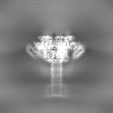
| Images |  emd_39544.png emd_39544.png | 158 KB | ||
| Filedesc metadata | emd-39544.cif.gz | 6.8 KB | ||
| Others | emd_39544_half_map_1.map.gzemd_39544_half_map_2.map.gz | 322.9 MB 322.9 MB | ||
| Archive directory |  http://ftp.pdbj.org/pub/emdb/structures/EMD-39544ftp://ftp.pdbj.org/pub/emdb/structures/EMD-39544 http://ftp.pdbj.org/pub/emdb/structures/EMD-39544ftp://ftp.pdbj.org/pub/emdb/structures/EMD-39544 | HTTPS FTP |
-Related structure data
| Related structure data |  8yrmMC M: atomic model generated by this map C: citing same article ( |
|---|---|
| Similar structure data |

-Links
| EMDB pages | EMDB (EBI/PDBe) / EMDataResource |
|---|---|
| Related items in Molecule of the Month |

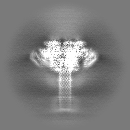
-Map
| File | Download / File: emd_39544.map.gz / Format: CCP4 / Size: 347.6 MB / Type: IMAGE STORED AS FLOATING POINT NUMBER (4 BYTES) | ||||||||||||||||||||||||||||||||||||
|---|---|---|---|---|---|---|---|---|---|---|---|---|---|---|---|---|---|---|---|---|---|---|---|---|---|---|---|---|---|---|---|---|---|---|---|---|---|
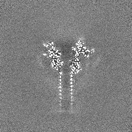
| Projections & slices | Image control
Images are generated by Spider. | ||||||||||||||||||||||||||||||||||||
| Voxel size | X=Y=Z: 0.675 Å | ||||||||||||||||||||||||||||||||||||
| Density |
| ||||||||||||||||||||||||||||||||||||
| Symmetry | Space group: 1 | ||||||||||||||||||||||||||||||||||||
| Details | EMDB XML:
|
 Z (Sec.)
 Y (Row.)
 X (Col.)


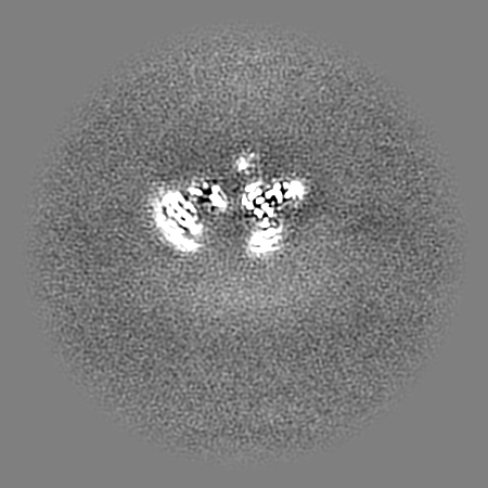


-Supplemental data
-Half map: #2
| File | emd_39544_half_map_1.map | ||||||||||||
|---|---|---|---|---|---|---|---|---|---|---|---|---|---|
| Projections & Slices |
| ||||||||||||
| Density Histograms |


-Half map: #1
| File | emd_39544_half_map_2.map | ||||||||||||
|---|---|---|---|---|---|---|---|---|---|---|---|---|---|
| Projections & Slices |
| ||||||||||||
| Density Histograms |


- Sample components
-Entire : Iota toxin Ib pore serine-clamp mutant
| Entire | Name: Iota toxin Ib pore serine-clamp mutant |
|---|---|
| Components |
|
-Supramolecule #1: Iota toxin Ib pore serine-clamp mutant
| Supramolecule | Name: Iota toxin Ib pore serine-clamp mutant / type: complex / ID: 1 / Parent: 0 / Macromolecule list: #1 |
|---|---|
| Source (natural) | Organism: Clostridium perfringens (bacteria) |
-Macromolecule #1: Iota toxin component Ib
| Macromolecule | Name: Iota toxin component Ib / type: protein_or_peptide / ID: 1 / Number of copies: 7 / Enantiomer: LEVO |
|---|---|
| Source (natural) | Organism: Clostridium perfringens (bacteria) |
| Molecular weight | Theoretical: 58.241887 KDa |
| Recombinant expression | Organism: |
| Sequence | String: EDLDTDNDNI PDAYEKNGYT IKDSIAVKWN DSFAEQGYKK YVSSYLESNT AGDPYTDYQK ASGSIDKAIK LEARDPLVAA YPVVGVGME NLIISTNEHA SSDQGKTVSR ATTNSKTDAN TVGVSISAGY QNGFTGNITT SYSHTTDNST AVQDSNGESW N TGLSINKG ...String: EDLDTDNDNI PDAYEKNGYT IKDSIAVKWN DSFAEQGYKK YVSSYLESNT AGDPYTDYQK ASGSIDKAIK LEARDPLVAA YPVVGVGME NLIISTNEHA SSDQGKTVSR ATTNSKTDAN TVGVSISAGY QNGFTGNITT SYSHTTDNST AVQDSNGESW N TGLSINKG ESAYINANVR YYNTGTAPMY KVTPTTNLVL DGETLATIKA QDNQIGNNLS PNETYPKKGL SPLALNTMDQ SN ARLIPIN YDQLKKLDSG KQIKLETTQV SGNYGTKNSQ GQIITEGNSW SNYISQIDSV SASIILDTGS QTFERRVAAK EQG NPEDKT PEITIGEAIK KAFSATKNGE LLYFNGIPID ESCVELIFDD NTSEIIKEQL KYLDDKKIYN VKLERGMNIL IKVP SYFTN FDEYNNFPAS WSNIDTKNQD GLQSVANKLS GETKIIIPMS KLKPYKRYVF SGYSKDPSTS NSITVNIKSK EQKTD YLVP EKDYTKFSYE FETTGKDSSD IEITLTSSGV IFLDNLSITE LNST UniProtKB: Iota toxin component Ib |
-Macromolecule #2: CALCIUM ION
| Macromolecule | Name: CALCIUM ION / type: ligand / ID: 2 / Number of copies: 14 / Formula: CA |
|---|---|
| Molecular weight | Theoretical: 40.078 Da |
-Experimental details
-Structure determination
| Method | cryo EM |
|---|---|
 Processing Processing | single particle reconstruction |
| Aggregation state | particle |
-Sample preparation
| Concentration | 0.669 mg/mL | ||||||||||||
|---|---|---|---|---|---|---|---|---|---|---|---|---|---|
| Buffer | pH: 7.5 Component:
Details: 10mM HEPES pH 7.5, 1mM CaCl2, 0.003% (w/v) LMNG | ||||||||||||
| Grid | Model: Quantifoil R1.2/1.3 / Material: COPPER / Mesh: 300 | ||||||||||||
| Vitrification | Cryogen name: ETHANE / Chamber humidity: 100 % / Chamber temperature: 277 K / Instrument: FEI VITROBOT MARK IV |
- Electron microscopy
| Microscope | FEI TITAN KRIOS |
|---|---|
| Image recording | Film or detector model: GATAN K3 (6k x 4k) / Number real images: 7080 / Average exposure time: 2.89 sec. / Average electron dose: 50.0 e/Å2 |
| Electron beam | Acceleration voltage: 300 kV / Electron source:  FIELD EMISSION GUN FIELD EMISSION GUN |
| Electron optics | Illumination mode: FLOOD BEAM / Imaging mode: BRIGHT FIELD / Cs: 0.0938 mm / Nominal defocus max: 1.8 µm / Nominal defocus min: 0.6 µm |
| Sample stage | Cooling holder cryogen: NITROGEN |
| Experimental equipment |  Model: Titan Krios / Image courtesy: FEI Company |
+Image processing

-Atomic model buiding 1
| Refinement | Space: REAL / Protocol: RIGID BODY FIT |
|---|---|
| Output model | PDB-8yrm: |
