 Movie
Movie Controller

[English] 日本語
 Yorodumi
- EMDB-3919: Stalled E. coli ribosomes (Fo-c SecM nascent chains) and native E... -
+ Open data

- Basic information
| Entry | Database: EMDB / ID: EMD-3919 | |||||||||
|---|---|---|---|---|---|---|---|---|---|---|
| Title | Stalled E. coli ribosomes (Fo-c SecM nascent chains) and native E. coli membranes containing recombinant YidC | |||||||||
 Map data Map data | Average of class 1 | |||||||||
 Sample Sample |
| |||||||||
| Biological species |  | |||||||||
| Method | subtomogram averaging / cryo EM / Resolution: 100.0 Å | |||||||||
 Authors Authors | Baker LA / Gruenewald K | |||||||||
 Citation Citation | Journal: Structure / Year: 2018 Title: Combined H-Detected Solid-State NMR Spectroscopy and Electron Cryotomography to Study Membrane Proteins across Resolutions in Native Environments. Authors: Lindsay A Baker / Tessa Sinnige / Pascale Schellenberger / Jeanine de Keyzer / C Alistair Siebert / Arnold J M Driessen / Marc Baldus / Kay Grünewald /   Abstract: Membrane proteins remain challenging targets for structural biology, despite much effort, as their native environment is heterogeneous and complex. Most methods rely on detergents to extract membrane ...Membrane proteins remain challenging targets for structural biology, despite much effort, as their native environment is heterogeneous and complex. Most methods rely on detergents to extract membrane proteins from their native environment, but this removal can significantly alter the structure and function of these proteins. Here, we overcome these challenges with a hybrid method to study membrane proteins in their native membranes, combining high-resolution solid-state nuclear magnetic resonance spectroscopy and electron cryotomography using the same sample. Our method allows the structure and function of membrane proteins to be studied in their native environments, across different spatial and temporal resolutions, and the combination is more powerful than each technique individually. We use the method to demonstrate that the bacterial membrane protein YidC adopts a different conformation in native membranes and that substrate binding to YidC in these native membranes differs from purified and reconstituted systems. | |||||||||
| History |
|
- Structure visualization
| Movie |
 Movie viewer Movie viewer |
|---|---|
| Structure viewer | EM map: SurfViewMolmilJmol/JSmol |
| Supplemental images |

- Downloads & links
-EMDB archive
| Map data | emd_3919.map.gz | 283.8 KB | EMDB map data format | |
|---|---|---|---|---|
| Header (meta data) | emd-3919-v30.xmlemd-3919.xml | 13.3 KB 13.3 KB | Display Display | EMDB header |
| Images | 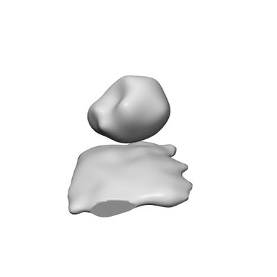 emd_3919.png emd_3919.png | 16.3 KB | ||
| Archive directory |  http://ftp.pdbj.org/pub/emdb/structures/EMD-3919ftp://ftp.pdbj.org/pub/emdb/structures/EMD-3919 http://ftp.pdbj.org/pub/emdb/structures/EMD-3919ftp://ftp.pdbj.org/pub/emdb/structures/EMD-3919 | HTTPS FTP |
-Validation report
| Summary document | emd_3919_validation.pdf.gz | 187.7 KB | Display | EMDB validaton report |
|---|---|---|---|---|
| Full document | emd_3919_full_validation.pdf.gz | 186.8 KB | Display | |
| Data in XML | emd_3919_validation.xml.gz | 4.4 KB | Display | |
| Arichive directory | https://ftp.pdbj.org/pub/emdb/validation_reports/EMD-3919ftp://ftp.pdbj.org/pub/emdb/validation_reports/EMD-3919 | HTTPS FTP |
-Related structure data
| Related structure data |  3909C  3920C  3921C  3922C  3923C  3924C C: citing same article ( |
|---|---|
| Similar structure data |
-Links
| EMDB pages | EMDB (EBI/PDBe) / EMDataResource |
|---|---|
| Related items in Molecule of the Month |

-Map
| File | Download / File: emd_3919.map.gz / Format: CCP4 / Size: 313.5 KB / Type: IMAGE STORED AS FLOATING POINT NUMBER (4 BYTES) | ||||||||||||||||||||||||||||||||||||||||||||||||||||||||||||
|---|---|---|---|---|---|---|---|---|---|---|---|---|---|---|---|---|---|---|---|---|---|---|---|---|---|---|---|---|---|---|---|---|---|---|---|---|---|---|---|---|---|---|---|---|---|---|---|---|---|---|---|---|---|---|---|---|---|---|---|---|---|
| Annotation | Average of class 1 | ||||||||||||||||||||||||||||||||||||||||||||||||||||||||||||
| Projections & slices | Image control
Images are generated by Spider. generated in cubic-lattice coordinate | ||||||||||||||||||||||||||||||||||||||||||||||||||||||||||||
| Voxel size | X=Y=Z: 9.04 Å | ||||||||||||||||||||||||||||||||||||||||||||||||||||||||||||
| Density |
| ||||||||||||||||||||||||||||||||||||||||||||||||||||||||||||
| Symmetry | Space group: 1 | ||||||||||||||||||||||||||||||||||||||||||||||||||||||||||||
| Details | EMDB XML:
CCP4 map header:
| ||||||||||||||||||||||||||||||||||||||||||||||||||||||||||||
 Z (Sec.)
 Y (Row.)
 X (Col.)


-Supplemental data
- Sample components
-Entire : Ribosome-nascent chains with native E.coli membranes containing YidC
| Entire | Name: Ribosome-nascent chains with native E.coli membranes containing YidC |
|---|---|
| Components |
|
-Supramolecule #1: Ribosome-nascent chains with native E.coli membranes containing YidC
| Supramolecule | Name: Ribosome-nascent chains with native E.coli membranes containing YidC type: organelle_or_cellular_component / ID: 1 / Parent: 0 |
|---|---|
| Source (natural) | Organism: |
| Recombinant expression | Organism: |
-Supramolecule #2: E. coli native membranes containing YidC
| Supramolecule | Name: E. coli native membranes containing YidC / type: organelle_or_cellular_component / ID: 2 / Parent: 1 |
|---|---|
| Source (natural) | Organism: |
| Recombinant expression | Organism: |
-Supramolecule #3: Ribosomes with SecM stalled nascent chains of ATP synthase subunit c
| Supramolecule | Name: Ribosomes with SecM stalled nascent chains of ATP synthase subunit c type: complex / ID: 3 / Parent: 1 |
|---|---|
| Source (natural) | Organism: |
| Recombinant expression | Organism: |
-Experimental details
-Structure determination
| Method | cryo EM |
|---|---|
 Processing Processing | subtomogram averaging |
| Aggregation state | particle |
-Sample preparation
| Buffer | pH: 7.4 |
|---|---|
| Grid | Model: Quantifoil R2/1 / Material: COPPER / Mesh: 200 / Support film - Material: CARBON / Support film - topology: HOLEY / Pretreatment - Type: GLOW DISCHARGE / Pretreatment - Atmosphere: AIR |
| Vitrification | Cryogen name: ETHANE-PROPANE / Instrument: HOMEMADE PLUNGER |
- Electron microscopy
| Microscope | FEI TITAN KRIOS |
|---|---|
| Specialist optics | Energy filter - Name: GIF Quantum LS / Energy filter - Lower energy threshold: 0 eV / Energy filter - Upper energy threshold: 20 eV |
| Image recording | Film or detector model: GATAN K2 QUANTUM (4k x 4k) / Detector mode: COUNTING / Digitization - Dimensions - Width: 3710 pixel / Digitization - Dimensions - Height: 3836 pixel / Average electron dose: 2.0 e/Å2 |
| Electron beam | Acceleration voltage: 300 kV / Electron source:  FIELD EMISSION GUN FIELD EMISSION GUN |
| Electron optics | C2 aperture diameter: 50.0 µm / Calibrated defocus max: 8.0 µm / Calibrated defocus min: 4.0 µm / Illumination mode: FLOOD BEAM / Imaging mode: BRIGHT FIELD / Cs: 2.7 mm / Nominal defocus max: 5.0 µm / Nominal defocus min: 2.5 µm |
| Sample stage | Specimen holder model: FEI TITAN KRIOS AUTOGRID HOLDER / Cooling holder cryogen: NITROGEN |
| Experimental equipment |  Model: Titan Krios / Image courtesy: FEI Company |
