 Movie
Movie Controller

[English] 日本語
 Yorodumi
- EMDB-38558: The structure determination of prokaryotic Glycerol-3-phosphate A... -
+ Open data

- Basic information
| Entry |  | |||||||||
|---|---|---|---|---|---|---|---|---|---|---|
| Title | The structure determination of prokaryotic Glycerol-3-phosphate Acyltransferase | |||||||||
 Map data Map data | ||||||||||
 Sample Sample |
| |||||||||
 Keywords Keywords | enzyme on the membrane / de newo synthetase of phospholipid / complex of enzyme ang ligands / LIPID TRANSPORT | |||||||||
| Biological species |  Thermomonas haemolytica (bacteria) Thermomonas haemolytica (bacteria) | |||||||||
| Method | single particle reconstruction / Resolution: 2.79 Å | |||||||||
 Authors Authors | Li YM / Liu ZF | |||||||||
| Funding support |  China, 1 items China, 1 items
| |||||||||
 Citation Citation | Journal: To Be Published Title: The phospholipid biosynthesis enzyme PlsB contains three distinct domains for membrane association, lysophosphatidic acid synthesis and dimerization Authors: Li YM / Liu ZF | |||||||||
| History |
|
- Structure visualization
| Supplemental images |
|---|

- Downloads & links
-EMDB archive
| Map data | emd_38558.map.gz | 7.1 MB |  EMDB map data format EMDB map data format | |
|---|---|---|---|---|
| Header (meta data) | emd-38558-v30.xmlemd-38558.xml | 14.9 KB 14.9 KB | Display Display | EMDB header |
| FSC (resolution estimation) | emd_38558_fsc.xml | 10.6 KB | Display | FSC data file |
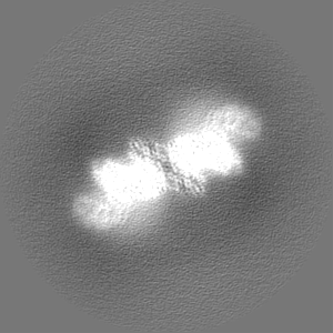
| Images |  emd_38558.png emd_38558.png | 78.1 KB | ||
| Masks | emd_38558_msk_1.map | 103 MB | Mask map | |
| Filedesc metadata | emd-38558.cif.gz | 5.6 KB | ||
| Others | emd_38558_half_map_1.map.gzemd_38558_half_map_2.map.gz | 79.7 MB 79.7 MB | ||
| Archive directory |  http://ftp.pdbj.org/pub/emdb/structures/EMD-38558ftp://ftp.pdbj.org/pub/emdb/structures/EMD-38558 http://ftp.pdbj.org/pub/emdb/structures/EMD-38558ftp://ftp.pdbj.org/pub/emdb/structures/EMD-38558 | HTTPS FTP |
-Links
| EMDB pages | EMDB (EBI/PDBe) / EMDataResource |
|---|
-Map
| File | Download / File: emd_38558.map.gz / Format: CCP4 / Size: 103 MB / Type: IMAGE STORED AS FLOATING POINT NUMBER (4 BYTES) | ||||||||||||||||||||||||||||||||||||
|---|---|---|---|---|---|---|---|---|---|---|---|---|---|---|---|---|---|---|---|---|---|---|---|---|---|---|---|---|---|---|---|---|---|---|---|---|---|
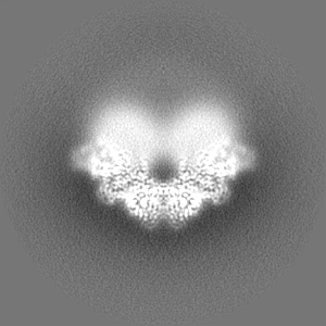
| Projections & slices | Image control
Images are generated by Spider. | ||||||||||||||||||||||||||||||||||||
| Voxel size | X=Y=Z: 1.04 Å | ||||||||||||||||||||||||||||||||||||
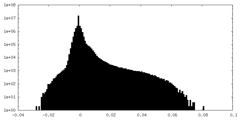
| Density |
| ||||||||||||||||||||||||||||||||||||
| Symmetry | Space group: 1 | ||||||||||||||||||||||||||||||||||||
| Details | EMDB XML:
|
 Z (Sec.)
 Y (Row.)
 X (Col.)


-Supplemental data
-Mask #1
| File | emd_38558_msk_1.map | ||||||||||||
|---|---|---|---|---|---|---|---|---|---|---|---|---|---|
| Projections & Slices |
| ||||||||||||
| Density Histograms |


-Half map: #2
| File | emd_38558_half_map_1.map | ||||||||||||
|---|---|---|---|---|---|---|---|---|---|---|---|---|---|
| Projections & Slices |
| ||||||||||||
| Density Histograms |


-Half map: #1
| File | emd_38558_half_map_2.map | ||||||||||||
|---|---|---|---|---|---|---|---|---|---|---|---|---|---|
| Projections & Slices |
| ||||||||||||
| Density Histograms |


- Sample components
-Entire : PlsB, Palmitoyl CoA, 1,2-dioleoyl-sn-glycero-3-phosphate
| Entire | Name: PlsB, Palmitoyl CoA, 1,2-dioleoyl-sn-glycero-3-phosphate |
|---|---|
| Components |
|
-Supramolecule #1: PlsB, Palmitoyl CoA, 1,2-dioleoyl-sn-glycero-3-phosphate
| Supramolecule | Name: PlsB, Palmitoyl CoA, 1,2-dioleoyl-sn-glycero-3-phosphate type: complex / ID: 1 / Parent: 0 / Macromolecule list: all Details: PlsB is a crucial enzyme catalyzing the first step of phospholipid synthesis by converting glycerol-3-phosphate and fatty acyl-coenzyme A (CoA)/acyl-carrier protein (ACP) to lysophosphatidic ...Details: PlsB is a crucial enzyme catalyzing the first step of phospholipid synthesis by converting glycerol-3-phosphate and fatty acyl-coenzyme A (CoA)/acyl-carrier protein (ACP) to lysophosphatidic acid and free CoA (CoASH)/ACP. |
|---|---|
| Source (natural) | Organism: Thermomonas haemolytica (bacteria) |
-Macromolecule #1: PlsB from Themomonas haemolytica (ThPlsB)
| Macromolecule | Name: PlsB from Themomonas haemolytica (ThPlsB) / type: protein_or_peptide / ID: 1 / Enantiomer: LEVO / EC number: glycerol-3-phosphate 1-O-acyltransferase |
|---|---|
| Source (natural) | Organism: Thermomonas haemolytica (bacteria) / Strain: Thermomonas haemolytica |
| Recombinant expression | Organism: |
| Sequence | String: MAAMPDRHSP DQPGLFDPPA AADDAATGPA GPAPLPLPGF LAADAATPPG PPSPPPAGKA RNPLWARLLG RLLAPWLRLE IEVDPAIAAD PRPICYVLED YGLSNALILQ RACREAALPP PLQPIAGDPL GRRRAYVALS RRHVNALGLL PGAEHKTHSG SLARLLQAHQ ...String: MAAMPDRHSP DQPGLFDPPA AADDAATGPA GPAPLPLPGF LAADAATPPG PPSPPPAGKA RNPLWARLLG RLLAPWLRLE IEVDPAIAAD PRPICYVLED YGLSNALILQ RACREAALPP PLQPIAGDPL GRRRAYVALS RRHVNALGLL PGAEHKTHSG SLARLLQAHQ QQPELDVHLV PVSIFVGQAP KRSSGWFSVL FSENWTLVGR FRRLLAILLN GRDTLVKFAA PVPVREIVAE GLEPERTVRK LSRILRTHFR RVREVVIGPD LSTRRMLADQ VLSSPLVKEA IADQARRDGS KPEAAWEKAN AYFWEIAADY SNTVVRSASF ALTFVWNRIY RGVLVHHLDQ FKQEAPGHEV VYVPSHRSHM DYLLVSYLLY THGVVPPHIF AGINLNLPVV GTLLRKGGAF FARRSFKGNA LYSAVFREYM AQLVAGGYSI EYFIEGGRSR TGRLLQPKGG SLAMTVRAYL RQPTRPVLFQ PVYIGYEKLM EGRSYLDELS GKPKEKESIW QLLAGIPKVL RSNYGQVVVN FGERIQLSQV LAELAPEWDG QPIGDDEKPA WFARTVDALA QRIQTNVNRA ADVNPINLLA LALLSTPKHA MGEADLRAQI ALSKTLLAEV PYSDWVTVTP HTPEQIIAHG EEIGLITRTA HPLGDVLGVE GDNAVLLSYF RNNVLHLFTA SSWIAVCFQN NRRMGRRQLQ QIGRTLYPFL QAELFLPWDE ETFAARIDRT IEVFVREGLL EQVSDEDGGI LQRNAGQSDE VFRLRALGHT LQQAFERYYI AIAILVKNGS GTLQAGELES LCQLTAQRLS LLYAPAAPEF FDKTLFRGFI QKLRELKLVW PDDTGRLAFD ERLKAWAKDA RVVLGRELRH TIEKISPAGS GRSSGEQPAL PPDPAATNGA S |
-Experimental details
-Structure determination
 Processing Processing | single particle reconstruction |
|---|---|
| Aggregation state | particle |
-Sample preparation
| Concentration | 6 mg/mL | |||||||||||||||
|---|---|---|---|---|---|---|---|---|---|---|---|---|---|---|---|---|
| Buffer | pH: 8 Component:
Details: 500 mM KCl, 2 mM DTT, 5 mM EDTA and 0.1% GDN | |||||||||||||||
| Sugar embedding | Material: GDN | |||||||||||||||
| Grid | Model: Quantifoil R1.2/1.3 / Material: GOLD / Mesh: 300 / Pretreatment - Type: GLOW DISCHARGE / Pretreatment - Time: 90 sec. / Pretreatment - Atmosphere: AIR | |||||||||||||||
| Details | The specimen was purified through Ni-TNA, and incubated overnight with the substrate. The secondary purification was performed by size-exclusion chromatography. |
- Electron microscopy
| Microscope | TFS KRIOS |
|---|---|
| Image recording | Film or detector model: FEI FALCON II (4k x 4k) / Average electron dose: 60.0 e/Å2 |
| Electron beam | Acceleration voltage: 300 kV / Electron source:  FIELD EMISSION GUN FIELD EMISSION GUN |
| Electron optics | Illumination mode: SPOT SCAN / Imaging mode: BRIGHT FIELD / Cs: 2.7 mm / Nominal defocus max: 1.8 µm / Nominal defocus min: 1.3 µm |
| Experimental equipment |  Model: Titan Krios / Image courtesy: FEI Company |

