 Movie
Movie Controller

[English] 日本語
 Yorodumi
- EMDB-38456: Cryo-EM structure of Ca2+-bound TMEM16A in complex with Tamsulosin -
+ Open data

- Basic information
| Entry |  | |||||||||
|---|---|---|---|---|---|---|---|---|---|---|
| Title | Cryo-EM structure of Ca2+-bound TMEM16A in complex with Tamsulosin | |||||||||
 Map data Map data | ||||||||||
 Sample Sample |
| |||||||||
 Keywords Keywords | Inhibitor / Modulator / Complex / Ion channel / LIPID BINDING PROTEIN | |||||||||
| Function / homology |  Function and homology information Function and homology informationglial cell projection elongation / trachea development / iodide transmembrane transporter activity / mucus secretion / iodide transport / intracellularly calcium-gated chloride channel activity / cellular response to peptide / Stimuli-sensing channels / voltage-gated chloride channel activity / calcium-activated cation channel activity ...glial cell projection elongation / trachea development / iodide transmembrane transporter activity / mucus secretion / iodide transport / intracellularly calcium-gated chloride channel activity / cellular response to peptide / Stimuli-sensing channels / voltage-gated chloride channel activity / calcium-activated cation channel activity / protein localization to membrane / chloride transport / chloride channel activity / detection of temperature stimulus involved in sensory perception of pain / chloride channel complex / chloride transmembrane transport / regulation of membrane potential / cellular response to heat / presynaptic membrane / phospholipase C-activating G protein-coupled receptor signaling pathway / apical plasma membrane / signaling receptor binding / external side of plasma membrane / glutamatergic synapse / protein homodimerization activity / nucleoplasm / metal ion binding / identical protein binding / plasma membrane Similarity search - Function | |||||||||
| Biological species |  | |||||||||
| Method | single particle reconstruction / Resolution: 2.93 Å | |||||||||
 Authors Authors | Li HL / Li SL / Li S | |||||||||
| Funding support |  China, 1 items China, 1 items
| |||||||||
 Citation Citation | Journal: Proc Natl Acad Sci U S A / Year: 2025 Title: Tamsulosin ameliorates bone loss by inhibiting the release of Cl through wedging into an allosteric site of TMEM16A. Authors: Shiliang Li / Weijia Sun / Shuang Li / Lili Zhu / Shuai Guo / Jiaqi He / Yuheng Li / Chaoquan Tian / Zhenjiang Zhao / Tao Yu / Jianwei Li / Yiqing Zhang / Youlong Hai / Jiawen Wang / Yongjun ...Authors: Shiliang Li / Weijia Sun / Shuang Li / Lili Zhu / Shuai Guo / Jiaqi He / Yuheng Li / Chaoquan Tian / Zhenjiang Zhao / Tao Yu / Jianwei Li / Yiqing Zhang / Youlong Hai / Jiawen Wang / Yongjun Zheng / Rui Wang / Xiaoyong Hu / Shukuan Ling / Honglin Li / Yingxian Li / Abstract: TMEM16A, a key calcium-activated chloride channel, is crucial for many physiological and pathological processes such as cancer, hypertension, and osteoporosis, etc. However, the regulatory mechanism ...TMEM16A, a key calcium-activated chloride channel, is crucial for many physiological and pathological processes such as cancer, hypertension, and osteoporosis, etc. However, the regulatory mechanism of TMEM16A is poorly understood, limiting the discovery of effective modulators. Here, we unveil an allosteric gating mechanism by presenting a high-resolution cryo-EM structure of TMEM16A in complex with a channel inhibitor that we identified, Tamsulosin, which is resolved at 2.93 Å. Tamsulosin wedges itself into a pocket within the extracellular domain of TMEM16A, surrounded by α1-α2, α5-α6, and α9-α10 loops. This binding stabilizes a transient preopen conformation of TMEM16A, which is activated by Ca ions while still preserving a closed pore to prevent Cl permeation. Validation of this binding site through computational, electrophysiological, and functional experiments, along with site-directed mutagenesis, confirmed the pivotal roles of the pocket-lining residues R605 and E624 on α5-α6 loop in modulating Tamsulosin binding and pore activity. Tamsulosin induces significant positional shifts in extracellular loops, particularly the α5-α6 loop, which moves toward the extracellular exit of the pore, leading to noticeable structural rearrangements in pore-lining helices. The hinges induced by P595 in α5 and G711 in α7 introduce flexibility to the transmembrane helices, orienting Y593 to collaborate with I641 in effectively gating the preopening pore. Notably, Tamsulosin demonstrates significant antiosteoporotic effects by inhibiting TMEM16A, suggesting potential for its repurposing in new therapeutic indications. Our study not only enhances our understanding of the gating mechanism of TMEM16A inhibition but also facilitates structure-based drug design targeting TMEM16A. | |||||||||
| History |
|
- Structure visualization
| Supplemental images |
|---|

- Downloads & links
-EMDB archive
| Map data | emd_38456.map.gz | 203.6 MB | EMDB map data format | |
|---|---|---|---|---|
| Header (meta data) | emd-38456-v30.xmlemd-38456.xml | 17.6 KB 17.6 KB | Display Display | EMDB header |
| FSC (resolution estimation) | emd_38456_fsc.xml | 12.7 KB | Display | FSC data file |
| Images |  emd_38456.png emd_38456.png | 165 KB | ||
| Filedesc metadata | emd-38456.cif.gz | 6.5 KB | ||
| Others | emd_38456_half_map_1.map.gzemd_38456_half_map_2.map.gz | 199.7 MB 199.7 MB | ||
| Archive directory |  http://ftp.pdbj.org/pub/emdb/structures/EMD-38456ftp://ftp.pdbj.org/pub/emdb/structures/EMD-38456 http://ftp.pdbj.org/pub/emdb/structures/EMD-38456ftp://ftp.pdbj.org/pub/emdb/structures/EMD-38456 | HTTPS FTP |
-Related structure data
| Related structure data |  8xlrMC M: atomic model generated by this map C: citing same article ( |
|---|---|
| Similar structure data |
-Links
| EMDB pages | EMDB (EBI/PDBe) / EMDataResource |
|---|
-Map
| File | Download / File: emd_38456.map.gz / Format: CCP4 / Size: 216 MB / Type: IMAGE STORED AS FLOATING POINT NUMBER (4 BYTES) | ||||||||||||||||||||||||||||||||||||
|---|---|---|---|---|---|---|---|---|---|---|---|---|---|---|---|---|---|---|---|---|---|---|---|---|---|---|---|---|---|---|---|---|---|---|---|---|---|
| Projections & slices | Image control
Images are generated by Spider. | ||||||||||||||||||||||||||||||||||||
| Voxel size | X=Y=Z: 0.86 Å | ||||||||||||||||||||||||||||||||||||
| Density |
| ||||||||||||||||||||||||||||||||||||
| Symmetry | Space group: 1 | ||||||||||||||||||||||||||||||||||||
| Details | EMDB XML:
|
 Z (Sec.)
 Y (Row.)
 X (Col.)


-Supplemental data
-Half map: #2
| File | emd_38456_half_map_1.map | ||||||||||||
|---|---|---|---|---|---|---|---|---|---|---|---|---|---|
| Projections & Slices |
| ||||||||||||
| Density Histograms |


-Half map: #1
| File | emd_38456_half_map_2.map | ||||||||||||
|---|---|---|---|---|---|---|---|---|---|---|---|---|---|
| Projections & Slices |
| ||||||||||||
| Density Histograms |


- Sample components
-Entire : Structure of Tamsulosin- and calcium-bound mTMEM16A chloride channel
| Entire | Name: Structure of Tamsulosin- and calcium-bound mTMEM16A chloride channel |
|---|---|
| Components |
|
-Supramolecule #1: Structure of Tamsulosin- and calcium-bound mTMEM16A chloride channel
| Supramolecule | Name: Structure of Tamsulosin- and calcium-bound mTMEM16A chloride channel type: complex / ID: 1 / Parent: 0 / Macromolecule list: #1 |
|---|---|
| Source (natural) | Organism: |
-Macromolecule #1: Anoctamin-1
| Macromolecule | Name: Anoctamin-1 / type: protein_or_peptide / ID: 1 / Number of copies: 2 / Enantiomer: LEVO |
|---|---|
| Source (natural) | Organism: |
| Molecular weight | Theoretical: 111.058992 KDa |
| Recombinant expression | Organism:  Homo sapiens (human) Homo sapiens (human) |
| Sequence | String: MRVPEKYSTL PAEDRSVHIV NICAIEDLGY LPSEGTLLNS LSVDPDAECK YGLYFRDGKR KVDYILVYHH KRASGSRTLA RRGLQNDMV LGTRSVRQDQ PLPGKGSPVD AGSPEVPMDY HEDDKRFRRE EYEGNLLEAG LELENDEDTK IHGVGFVKIH A PWHVLCRE ...String: MRVPEKYSTL PAEDRSVHIV NICAIEDLGY LPSEGTLLNS LSVDPDAECK YGLYFRDGKR KVDYILVYHH KRASGSRTLA RRGLQNDMV LGTRSVRQDQ PLPGKGSPVD AGSPEVPMDY HEDDKRFRRE EYEGNLLEAG LELENDEDTK IHGVGFVKIH A PWHVLCRE AEFLKLKMPT KKVYHISETR GLLKTINSVL QKITDPIQPK VAEHRPQTTK RLSYPFSREK QHLFDLTDRD SF FDSKTRS TIVYEILKRT TCTKAKYSMG ITSLLANGVY SAAYPLHDGD YEGDNVEFND RKLLYEEWAS YGVFYKYQPI DLV RKYFGE KVGLYFAWLG AYTQMLIPAS IVGVIVFLYG CATVDENIPS MEMCDQRYNI TMCPLCDKTC SYWKMSSACA TARA SHLFD NPATVFFSVF MALWAATFME HWKRKQMRLN YRWDLTGFEE EEEAVKDHPR AEYEARVLEK SLRKESRNKE TDKVK LTWR DRFPAYFTNL VSIIFMIAVT FAIVLGVIIY RISTAAALAM NSSPSVRSNI RVTVTATAVI INLVVIILLD EVYGCI ARW LTKIEVPKTE KSFEERLTFK AFLLKFVNSY TPIFYVAFFK GRFVGRPGDY VYIFRSFRME ECAPGGCLME LCIQLSI IM LGKQLIQNNL FEIGIPKMKK FIRYLKLRRQ SPSDREEYVK RKQRYEVDFN LEPFAGLTPE YMEMIIQFGF VTLFVASF P LAPLFALLNN IIEIRLDAKK FVTELRRPVA IRAKDIGIWY NILRGVGKLA VIINAFVISF TSDFIPRLVY LYMYSQNGT MHGFVNHTLS SFNVSDFQNG TAPNDPLDLG YEVQICRYKD YREPPWSEHK YDISKDFWAV LAARLAFVIV FQNLVMFMSD FVDWVIPDI PKDISQQIHK EKVLMVELFM REEQGKQQLL DTWMEKEKPR DVPCNNHSPT THPEAGDGSP VPSYEYHGDA L UniProtKB: Anoctamin-1 |
-Macromolecule #2: 2-acetamido-2-deoxy-beta-D-glucopyranose
| Macromolecule | Name: 2-acetamido-2-deoxy-beta-D-glucopyranose / type: ligand / ID: 2 / Number of copies: 8 / Formula: NAG |
|---|---|
| Molecular weight | Theoretical: 221.208 Da |
| Chemical component information |  ChemComp-NAG: |
-Macromolecule #3: TETRADECANE
| Macromolecule | Name: TETRADECANE / type: ligand / ID: 3 / Number of copies: 50 / Formula: C14 |
|---|---|
| Molecular weight | Theoretical: 198.388 Da |
| Chemical component information |  ChemComp-C14: |
-Macromolecule #4: CALCIUM ION
| Macromolecule | Name: CALCIUM ION / type: ligand / ID: 4 / Number of copies: 4 / Formula: CA |
|---|---|
| Molecular weight | Theoretical: 40.078 Da |
-Macromolecule #5: Tamsulosin
| Macromolecule | Name: Tamsulosin / type: ligand / ID: 5 / Number of copies: 2 / Formula: JGX |
|---|---|
| Molecular weight | Theoretical: 408.512 Da |
| Chemical component information |  ChemComp-JGX: |
-Macromolecule #6: HEXANE
| Macromolecule | Name: HEXANE / type: ligand / ID: 6 / Number of copies: 2 / Formula: HEX |
|---|---|
| Molecular weight | Theoretical: 86.175 Da |
| Chemical component information | 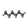 ChemComp-HEX: |
-Experimental details
-Structure determination
 Processing Processing | single particle reconstruction |
|---|---|
| Aggregation state | particle |
-Sample preparation
| Concentration | 4 mg/mL | ||||||||||||
|---|---|---|---|---|---|---|---|---|---|---|---|---|---|
| Buffer | pH: 7.5 Component:
| ||||||||||||
| Grid | Model: Quantifoil R1.2/1.3 / Mesh: 300 |
- Electron microscopy
| Microscope | FEI TITAN KRIOS |
|---|---|
| Image recording | Film or detector model: GATAN K3 BIOQUANTUM (6k x 4k) / Average electron dose: 50.0 e/Å2 |
| Electron beam | Acceleration voltage: 300 kV / Electron source:  FIELD EMISSION GUN FIELD EMISSION GUN |
| Electron optics | Illumination mode: FLOOD BEAM / Imaging mode: BRIGHT FIELD / Nominal defocus max: 2.0 µm / Nominal defocus min: 1.0 µm |
| Experimental equipment |  Model: Titan Krios / Image courtesy: FEI Company |
+Image processing

-Atomic model buiding 1
| Refinement | Protocol: AB INITIO MODEL |
|---|---|
| Output model | PDB-8xlr: |
