 Movie
Movie Controller


+ Open data

- Basic information
| Entry |  | |||||||||
|---|---|---|---|---|---|---|---|---|---|---|
| Title | Cryo-EM structure of human GPR1 bound to chemerin | |||||||||
 Map data Map data | ||||||||||
 Sample Sample |
| |||||||||
 Keywords Keywords | MEMBRANE PROTEIN | |||||||||
| Function / homology |  Function and homology information Function and homology informationadipokinetic hormone binding / adipokinetic hormone receptor activity / platelet dense granule lumen / regulation of lipid catabolic process / antifungal innate immune response / embryonic digestive tract development / antifungal humoral response / positive regulation of chemotaxis / neuropeptide binding / positive regulation of macrophage chemotaxis ...adipokinetic hormone binding / adipokinetic hormone receptor activity / platelet dense granule lumen / regulation of lipid catabolic process / antifungal innate immune response / embryonic digestive tract development / antifungal humoral response / positive regulation of chemotaxis / neuropeptide binding / positive regulation of macrophage chemotaxis / positive regulation of fat cell differentiation / neuropeptide signaling pathway / retinoid metabolic process / positive regulation of protein localization to cell cortex / Adenylate cyclase inhibitory pathway / T cell migration / D2 dopamine receptor binding / response to prostaglandin E / G protein-coupled serotonin receptor binding / adenylate cyclase regulator activity / adenylate cyclase-inhibiting serotonin receptor signaling pathway / cellular response to forskolin / extracellular matrix / regulation of mitotic spindle organization / Regulation of insulin secretion / positive regulation of cholesterol biosynthetic process / G protein-coupled receptor binding / adenylate cyclase-inhibiting G protein-coupled receptor signaling pathway / G protein-coupled receptor activity / adenylate cyclase-modulating G protein-coupled receptor signaling pathway / response to peptide hormone / G-protein beta/gamma-subunit complex binding / centriolar satellite / Olfactory Signaling Pathway / Activation of the phototransduction cascade / G beta:gamma signalling through PLC beta / Presynaptic function of Kainate receptors / Thromboxane signalling through TP receptor / G protein-coupled acetylcholine receptor signaling pathway / G-protein activation / Activation of G protein gated Potassium channels / Inhibition of voltage gated Ca2+ channels via Gbeta/gamma subunits / Prostacyclin signalling through prostacyclin receptor / G beta:gamma signalling through CDC42 / Glucagon signaling in metabolic regulation / G beta:gamma signalling through BTK / Synthesis, secretion, and inactivation of Glucagon-like Peptide-1 (GLP-1) / ADP signalling through P2Y purinoceptor 12 / Sensory perception of sweet, bitter, and umami (glutamate) taste / chemotaxis / photoreceptor disc membrane / Glucagon-type ligand receptors / Adrenaline,noradrenaline inhibits insulin secretion / Vasopressin regulates renal water homeostasis via Aquaporins / GDP binding / G alpha (z) signalling events / Glucagon-like Peptide-1 (GLP1) regulates insulin secretion / cellular response to catecholamine stimulus / ADORA2B mediated anti-inflammatory cytokines production / ADP signalling through P2Y purinoceptor 1 / G beta:gamma signalling through PI3Kgamma / Cooperation of PDCL (PhLP1) and TRiC/CCT in G-protein beta folding / adenylate cyclase-activating dopamine receptor signaling pathway / GPER1 signaling / Inactivation, recovery and regulation of the phototransduction cascade / cellular response to prostaglandin E stimulus / insulin receptor signaling pathway / G-protein beta-subunit binding / glucose homeostasis / heterotrimeric G-protein complex / Platelet degranulation / G alpha (12/13) signalling events / sensory perception of taste / extracellular vesicle / signaling receptor complex adaptor activity / Thrombin signalling through proteinase activated receptors (PARs) / positive regulation of protein phosphorylation / G protein activity / GTPase binding / Ca2+ pathway / retina development in camera-type eye / midbody / cell cortex / High laminar flow shear stress activates signaling by PIEZO1 and PECAM1:CDH5:KDR in endothelial cells / : / G alpha (i) signalling events / defense response to Gram-negative bacterium / Hydrolases; Acting on acid anhydrides; Acting on GTP to facilitate cellular and subcellular movement / G alpha (s) signalling events / phospholipase C-activating G protein-coupled receptor signaling pathway / G alpha (q) signalling events / Ras protein signal transduction / cell differentiation / Extra-nuclear estrogen signaling / cell population proliferation / defense response to Gram-positive bacterium / neuron projection / ciliary basal body / G protein-coupled receptor signaling pathway / inflammatory response Similarity search - Function | |||||||||
| Biological species |  Homo sapiens (human) / Homo sapiens (human) /  | |||||||||
| Method | single particle reconstruction / cryo EM / Resolution: 3.29 Å | |||||||||
 Authors Authors | Liu AJ / Liu YZ / Ye RD | |||||||||
| Funding support | 1 items
| |||||||||
 Citation Citation | Journal: PLoS Biol / Year: 2024 Title: Structure of G protein-coupled receptor GPR1 bound to full-length chemerin adipokine reveals a chemokine-like reverse binding mode. Authors: Aijun Liu / Yezhou Liu / Geng Chen / Wenping Lyu / Fang Ye / Junlin Wang / Qiwen Liao / Lizhe Zhu / Yang Du / Richard D Ye /  Abstract: Chemerin is an adipokine with chemotactic activity to a subset of leukocytes. Chemerin binds to 3 G protein-coupled receptors, including chemokine-like receptor 1 (CMKLR1), G protein-coupled receptor ...Chemerin is an adipokine with chemotactic activity to a subset of leukocytes. Chemerin binds to 3 G protein-coupled receptors, including chemokine-like receptor 1 (CMKLR1), G protein-coupled receptor 1 (GPR1), and C-C chemokine receptor-like 2 (CCRL2). Here, we report that GPR1 is capable of Gi signaling when stimulated with full-length chemerin or its C-terminal nonapeptide (C9, YFPGQFAFS). We present high-resolution cryo-EM structures of Gi-coupled GPR1 bound to full-length chemerin and to the C9 peptide, respectively. C9 insertion into the transmembrane (TM) binding pocket is both necessary and sufficient for GPR1 signaling, whereas the full-length chemerin uses its bulky N-terminal core for interaction with a β-strand located at the N-terminus of GPR1. This interaction involves multiple β-strands of full-length chemerin, forming a β-sheet that serves as a "lid" for the TM binding pocket and is energetically expensive to remove as indicated by molecular dynamics simulations with free energy landscape analysis. Combining results from functional assays, our structural model explains why C9 is an activating peptide at GPR1 and how the full-length chemerin uses a "two-site" model for enhanced interaction with GPR1. | |||||||||
| History |
|
- Structure visualization
| Supplemental images |
|---|

- Downloads & links
-EMDB archive
| Map data | emd_38328.map.gz | 117.8 MB | EMDB map data format | |
|---|---|---|---|---|
| Header (meta data) | emd-38328-v30.xmlemd-38328.xml | 23.4 KB 23.4 KB | Display Display | EMDB header |
| Images |  emd_38328.png emd_38328.png | 31.8 KB | ||
| Filedesc metadata | emd-38328.cif.gz | 7.1 KB | ||
| Others | emd_38328_half_map_1.map.gzemd_38328_half_map_2.map.gz | 116 MB 115.9 MB | ||
| Archive directory |  http://ftp.pdbj.org/pub/emdb/structures/EMD-38328ftp://ftp.pdbj.org/pub/emdb/structures/EMD-38328 http://ftp.pdbj.org/pub/emdb/structures/EMD-38328ftp://ftp.pdbj.org/pub/emdb/structures/EMD-38328 | HTTPS FTP |
-Related structure data
| Related structure data |  8xgmMC 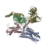 8jjpC M: atomic model generated by this map C: citing same article ( |
|---|---|
| Similar structure data |

-Links
| EMDB pages | EMDB (EBI/PDBe) / EMDataResource |
|---|---|
| Related items in Molecule of the Month |


-Map
| File | Download / File: emd_38328.map.gz / Format: CCP4 / Size: 125 MB / Type: IMAGE STORED AS FLOATING POINT NUMBER (4 BYTES) | ||||||||||||||||||||||||||||||||||||
|---|---|---|---|---|---|---|---|---|---|---|---|---|---|---|---|---|---|---|---|---|---|---|---|---|---|---|---|---|---|---|---|---|---|---|---|---|---|
| Projections & slices | Image control
Images are generated by Spider. | ||||||||||||||||||||||||||||||||||||
| Voxel size | X=Y=Z: 0.85 Å | ||||||||||||||||||||||||||||||||||||
| Density |
| ||||||||||||||||||||||||||||||||||||
| Symmetry | Space group: 1 | ||||||||||||||||||||||||||||||||||||
| Details | EMDB XML:
|
 Z (Sec.)
 Y (Row.)
 X (Col.)


-Supplemental data
-Half map: #2
| File | emd_38328_half_map_1.map | ||||||||||||
|---|---|---|---|---|---|---|---|---|---|---|---|---|---|
| Projections & Slices |
| ||||||||||||
| Density Histograms |


-Half map: #1
| File | emd_38328_half_map_2.map | ||||||||||||
|---|---|---|---|---|---|---|---|---|---|---|---|---|---|
| Projections & Slices |
| ||||||||||||
| Density Histograms |


- Sample components
-Entire : Human GPR1 in complex with chemerin and heterotrimeric Gi proteins
| Entire | Name: Human GPR1 in complex with chemerin and heterotrimeric Gi proteins |
|---|---|
| Components |
|
-Supramolecule #1: Human GPR1 in complex with chemerin and heterotrimeric Gi proteins
| Supramolecule | Name: Human GPR1 in complex with chemerin and heterotrimeric Gi proteins type: complex / ID: 1 / Parent: 0 / Macromolecule list: #1-#6 |
|---|---|
| Source (natural) | Organism: Homo sapiens (human) |
-Macromolecule #1: scFV16
| Macromolecule | Name: scFV16 / type: protein_or_peptide / ID: 1 / Number of copies: 1 / Enantiomer: LEVO |
|---|---|
| Source (natural) | Organism: |
| Molecular weight | Theoretical: 26.337307 KDa |
| Recombinant expression | Organism:   Spodoptera frugiperda (fall armyworm) Spodoptera frugiperda (fall armyworm) |
| Sequence | String: DVQLVESGGG LVQPGGSRKL SCSASGFAFS SFGMHWVRQA PEKGLEWVAY ISSGSGTIYY ADTVKGRFTI SRDDPKNTLF LQMTSLRSE DTAMYYCVRS IYYYGSSPFD FWGQGTTLTV SSGGGGSGGG GSGGGGSDIV MTQATSSVPV TPGESVSISC R SSKSLLHS ...String: DVQLVESGGG LVQPGGSRKL SCSASGFAFS SFGMHWVRQA PEKGLEWVAY ISSGSGTIYY ADTVKGRFTI SRDDPKNTLF LQMTSLRSE DTAMYYCVRS IYYYGSSPFD FWGQGTTLTV SSGGGGSGGG GSGGGGSDIV MTQATSSVPV TPGESVSISC R SSKSLLHS NGNTYLYWFL QRPGQSPQLL IYRMSNLASG VPDRFSGSGS GTAFTLTISR LEAEDVGVYY CMQHLEYPLT FG AGTKLEL |
-Macromolecule #2: G-protein coupled receptor 1
| Macromolecule | Name: G-protein coupled receptor 1 / type: protein_or_peptide / ID: 2 / Number of copies: 1 / Enantiomer: LEVO |
|---|---|
| Source (natural) | Organism: Homo sapiens (human) |
| Molecular weight | Theoretical: 35.785262 KDa |
| Recombinant expression | Organism: Spodoptera frugiperda (fall armyworm) |
| Sequence | String: NYSYDLDYYS LESDLEEKVQ LGVVHWVSLV LYCLAFVLGI PGNAIVIWFT GFKWKKTVTT LWFLNLAIAD FIFLLFLPLY ISYVAMNFH WPFGIWLCKA NSFTAQLNMF ASVFFLTVIS LDHYIHLIHP VLSHRHRTLK NSLIVIIFIW LLASLIGGPA L YFRDTVEF ...String: NYSYDLDYYS LESDLEEKVQ LGVVHWVSLV LYCLAFVLGI PGNAIVIWFT GFKWKKTVTT LWFLNLAIAD FIFLLFLPLY ISYVAMNFH WPFGIWLCKA NSFTAQLNMF ASVFFLTVIS LDHYIHLIHP VLSHRHRTLK NSLIVIIFIW LLASLIGGPA L YFRDTVEF NNHTLCYNNF QKHDPDLTLI RHHVLTWVKF IIGYLFPLLT MSICYLCLIF KVKKRSILIS SRHFWTILVV VV AFVVCWT PYHLFSIWEL TIHHNSYSHH VMQAGIPLST GLAFLNSCLN PILYVLISKK FQARFRSSV UniProtKB: Chemerin-like receptor 2 |
-Macromolecule #3: Guanine nucleotide-binding protein G(I)/G(S)/G(T) subunit beta-1
| Macromolecule | Name: Guanine nucleotide-binding protein G(I)/G(S)/G(T) subunit beta-1 type: protein_or_peptide / ID: 3 / Number of copies: 1 / Enantiomer: LEVO |
|---|---|
| Source (natural) | Organism: Homo sapiens (human) |
| Molecular weight | Theoretical: 37.41693 KDa |
| Recombinant expression | Organism: Spodoptera frugiperda (fall armyworm) |
| Sequence | String: MSELDQLRQE AEQLKNQIRD ARKACADATL SQITNNIDPV GRIQMRTRRT LRGHLAKIYA MHWGTDSRLL VSASQDGKLI IWDSYTTNK VHAIPLRSSW VMTCAYAPSG NYVACGGLDN ICSIYNLKTR EGNVRVSREL AGHTGYLSCC RFLDDNQIVT S SGDTTCAL ...String: MSELDQLRQE AEQLKNQIRD ARKACADATL SQITNNIDPV GRIQMRTRRT LRGHLAKIYA MHWGTDSRLL VSASQDGKLI IWDSYTTNK VHAIPLRSSW VMTCAYAPSG NYVACGGLDN ICSIYNLKTR EGNVRVSREL AGHTGYLSCC RFLDDNQIVT S SGDTTCAL WDIETGQQTT TFTGHTGDVM SLSLAPDTRL FVSGACDASA KLWDVREGMC RQTFTGHESD INAICFFPNG NA FATGSDD ATCRLFDLRA DQELMTYSHD NIICGITSVS FSKSGRLLLA GYDDFNCNVW DALKADRAGV LAGHDNRVSC LGV TDDGMA VATGSWDSFL KIWN UniProtKB: Guanine nucleotide-binding protein G(I)/G(S)/G(T) subunit beta-1 |
-Macromolecule #4: Guanine nucleotide-binding protein G(i) subunit alpha-1
| Macromolecule | Name: Guanine nucleotide-binding protein G(i) subunit alpha-1 type: protein_or_peptide / ID: 4 / Number of copies: 1 / Enantiomer: LEVO |
|---|---|
| Source (natural) | Organism: Homo sapiens (human) |
| Molecular weight | Theoretical: 40.445059 KDa |
| Recombinant expression | Organism: Spodoptera frugiperda (fall armyworm) |
| Sequence | String: MGCTLSAEDK AAVERSKMID RNLREDGEKA AREVKLLLLG AGESGKSTIV KQMKIIHEAG YSEEECKQYK AVVYSNTIQS IIAIIRAMG RLKIDFGDSA RADDARQLFV LAGAAEEGFM TAELAGVIKR LWKDSGVQAC FNRSREYQLN DSAAYYLNDL D RIAQPNYI ...String: MGCTLSAEDK AAVERSKMID RNLREDGEKA AREVKLLLLG AGESGKSTIV KQMKIIHEAG YSEEECKQYK AVVYSNTIQS IIAIIRAMG RLKIDFGDSA RADDARQLFV LAGAAEEGFM TAELAGVIKR LWKDSGVQAC FNRSREYQLN DSAAYYLNDL D RIAQPNYI PTQQDVLRTR VKTTGIVETH FTFKDLHFKM FDVGAQRSER KKWIHCFEGV TAIIFCVALS DYDLVLAEDE EM NRMHESM KLFDSICNNK WFTDTSIILF LNKKDLFEEK IKKSPLTICY PEYAGSNTYE EAAAYIQCQF EDLNKRKDTK EIY THFTCS TDTKNVQFVF DAVTDVIIKN NLKDCGLF UniProtKB: Guanine nucleotide-binding protein G(i) subunit alpha-1 |
-Macromolecule #5: Guanine nucleotide-binding protein subunit gamma
| Macromolecule | Name: Guanine nucleotide-binding protein subunit gamma / type: protein_or_peptide / ID: 5 / Number of copies: 1 / Enantiomer: LEVO |
|---|---|
| Source (natural) | Organism: Homo sapiens (human) |
| Molecular weight | Theoretical: 6.089048 KDa |
| Recombinant expression | Organism: Spodoptera frugiperda (fall armyworm) |
| Sequence | String: SIAQARKLVE QLKMEANIDR IKVSKAAADL MAYCEAHAKE DPLLTPVPAS ENPFR UniProtKB: Guanine nucleotide-binding protein subunit gamma |
-Macromolecule #6: Retinoic acid receptor responder protein 2
| Macromolecule | Name: Retinoic acid receptor responder protein 2 / type: protein_or_peptide / ID: 6 / Number of copies: 1 / Enantiomer: LEVO |
|---|---|
| Source (natural) | Organism: Homo sapiens (human) |
| Molecular weight | Theoretical: 15.904175 KDa |
| Recombinant expression | Organism: Spodoptera frugiperda (fall armyworm) |
| Sequence | String: ELTEAQRRGL QVALEEFHKH PPVQWAFQET SVESAVDTPF PAGIFVRLEF KLQQTSCRKR DWKKPECKVR PNGRKRKCLA CIKLGSEDK VLGRLVHCPI ETQVLREAEE HQETQCLRVQ RAGEDPHSFY FPGQFAFS UniProtKB: Retinoic acid receptor responder protein 2 |
-Macromolecule #7: CHOLESTEROL
| Macromolecule | Name: CHOLESTEROL / type: ligand / ID: 7 / Number of copies: 1 / Formula: CLR |
|---|---|
| Molecular weight | Theoretical: 386.654 Da |
| Chemical component information |  ChemComp-CLR: |
-Experimental details
-Structure determination
| Method | cryo EM |
|---|---|
 Processing Processing | single particle reconstruction |
| Aggregation state | particle |
-Sample preparation
| Buffer | pH: 7.4 |
|---|---|
| Vitrification | Cryogen name: ETHANE |
- Electron microscopy
| Microscope | FEI TITAN KRIOS |
|---|---|
| Image recording | Film or detector model: GATAN K3 BIOQUANTUM (6k x 4k) / Average electron dose: 52.0 e/Å2 |
| Electron beam | Acceleration voltage: 300 kV / Electron source:  FIELD EMISSION GUN FIELD EMISSION GUN |
| Electron optics | Illumination mode: FLOOD BEAM / Imaging mode: BRIGHT FIELD / Nominal defocus max: 2.0 µm / Nominal defocus min: 1.2 µm |
| Experimental equipment |  Model: Titan Krios / Image courtesy: FEI Company |
-Image processing
| Startup model | Type of model: PDB ENTRY PDB model - PDB ID: |
|---|---|
| Final reconstruction | Resolution.type: BY AUTHOR / Resolution: 3.29 Å / Resolution method: FSC 0.143 CUT-OFF / Number images used: 107273 |
| Initial angle assignment | Type: MAXIMUM LIKELIHOOD |
| Final angle assignment | Type: MAXIMUM LIKELIHOOD |

