 Movie
Movie Controller

[English] 日本語
 Yorodumi
- EMDB-38263: Cryo-EM structure of Glutamate dehydrogenase from Thermococcus pr... -
+ Open data

- Basic information
| Entry |  | |||||||||||||||||||||||||||||||||||||||||||||
|---|---|---|---|---|---|---|---|---|---|---|---|---|---|---|---|---|---|---|---|---|---|---|---|---|---|---|---|---|---|---|---|---|---|---|---|---|---|---|---|---|---|---|---|---|---|---|
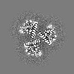
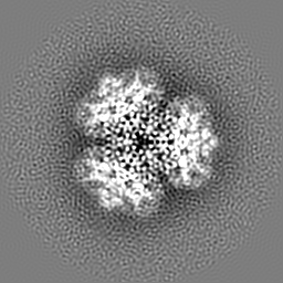
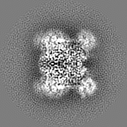
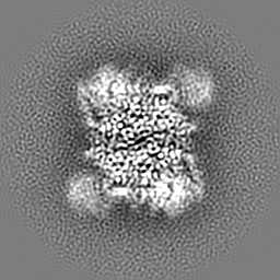
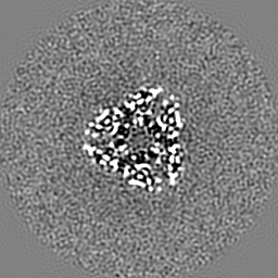
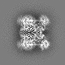
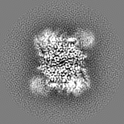
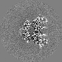
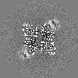
| Title | Cryo-EM structure of Glutamate dehydrogenase from Thermococcus profundus incorporating NADP and GLU in the steady stage of reaction | |||||||||||||||||||||||||||||||||||||||||||||
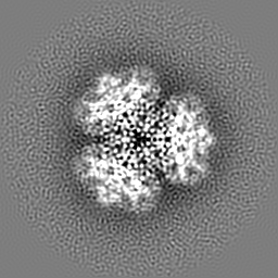
 Map data Map data | ||||||||||||||||||||||||||||||||||||||||||||||
 Sample Sample |
| |||||||||||||||||||||||||||||||||||||||||||||
 Keywords Keywords | Complex / Coenzyme / NADP / Glutamate / OXIDOREDUCTASE | |||||||||||||||||||||||||||||||||||||||||||||
| Function / homology |  Function and homology information Function and homology informationglutamate dehydrogenase [NAD(P)+] / L-glutamate dehydrogenase (NADP+) activity / L-glutamate dehydrogenase (NAD+) activity / L-glutamate catabolic process Similarity search - Function | |||||||||||||||||||||||||||||||||||||||||||||
| Biological species |   Thermococcus profundus (archaea) Thermococcus profundus (archaea) | |||||||||||||||||||||||||||||||||||||||||||||
| Method | single particle reconstruction / cryo EM / Resolution: 2.64 Å | |||||||||||||||||||||||||||||||||||||||||||||
 Authors Authors | Wakabayashi T / Oide M / Nakasako M | |||||||||||||||||||||||||||||||||||||||||||||
| Funding support |  Japan, 14 items Japan, 14 items
| |||||||||||||||||||||||||||||||||||||||||||||
 Citation Citation | Journal: Sci Rep / Year: 2024 Title: CryoEM-sampling of metastable conformations appearing in cofactor-ligand association and catalysis of glutamate dehydrogenase. Authors: Taiki Wakabayashi / Mao Oide / Masayoshi Nakasako / Abstract: Kinetic aspects of enzymatic reactions are described by equations based on the Michaelis-Menten theory for the initial stage. However, the kinetic parameters provide little information on the atomic ...Kinetic aspects of enzymatic reactions are described by equations based on the Michaelis-Menten theory for the initial stage. However, the kinetic parameters provide little information on the atomic mechanism of the reaction. In this study, we analyzed structures of glutamate dehydrogenase in the initial and steady stages of the reaction using cryoEM at near-atomic resolution. In the initial stage, four metastable conformations displayed different domain motions and cofactor/ligand association modes. The most striking finding was that the enzyme-cofactor-substrate complex, treated as a single state in the enzyme kinetic theory, comprised at least three different metastable conformations. In the steady stage, seven conformations, including derivatives from the four conformations in the initial stage, made the reaction pathway complicated. Based on the visualized conformations, we discussed stage-dependent pathways to illustrate the dynamics of the enzyme in action. | |||||||||||||||||||||||||||||||||||||||||||||
| History |
|
- Structure visualization
| Supplemental images |
|---|

- Downloads & links
-EMDB archive
| Map data | emd_38263.map.gz | 26.9 MB | EMDB map data format | |
|---|---|---|---|---|
| Header (meta data) | emd-38263-v30.xmlemd-38263.xml | 21.3 KB 21.3 KB | Display Display | EMDB header |
| FSC (resolution estimation) | emd_38263_fsc.xml | 9.2 KB | Display | FSC data file |
| Images | 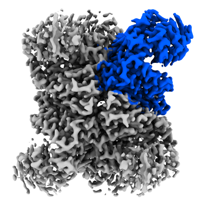 emd_38263.png emd_38263.png | 151.9 KB | ||
| Masks | emd_38263_msk_1.map | 64 MB | Mask map | |
| Filedesc metadata | emd-38263.cif.gz | 6.6 KB | ||
| Others | emd_38263_half_map_1.map.gzemd_38263_half_map_2.map.gz | 60 MB 60 MB | ||
| Archive directory |  http://ftp.pdbj.org/pub/emdb/structures/EMD-38263ftp://ftp.pdbj.org/pub/emdb/structures/EMD-38263 http://ftp.pdbj.org/pub/emdb/structures/EMD-38263ftp://ftp.pdbj.org/pub/emdb/structures/EMD-38263 | HTTPS FTP |
-Related structure data
| Related structure data | 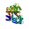 8xd2MC 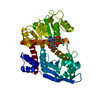 8xcoC 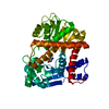 8xcpC 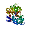 8xcqC 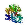 8xcrC  8xcsC 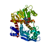 8xctC  8xcuC  8xcvC 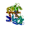 8xcwC  8xcxC 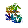 8xcyC 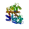 8xczC 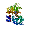 8xd0C  8xd1C 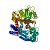 8xd3C 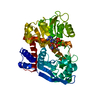 8xd4C  8xd5C 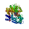 8xd6C M: atomic model generated by this map C: citing same article ( |
|---|---|
| Similar structure data |


-Links
| EMDB pages | EMDB (EBI/PDBe) / EMDataResource |
|---|
-Map
| File | Download / File: emd_38263.map.gz / Format: CCP4 / Size: 64 MB / Type: IMAGE STORED AS FLOATING POINT NUMBER (4 BYTES) | ||||||||||||||||||||||||||||||||||||
|---|---|---|---|---|---|---|---|---|---|---|---|---|---|---|---|---|---|---|---|---|---|---|---|---|---|---|---|---|---|---|---|---|---|---|---|---|---|
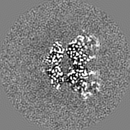
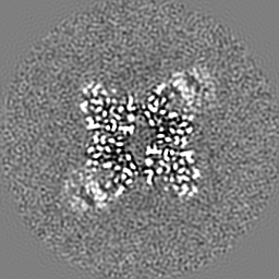
| Projections & slices | Image control
Images are generated by Spider. | ||||||||||||||||||||||||||||||||||||
| Voxel size | X=Y=Z: 0.752 Å | ||||||||||||||||||||||||||||||||||||
| Density |
| ||||||||||||||||||||||||||||||||||||
| Symmetry | Space group: 1 | ||||||||||||||||||||||||||||||||||||
| Details | EMDB XML:
|
 Z (Sec.)
 Y (Row.)
 X (Col.)


-Supplemental data
-Mask #1
| File | emd_38263_msk_1.map | ||||||||||||
|---|---|---|---|---|---|---|---|---|---|---|---|---|---|
| Projections & Slices |
| ||||||||||||
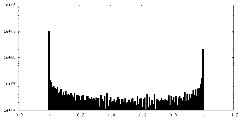
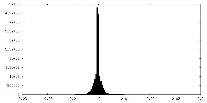
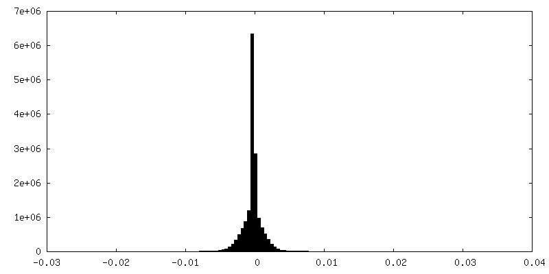
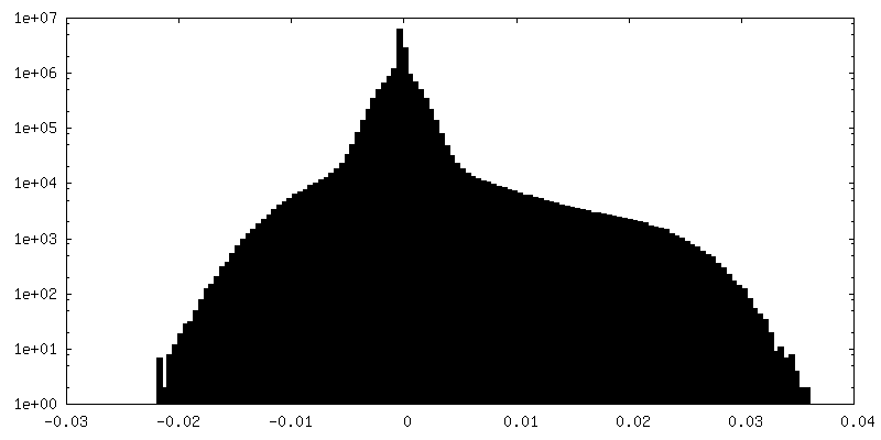
| Density Histograms |


-Half map: #2
| File | emd_38263_half_map_1.map | ||||||||||||
|---|---|---|---|---|---|---|---|---|---|---|---|---|---|
| Projections & Slices |
| ||||||||||||
| Density Histograms |

-Half map: #1
| File | emd_38263_half_map_2.map | ||||||||||||
|---|---|---|---|---|---|---|---|---|---|---|---|---|---|
| Projections & Slices |
| ||||||||||||
| Density Histograms |

- Sample components
-Entire : Hexamer of glutamate dehydrogenase in the presence of NADP and gl...
| Entire | Name: Hexamer of glutamate dehydrogenase in the presence of NADP and glutamate |
|---|---|
| Components |
|
-Supramolecule #1: Hexamer of glutamate dehydrogenase in the presence of NADP and gl...
| Supramolecule | Name: Hexamer of glutamate dehydrogenase in the presence of NADP and glutamate type: complex / ID: 1 / Parent: 0 / Macromolecule list: #1 |
|---|---|
| Source (natural) | Organism: Thermococcus profundus (archaea) |
| Molecular weight | Theoretical: 280 KDa |
-Macromolecule #1: Glutamate dehydrogenase
| Macromolecule | Name: Glutamate dehydrogenase / type: protein_or_peptide / ID: 1 / Number of copies: 1 / Enantiomer: LEVO / EC number: glutamate dehydrogenase [NAD(P)+] |
|---|---|
| Source (natural) | Organism: Thermococcus profundus (archaea) |
| Molecular weight | Theoretical: 46.758477 KDa |
| Recombinant expression | Organism:  |
| Sequence | String: MVEIDPFEMA VKQLERAAQY MDISEEALEW LKKPMRIVEV SVPIEMDDGS VKVFTGFRVQ HNWARGPTKG GIRWHPAETL STVKALATW MTWKVAVVDL PYGGGKGGII VNPKELSERE QERLARAYIR AVYDVIGPWT DIPAPDVYTN PKIMGWMMDE Y ETIMRRKG ...String: MVEIDPFEMA VKQLERAAQY MDISEEALEW LKKPMRIVEV SVPIEMDDGS VKVFTGFRVQ HNWARGPTKG GIRWHPAETL STVKALATW MTWKVAVVDL PYGGGKGGII VNPKELSERE QERLARAYIR AVYDVIGPWT DIPAPDVYTN PKIMGWMMDE Y ETIMRRKG PAFGVITGKP LSIGGSLGRG TATAQGAIFT IREAAKALGI DLKGKKIAVQ GYGNAGYYTA KLAKEQLGMT VV AVSDSRG GIYNPDGLDP DEVLKWKREH GSVKDFPGAT NITNEELLEL EVDVLAPAAI EEVITEKNAD NIKAKIVAEV ANG PVTPEA DDILREKGIL QIPDFLCNAG GVTVSYFEWV QNINGYYWTE EEVREKLDKK MTKAFWEVYN THKDKNIHMR DAAY VVAVS RVYQAMKDRG WVKK UniProtKB: Glutamate dehydrogenase |
-Macromolecule #2: NADP NICOTINAMIDE-ADENINE-DINUCLEOTIDE PHOSPHATE
| Macromolecule | Name: NADP NICOTINAMIDE-ADENINE-DINUCLEOTIDE PHOSPHATE / type: ligand / ID: 2 / Number of copies: 1 / Formula: NAP |
|---|---|
| Molecular weight | Theoretical: 743.405 Da |
| Chemical component information |  ChemComp-NAP: |
-Macromolecule #3: GAMMA-L-GLUTAMIC ACID
| Macromolecule | Name: GAMMA-L-GLUTAMIC ACID / type: ligand / ID: 3 / Number of copies: 1 / Formula: GGL |
|---|---|
| Molecular weight | Theoretical: 147.129 Da |
| Chemical component information |  ChemComp-GGL: |
-Macromolecule #4: water
| Macromolecule | Name: water / type: ligand / ID: 4 / Number of copies: 9 / Formula: HOH |
|---|---|
| Molecular weight | Theoretical: 18.015 Da |
| Chemical component information |  ChemComp-HOH: |
-Experimental details
-Structure determination
| Method | cryo EM |
|---|---|
 Processing Processing | single particle reconstruction |
| Aggregation state | particle |
-Sample preparation
| Concentration | 4.8 mg/mL | ||||||||||||
|---|---|---|---|---|---|---|---|---|---|---|---|---|---|
| Buffer | pH: 7.5 Component:
| ||||||||||||
| Vitrification | Cryogen name: ETHANE / Chamber humidity: 100 % / Chamber temperature: 281 K / Instrument: FEI VITROBOT MARK IV Details: The sample solution kept at room temperature was flash-frozen 1-h after mixing GDH, NADP and glutamate solutions.. |
- Electron microscopy
| Microscope | JEOL CRYO ARM 300 |
|---|---|
| Details | Grid information as following: Company/model: Quantifoil Cu 1.2/1.3 Material:Cu Grid mesh: 200 |
| Image recording | Film or detector model: GATAN K3 (6k x 4k) / Number real images: 7075 / Average electron dose: 1.0 e/Å2 |
| Electron beam | Acceleration voltage: 300 kV / Electron source:  FIELD EMISSION GUN FIELD EMISSION GUN |
| Electron optics | Illumination mode: OTHER / Imaging mode: BRIGHT FIELD / Nominal defocus max: 47.0 µm / Nominal defocus min: 0.4 µm |
| Sample stage | Cooling holder cryogen: NITROGEN |
+Image processing

-Atomic model buiding 1
| Refinement | Protocol: RIGID BODY FIT |
|---|---|
| Output model | PDB-8xd2: |
