[English] 日本語
Yorodumi
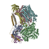
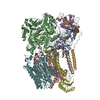
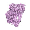
- EMDB-38012: Cryo-EM structure of the photosynthetic alternative complex III w... -

+
Open data


ID or keywords:

Loading...

-
Basic information

Entry
Database: EMDB / ID: EMD-38012
TitleCryo-EM structure of the photosynthetic alternative complex III with a quinone inhibitor HQNO from Chloroflexus aurantiacus
Map data
Sample
  • Complex: Alternative complex III
    • Protein or peptide: x 8 types
  • Ligand: x 7 types
KeywordsPhotosynthetic alternative complex III / MEMBRANE PROTEIN
Function / homology
Function and homology information


electron transfer activity / heme binding / membrane / metal ion binding / plasma membrane
Similarity search - Function
NrfD family / Alternative complex III, ActD subunit / MoCo/4Fe-4S cofactor protein extended Tat translocation domain / Polysulphide reductase, NrfD / Alternative complex III, ActD subunit / Cytochrome c7-like / Cytochrome c7 and related cytochrome c / Cytochrome C oxidase, cbb3-type, subunit III / Multiheme cytochrome superfamily / 4Fe-4S dicluster domain ...NrfD family / Alternative complex III, ActD subunit / MoCo/4Fe-4S cofactor protein extended Tat translocation domain / Polysulphide reductase, NrfD / Alternative complex III, ActD subunit / Cytochrome c7-like / Cytochrome c7 and related cytochrome c / Cytochrome C oxidase, cbb3-type, subunit III / Multiheme cytochrome superfamily / 4Fe-4S dicluster domain / Cytochrome c family profile. / Cytochrome c-like domain / Cytochrome c-like domain superfamily / Twin arginine translocation (Tat) signal profile. / Twin-arginine translocation pathway, signal sequence / 4Fe-4S ferredoxin-type iron-sulfur binding domain profile. / 4Fe-4S ferredoxin-type, iron-sulphur binding domain / Prokaryotic membrane lipoprotein lipid attachment site profile.
Similarity search - Domain/homology
Cytochrome c7-like domain-containing protein / Fe-S-cluster-containing hydrogenase components 1-like protein / Polysulphide reductase NrfD / Quinol:cytochrome c oxidoreductase membrane protein / Cytochrome c domain-containing protein / Quinol:cytochrome c oxidoreductase quinone-binding subunit 2 / Uncharacterized protein
Similarity search - Component
Biological speciesChloroflexus aurantiacus (strain ATCC 29366 / DSM 635 / J-10-fl) (bacteria)
Methodsingle particle reconstruction / cryo EM / Resolution: 2.7 Å
AuthorsXu X
Funding support China, 1 items
OrganizationGrant numberCountry
National Natural Science Foundation of China (NSFC) China
CitationJournal: Plant Cell / Year: 2024
Title: Cryo-EM structure of HQNO-bound alternative complex III from the anoxygenic phototrophic bacterium Chloroflexus aurantiacus.
Authors: Jiyu Xin / Zhenzhen Min / Lu Yu / Xinyi Yuan / Aokun Liu / Wenping Wu / Xin Zhang / Huimin He / Jingyi Wu / Yueyong Xin / Robert E Blankenship / Changlin Tian / Xiaoling Xu /
Abstract: Alternative complex III (ACIII) couples quinol oxidation and electron acceptor reduction with potential transmembrane proton translocation. It is compositionally and structurally different from the ...Alternative complex III (ACIII) couples quinol oxidation and electron acceptor reduction with potential transmembrane proton translocation. It is compositionally and structurally different from the cytochrome bc1/b6f complexes but functionally replaces these enzymes in the photosynthetic and/or respiratory electron transport chains (ETCs) of many bacteria. However, the true compositions and architectures of ACIIIs remain unclear, as do their structural and functional relevance in mediating the ETCs. We here determined cryogenic electron microscopy structures of photosynthetic ACIII isolated from Chloroflexus aurantiacus (CaACIIIp), in apo-form and in complexed form bound to a menadiol analog 2-heptyl-4-hydroxyquinoline-N-oxide. Besides 6 canonical subunits (ActABCDEF), the structures revealed conformations of 2 previously unresolved subunits, ActG and I, which contributed to the complex stability. We also elucidated the structural basis of menaquinol oxidation and subsequent electron transfer along the [3Fe-4S]-6 hemes wire to its periplasmic electron acceptors, using electron paramagnetic resonance, spectroelectrochemistry, enzymatic analyses, and molecular dynamics simulations. A unique insertion loop in ActE was shown to function in determining the binding specificity of CaACIIIp for downstream electron acceptors. This study broadens our understanding of the structural diversity and molecular evolution of ACIIIs, enabling further investigation of the (mena)quinol oxidoreductases-evolved coupling mechanism in bacterial energy conservation.
History
DepositionNov 9, 2023-
Header (metadata) releaseMar 6, 2024-
Map releaseMar 6, 2024-
UpdateMar 6, 2024-
Current statusMar 6, 2024Processing site: PDBc / Status: Released

-
Structure visualization

Supplemental images

Downloads & links

-
Map

FileDownload / File: emd_38012.map.gz / Format: CCP4 / Size: 38.4 MB / Type: IMAGE STORED AS FLOATING POINT NUMBER (4 BYTES)
Projections & slices

Image control

Size
Brightness
Contrast
Others
AxesZ (Sec.)Y (Row.)X (Col.)
0.93 Å/pix.
x 216 pix.
= 200.88 Å
0.93 Å/pix.
x 216 pix.
= 200.88 Å
0.93 Å/pix.
x 216 pix.
= 200.88 Å

Surface

Projections

Slices (1/3)

Slices (1/2)

Slices (2/3)

Images are generated by Spider.

Voxel sizeX=Y=Z: 0.93 Å
Density
Contour LevelBy AUTHOR: 0.13
Minimum - Maximum-1.4348722 - 1.6940205
Average (Standard dev.)0.0038035687 (±0.0717799)
SymmetrySpace group: 1
Details

EMDB XML:

Map geometry
Axis orderXYZ
Origin000
Dimensions216216216
Spacing216216216
CellA=B=C: 200.88 Å
α=β=γ: 90.0 °

-
Supplemental data

-
Half map: #2

Fileemd_38012_half_map_1.map
Projections & Slices
AxesZYX

Projections

Slices (1/2)
Density Histograms

-
Half map: #1

Fileemd_38012_half_map_2.map
Projections & Slices
AxesZYX

Projections

Slices (1/2)
Density Histograms

-
Sample components

+
Entire : Alternative complex III

EntireName: Alternative complex III
Components
  • Complex: Alternative complex III
    • Protein or peptide: Cytochrome c7-like domain-containing protein
    • Protein or peptide: Fe-S-cluster-containing hydrogenase components 1-like protein
    • Protein or peptide: Polysulphide reductase NrfD
    • Protein or peptide: Quinol:cytochrome c oxidoreductase membrane protein
    • Protein or peptide: Cytochrome c domain-containing protein
    • Protein or peptide: Quinol:cytochrome c oxidoreductase quinone-binding subunit 2
    • Protein or peptide: Uncharacterized protein
    • Protein or peptide: unknown
  • Ligand: HEME C
  • Ligand: IRON/SULFUR CLUSTER
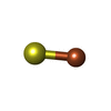
  • Ligand: FE3-S4 CLUSTER
  • Ligand: [(2~{R})-3-[2-azanylethoxy(oxidanyl)phosphoryl]oxy-2-tetradecanoyloxy-propyl] hexadecanoate
  • Ligand: 2-HEPTYL-4-HYDROXY QUINOLINE N-OXIDE
  • Ligand: [(2~{R})-3-[2-azanylethoxy(oxidanyl)phosphoryl]oxy-2-pentadecanoyloxy-propyl] pentadecanoate
  • Ligand: 1,3-bis(13-methyltetradecanoyloxy)propan-2-yl pentadecanoate

+
Supramolecule #1: Alternative complex III

SupramoleculeName: Alternative complex III / type: complex / ID: 1 / Parent: 0 / Macromolecule list: #1-#8
Source (natural)Organism: Chloroflexus aurantiacus (strain ATCC 29366 / DSM 635 / J-10-fl) (bacteria)

+
Macromolecule #1: Cytochrome c7-like domain-containing protein

MacromoleculeName: Cytochrome c7-like domain-containing protein / type: protein_or_peptide / ID: 1 / Number of copies: 1 / Enantiomer: LEVO
Source (natural)Organism: Chloroflexus aurantiacus (strain ATCC 29366 / DSM 635 / J-10-fl) (bacteria)
Molecular weightTheoretical: 25.247039 KDa
SequenceString: MAQIFPRNAN LLSRLSIFAL VLLVVEGILI LGVYFRSNYF RQVNVAIEQP VAFSHQLHVN VVGIDCRYCH TSVDQSYFAN IPATETCMT CHSQIKTYSP LLEKVRESYA TGKPIEWVKV YDLPNFVYFN HSIHVNKGIG CSTCHGQVNN MPVVWQQQAL Y MGWCLNCH ...String:
MAQIFPRNAN LLSRLSIFAL VLLVVEGILI LGVYFRSNYF RQVNVAIEQP VAFSHQLHVN VVGIDCRYCH TSVDQSYFAN IPATETCMT CHSQIKTYSP LLEKVRESYA TGKPIEWVKV YDLPNFVYFN HSIHVNKGIG CSTCHGQVNN MPVVWQQQAL Y MGWCLNCH RNPELYVRPR EEVYNMDYVP PSNQLEIGRQ LVAEYGIMPP DQLTNCYVCH R

UniProtKB: Cytochrome c7-like domain-containing protein

+
Macromolecule #2: Fe-S-cluster-containing hydrogenase components 1-like protein

MacromoleculeName: Fe-S-cluster-containing hydrogenase components 1-like protein
type: protein_or_peptide / ID: 2 / Number of copies: 1 / Enantiomer: LEVO
Source (natural)Organism: Chloroflexus aurantiacus (strain ATCC 29366 / DSM 635 / J-10-fl) (bacteria)
Molecular weightTheoretical: 113.115359 KDa
SequenceString: MTQQQPDLEA IRAQLRDARG PQFWRSLDQL ADAPAFRELI EREFPRGASE LEDGISRRTF LKLMGASLAL AGVTACTYQP RQYIAPFDR QPEGRVPGIP QYFASTLTLG GYGTGVLVRS NEGRPTKVEG NPRHPASLGG TDLFAQAEIL TMYDPDRSTT V LRQGVPST ...String:
MTQQQPDLEA IRAQLRDARG PQFWRSLDQL ADAPAFRELI EREFPRGASE LEDGISRRTF LKLMGASLAL AGVTACTYQP RQYIAPFDR QPEGRVPGIP QYFASTLTLG GYGTGVLVRS NEGRPTKVEG NPRHPASLGG TDLFAQAEIL TMYDPDRSTT V LRQGVPST WAEFTTTLGN ALTAARATQG AGVRLLTTTI TSPSLAAQIE QFLQAYPQAR WYQYEPINRD NVVAGARLAF GR DVTTRYD LSAAQVVVSL DADFLAPGPG FVAYARAFAE RRKVRKDSTT MNRLYVVEAS PSTTGTAADH RLPLRADAIA AFT GALANE LGVGGAPATL SPKAEEFLRA IARDLEEHRG QSVVIAGDQQ PPIVHALAHL INAELGNVGQ TVFYHEPVEA RPTN QTEEL VALVSEMAAG RVETLIMIGG NPVYNAPGDL RFADRMASVP LTIHLSQFVD ETSARATWHI PQAHPLESWG DARAF DGTA SIVQPLIEPL YGGKTANELL AAMLGQPEAE SYDLVRSFWL EQIGETGWQV ALANGVIAET VAPVIEPTLN EGAIRA TPI PQPGDGVEIV FRPDPSLFDG FYANNGWLQE LPRPLTKLVW DNAALMSPRT AIKLLGLPFN ADRLIGTEAD DRERQQY LE QLSKVNGTIA RIEYRGGIIE IPIWLLPGHA EDSITLNLGY GRTHAGRVGN NVGIDVYPIR TSDSPWFGAG ARVTNTGR T YLLVSTQDHW TLEGRDIYRV GEFKKFKEDP KYIAKEVYQE EYGRETPNYQ SLQPGDDYTG RNAWGMTINL NACIGCNAC VVACQAENNI AVVGKDQVSR GREMHWIRID RYFAGEDLDN PSIYMMPVNC MQCEKAPCEV VCPVAATVHD YEGLNNMVYN RCVGTKYCS NNCPYKVRRF NFLQYSDTTT ETFKLAFNPD VTVRIRGVME KCTYCVQRIS GARIAAKRAA VQAGQSSYVI S DGAIQTAC EQACPTGAIV FGDINDSNSR VAKWKAEGHN YGLLGFLNTV PRTTYLARVR NPSEELEKVE G

UniProtKB: Fe-S-cluster-containing hydrogenase components 1-like protein

+
Macromolecule #3: Polysulphide reductase NrfD

MacromoleculeName: Polysulphide reductase NrfD / type: protein_or_peptide / ID: 3 / Number of copies: 1 / Enantiomer: LEVO
Source (natural)Organism: Chloroflexus aurantiacus (strain ATCC 29366 / DSM 635 / J-10-fl) (bacteria)
Molecular weightTheoretical: 55.22352 KDa
SequenceString: MAQAQPLRTR PQDDGEAYLL PGETYTSISA KIGDVPLTPP LKTPKGWLAG FSVAFFMLMI FFVSVTWLFI RGVGIWGINI PVGWGMDII NFVWWIGIGH AGTLISAILL LLNQGWRNSI NRFAEAMTLF AVACAGLYPI LHLGRPWLFY WLIPYPNTHG M WPQFRSAL ...String:
MAQAQPLRTR PQDDGEAYLL PGETYTSISA KIGDVPLTPP LKTPKGWLAG FSVAFFMLMI FFVSVTWLFI RGVGIWGINI PVGWGMDII NFVWWIGIGH AGTLISAILL LLNQGWRNSI NRFAEAMTLF AVACAGLYPI LHLGRPWLFY WLIPYPNTHG M WPQFRSAL AWDVFAISTY ATVSLVFWLV GLIPDFATLR DRAKNIWVKR LYGIAALGWR GSARHWHRYE MASILLAGLS TP LVVSVHS IISLDFAISQ VPGWQVTVFP PYFVAGAVFA GFAMVLLLMI PVRTFYGFEN YITLHHLDVM AKVMLTTGMI VVY GYFMEV FASLYSGNEF EEYLLYNRLF GPSSWAYWGL LFCNAVAIQP LWFKKVRQNI PALLIISLIV SVGMWLERYV IIVI SLERD FLPSSWDIYI PTIWDWSLYI GTFGLFFTLL FLFIRVLPMI NIFEMRLFLY QETEKAKQRA GHGAHGHGHE QSPAH GAAT AD

UniProtKB: Polysulphide reductase NrfD

+
Macromolecule #4: Quinol:cytochrome c oxidoreductase membrane protein

MacromoleculeName: Quinol:cytochrome c oxidoreductase membrane protein / type: protein_or_peptide / ID: 4 / Number of copies: 1 / Enantiomer: LEVO
Source (natural)Organism: Chloroflexus aurantiacus (strain ATCC 29366 / DSM 635 / J-10-fl) (bacteria)
Molecular weightTheoretical: 19.64857 KDa
SequenceString:
MRNDVYGVMA EFPTPEALIE ATRKAKAAGY TKMDAFSPFP IEEVIEEIAH GDTGVPRLVL LFGLIGAASG FILQYIGNLV DYPLNVGGR PLDITNWPAM IPITFESGIL LASFAAAIGM IVLNGLPSPY HPVFNVPRFQ YASQDAFFLC IEATDPLFDR S RTSQFLRS LNPMQVSEVA Y

UniProtKB: Quinol:cytochrome c oxidoreductase membrane protein

+
Macromolecule #5: Cytochrome c domain-containing protein

MacromoleculeName: Cytochrome c domain-containing protein / type: protein_or_peptide / ID: 5 / Number of copies: 1 / Enantiomer: LEVO
Source (natural)Organism: Chloroflexus aurantiacus (strain ATCC 29366 / DSM 635 / J-10-fl) (bacteria)
Molecular weightTheoretical: 23.017016 KDa
SequenceString: MQKPRLTSRM IRFGWVGLLV LLLTACHQDM YDQQKYTTYE PSSFFADGRS SRPNVPGTTP FEVVKTDEFL YTGLIDGQEV DAMPFPVTK DLLLRGQLKY NIYCAVCHGE AGYGASMVAE RGGIVPANFH QQRLREAPLS HFFVVITNGV YRGDPENGGY Q SMYGYASR ...String:
MQKPRLTSRM IRFGWVGLLV LLLTACHQDM YDQQKYTTYE PSSFFADGRS SRPNVPGTTP FEVVKTDEFL YTGLIDGQEV DAMPFPVTK DLLLRGQLKY NIYCAVCHGE AGYGASMVAE RGGIVPANFH QQRLREAPLS HFFVVITNGV YRGDPENGGY Q SMYGYASR ITPEDRWAIA AYIRALQLSQ NATIDDVPPD QRAQLGN

UniProtKB: Cytochrome c domain-containing protein

+
Macromolecule #6: Quinol:cytochrome c oxidoreductase quinone-binding subunit 2

MacromoleculeName: Quinol:cytochrome c oxidoreductase quinone-binding subunit 2
type: protein_or_peptide / ID: 6 / Number of copies: 1 / Enantiomer: LEVO
Source (natural)Organism: Chloroflexus aurantiacus (strain ATCC 29366 / DSM 635 / J-10-fl) (bacteria)
Molecular weightTheoretical: 45.721672 KDa
SequenceString: MATTSISQTR IPQLGQVQML GLAAAVIGIG VLAAGYFLSP TSFFESYIYG YYVAMTIPLG CLGFLMVQHL TGGAWGVTVR RMLEAGAAT LPIMGLLFIP IALGYFDTYK ALGLEHPLYE WANPEVVTPG GAEFDPIIAH KVPWLSPLWV TARIAIFFII W SALALTLR ...String:
MATTSISQTR IPQLGQVQML GLAAAVIGIG VLAAGYFLSP TSFFESYIYG YYVAMTIPLG CLGFLMVQHL TGGAWGVTVR RMLEAGAAT LPIMGLLFIP IALGYFDTYK ALGLEHPLYE WANPEVVTPG GAEFDPIIAH KVPWLSPLWV TARIAIFFII W SALALTLR AWSRQQDAGG DAKKLATRMR RLSGIGVALF VITVTFFSFD VAMSLDPHWF STIYGAHYMA NAGLMTLAFL AL MMSRVRD AALFREYVSV KPIHDIGKLI FAFTVLWTYM SYGQLVIIWS GDVAEFTPWY VHRTQHGWVF VALALMLFAF ALP FFVLLF RGTKRNLNTL ATIAGWIVVM RFVDMAWIIL PEFREHLWDI AITDVAAPIG LIGLVIALFA ANVQQAPLLP LRDP NMEQL QNSGHH

UniProtKB: Quinol:cytochrome c oxidoreductase quinone-binding subunit 2

+
Macromolecule #7: Uncharacterized protein

MacromoleculeName: Uncharacterized protein / type: protein_or_peptide / ID: 7 / Number of copies: 1 / Enantiomer: LEVO
Source (natural)Organism: Chloroflexus aurantiacus (strain ATCC 29366 / DSM 635 / J-10-fl) (bacteria)
Molecular weightTheoretical: 12.485551 KDa
SequenceString:
MSYRPNYSAS RYTAGRPAQP VRTARTMAEP SLSRLMIAGL MVFLVLSLVV LLAGRLPFTP QPAPVTGNTY RTYVNDARTL LNSYGYTME GKVHIPIDRA MDLIVERGLP VRE

UniProtKB: Uncharacterized protein

+
Macromolecule #8: unknown

MacromoleculeName: unknown / type: protein_or_peptide / ID: 8 / Number of copies: 1 / Enantiomer: LEVO
Source (natural)Organism: Chloroflexus aurantiacus (strain ATCC 29366 / DSM 635 / J-10-fl) (bacteria)
Molecular weightTheoretical: 4.322134 KDa
SequenceString:
MQPEWSGDPE VKPVFLAVTL TGMVAFLLMV WLFAFYW

+
Macromolecule #9: HEME C

MacromoleculeName: HEME C / type: ligand / ID: 9 / Number of copies: 6 / Formula: HEC
Molecular weightTheoretical: 618.503 Da
Chemical component information

ChemComp-HEC:
HEME C

+
Macromolecule #10: IRON/SULFUR CLUSTER

MacromoleculeName: IRON/SULFUR CLUSTER / type: ligand / ID: 10 / Number of copies: 3 / Formula: SF4
Molecular weightTheoretical: 351.64 Da
Chemical component information

ChemComp-FS1:
IRON/SULFUR CLUSTER

+
Macromolecule #11: FE3-S4 CLUSTER

MacromoleculeName: FE3-S4 CLUSTER / type: ligand / ID: 11 / Number of copies: 1 / Formula: F3S
Molecular weightTheoretical: 295.795 Da
Chemical component information

ChemComp-F3S:
FE3-S4 CLUSTER

+
Macromolecule #12: [(2~{R})-3-[2-azanylethoxy(oxidanyl)phosphoryl]oxy-2-tetradecanoy...

MacromoleculeName: [(2~{R})-3-[2-azanylethoxy(oxidanyl)phosphoryl]oxy-2-tetradecanoyloxy-propyl] hexadecanoate
type: ligand / ID: 12 / Number of copies: 1 / Formula: JLQ
Molecular weightTheoretical: 663.906 Da

+
Macromolecule #13: 2-HEPTYL-4-HYDROXY QUINOLINE N-OXIDE

MacromoleculeName: 2-HEPTYL-4-HYDROXY QUINOLINE N-OXIDE / type: ligand / ID: 13 / Number of copies: 1 / Formula: HQO
Molecular weightTheoretical: 259.343 Da
Chemical component information

ChemComp-HQO:
2-HEPTYL-4-HYDROXY QUINOLINE N-OXIDE

+
Macromolecule #14: [(2~{R})-3-[2-azanylethoxy(oxidanyl)phosphoryl]oxy-2-pentadecanoy...

MacromoleculeName: [(2~{R})-3-[2-azanylethoxy(oxidanyl)phosphoryl]oxy-2-pentadecanoyloxy-propyl] pentadecanoate
type: ligand / ID: 14 / Number of copies: 1 / Formula: JL3
Molecular weightTheoretical: 663.906 Da

+
Macromolecule #15: 1,3-bis(13-methyltetradecanoyloxy)propan-2-yl pentadecanoate

MacromoleculeName: 1,3-bis(13-methyltetradecanoyloxy)propan-2-yl pentadecanoate
type: ligand / ID: 15 / Number of copies: 1 / Formula: JM9
Molecular weightTheoretical: 765.24 Da

-
Experimental details

-
Structure determination

Methodcryo EM
Processingsingle particle reconstruction
Aggregation stateparticle

-
Sample preparation

BufferpH: 8
VitrificationCryogen name: ETHANE

-
Electron microscopy

MicroscopeFEI TITAN KRIOS
Image recordingFilm or detector model: FEI FALCON IV (4k x 4k) / Average electron dose: 60.0 e/Å2
Electron beamAcceleration voltage: 300 kV / Electron source: FIELD EMISSION GUN
Electron opticsIllumination mode: FLOOD BEAM / Imaging mode: BRIGHT FIELD / Nominal defocus max: 1.6 µm / Nominal defocus min: 1.0 µm
Experimental equipment
Model: Titan Krios / Image courtesy: FEI Company

-
Image processing

Startup modelType of model: NONE
Final reconstructionResolution.type: BY AUTHOR / Resolution: 2.7 Å / Resolution method: FSC 0.143 CUT-OFF / Number images used: 160264
Initial angle assignmentType: PROJECTION MATCHING
Final angle assignmentType: PROJECTION MATCHING

+
About Yorodumi

-
News

-
Feb 9, 2022. New format data for meta-information of EMDB entries

New format data for meta-information of EMDB entries

  • Version 3 of the EMDB header file is now the official format.
  • The previous official version 1.9 will be removed from the archive.

Related info.:EMDB header

External links:wwPDB to switch to version 3 of the EMDB data model

-
Aug 12, 2020. Covid-19 info

Covid-19 info

URL: https://pdbj.org/emnavi/covid19.php

New page: Covid-19 featured information page in EM Navigator.

Related info.:Covid-19 info / Mar 5, 2020. Novel coronavirus structure data

+
Mar 5, 2020. Novel coronavirus structure data

Novel coronavirus structure data

Related info.:Yorodumi Speices / Aug 12, 2020. Covid-19 info

External links:COVID-19 featured content - PDBj / Molecule of the Month (242):Coronavirus Proteases

+
Jan 31, 2019. EMDB accession codes are about to change! (news from PDBe EMDB page)
EMDB accession codes are about to change! (news from PDBe EMDB page)

  • The allocation of 4 digits for EMDB accession codes will soon come to an end. Whilst these codes will remain in use, new EMDB accession codes will include an additional digit and will expand incrementally as the available range of codes is exhausted. The current 4-digit format prefixed with “EMD-” (i.e. EMD-XXXX) will advance to a 5-digit format (i.e. EMD-XXXXX), and so on. It is currently estimated that the 4-digit codes will be depleted around Spring 2019, at which point the 5-digit format will come into force.
  • The EM Navigator/Yorodumi systems omit the EMD- prefix.

Related info.:Q: What is EMD? / ID/Accession-code notation in Yorodumi/EM Navigator

External links:EMDB Accession Codes are Changing Soon! / Contact to PDBj

+
Jul 12, 2017. Major update of PDB

Major update of PDB

  • wwPDB released updated PDB data conforming to the new PDBx/mmCIF dictionary.
  • This is a major update changing the version number from 4 to 5, and with Remediation, in which all the entries are updated.
  • In this update, many items about electron microscopy experimental information are reorganized (e.g. em_software).
  • Now, EM Navigator and Yorodumi are based on the updated data.

External links:wwPDB Remediation / Enriched Model Files Conforming to OneDep Data Standards Now Available in the PDB FTP Archive

-
Yorodumi

Thousand views of thousand structures

  • Yorodumi is a browser for structure data from EMDB, PDB, SASBDB, etc.
  • This page is also the successor to EM Navigator detail page, and also detail information page/front-end page for Omokage search.
  • The word "yorodu" (or yorozu) is an old Japanese word meaning "ten thousand". "mi" (miru) is to see.

Related info.:EMDB / PDB / SASBDB / Comparison of 3 databanks / Yorodumi Search / Aug 31, 2016. New EM Navigator & Yorodumi / Yorodumi Papers / Jmol/JSmol / Function and homology information / Changes in new EM Navigator and Yorodumi

Read more