- EMDB-37963: Cryo-EM structure of the hamster prion 23-144 fibril at pH 3.7 -
+
データを開く
IDまたはキーワード:
読み込み中...
-
基本情報
登録情報
データベース: EMDB / ID: EMD-37963
タイトル
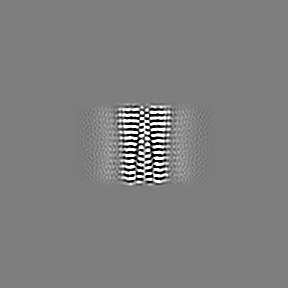
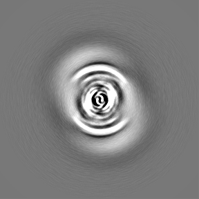
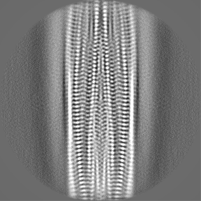
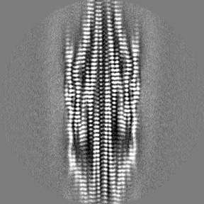
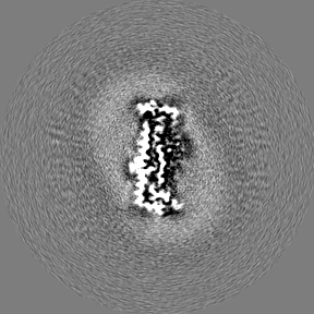
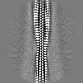
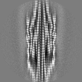
Cryo-EM structure of the hamster prion 23-144 fibril at pH 3.7
マップデータ
試料
複合体: hamster prion 23-144 fibril
タンパク質・ペプチド: Major prion protein
キーワード
prion / 23-144 / hamster / PrP / PROTEIN FIBRIL
機能・相同性
機能・相同性情報
aspartic-type endopeptidase inhibitor activity / regulation of calcium ion import across plasma membrane / positive regulation of glutamate receptor signaling pathway / glycosaminoglycan binding / type 5 metabotropic glutamate receptor binding / negative regulation of interleukin-17 production / cupric ion binding / regulation of potassium ion transmembrane transport / negative regulation of dendritic spine maintenance / negative regulation of calcineurin-NFAT signaling cascade ...aspartic-type endopeptidase inhibitor activity / regulation of calcium ion import across plasma membrane / positive regulation of glutamate receptor signaling pathway / glycosaminoglycan binding / type 5 metabotropic glutamate receptor binding / negative regulation of interleukin-17 production / cupric ion binding / regulation of potassium ion transmembrane transport / negative regulation of dendritic spine maintenance / negative regulation of calcineurin-NFAT signaling cascade / negative regulation of interleukin-2 production / negative regulation of T cell receptor signaling pathway / negative regulation of activated T cell proliferation / negative regulation of amyloid-beta formation / negative regulation of type II interferon production / cuprous ion binding / positive regulation of protein targeting to membrane / side of membrane / inclusion body / neuron projection maintenance / positive regulation of calcium-mediated signaling / molecular function activator activity / cellular response to copper ion / positive regulation of protein localization to plasma membrane / molecular condensate scaffold activity / protein homooligomerization / protein destabilization / cellular response to xenobiotic stimulus / cellular response to amyloid-beta / terminal bouton / positive regulation of neuron apoptotic process / signaling receptor activity / presynapse / amyloid-beta binding / response to oxidative stress / protease binding / nuclear membrane / microtubule binding / amyloid fibril formation / learning or memory / regulation of cell cycle / intracellular signal transduction / membrane raft / copper ion binding / dendrite / negative regulation of apoptotic process / protein-containing complex binding / cell surface / endoplasmic reticulum / negative regulation of transcription by RNA polymerase II / Golgi apparatus / identical protein binding / plasma membrane / cytosol 類似検索 - 分子機能
Prion, copper binding octapeptide repeat / Copper binding octapeptide repeat region / Major prion protein N-terminal domain / Major prion protein bPrPp - N terminal / Prion protein signature 1. / Prion protein signature 2. / Prion protein / Major prion protein / Prion/Doppel protein, beta-ribbon domain / Prion/Doppel beta-ribbon domain superfamily / Prion/Doppel alpha-helical domain 類似検索 - ドメイン・相同性
ジャーナル: J Mol Biol / 年: 2024 タイトル: The Positively Charged Cluster in the N-terminal Disordered Region may Affect Prion Protein Misfolding: Cryo-EM Structure of Hamster PrP(23-144) Fibrils. 著者: Chih-Hsuan Lee / Jing-Ee Saw / Eric H-L Chen / Chun-Hsiung Wang / Takayuki Uchihashi / Rita P-Y Chen / 要旨: Prions, the misfolding form of prion proteins, are contagious proteinaceous macromolecules. Recent studies have shown that infectious prion fibrils formed in the brain and non-infectious fibrils ...Prions, the misfolding form of prion proteins, are contagious proteinaceous macromolecules. Recent studies have shown that infectious prion fibrils formed in the brain and non-infectious fibrils formed from recombinant prion protein in a partially denaturing condition have distinct structures. The amyloid core of the in vitro-prepared non-infectious fibrils starts at about residue 160, while that of infectious prion fibrils formed in the brain involves a longer sequence (residues ∼90-230) of structural conversion. The C-terminal truncated prion protein PrP(23-144) can form infectious fibrils under certain conditions and cause disease in animals. In this study, we used cryogenic electron microscopy (cryo-EM) to resolve the structure of hamster sHaPrP(23-144) fibrils prepared at pH 3.7. This 2.88 Å cryo-EM structure has an amyloid core covering residues 94-144. It comprises two protofilaments, each containing five β-strands arranged as a long hairpin plus an N-terminal β-strand. This N-terminal β-strand resides in a positively charged cluster region (named PCC2; sequence 96-111), which interacts with the turn region of the opposite protofilaments' hairpin to stabilize the fibril structure. Interestingly, this sHaPrP(23-144) fibril structure differs from a recently reported structure formed by the human or mouse counterpart at pH 6.5. Moreover, sHaPrP(23-144) fibrils have many structural features in common with infectious prions. Whether this structure is infectious remains to be determined. More importantly, the sHaPrP(23-144) structure is different from the sHaPrP(108-144) fibrils prepared in the same fibrillization buffer, indicating that the N-terminal disordered region, possibly the positively charged cluster, influences the misfolding pathway of the prion protein.
 ムービー
ムービー コントローラー


 データを開く
 基本情報
基本情報
 マップデータ
 試料
試料 キーワード
キーワード 機能・相同性情報
 Mesocricetus auratus (ネズミ)
 データ登録者
 台湾, 3件
  引用
引用
 構造の表示
構造の表示
 ダウンロードとリンク
 emd_37963.png
 http://ftp.pdbj.org/pub/emdb/structures/EMD-37963

 Z (Sec.)
 Y (Row.)
 X (Col.)


 試料の構成要素
 解析
解析 電子顕微鏡法
 FIELD EMISSION GUN


