 Movie
Movie Controller


+ Open data

- Basic information
| Entry | Database: EMDB / ID: EMD-3792 | |||||||||
|---|---|---|---|---|---|---|---|---|---|---|
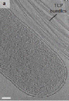
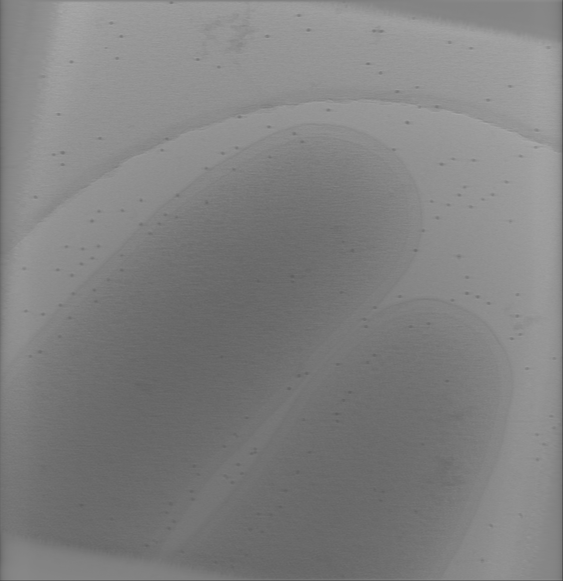
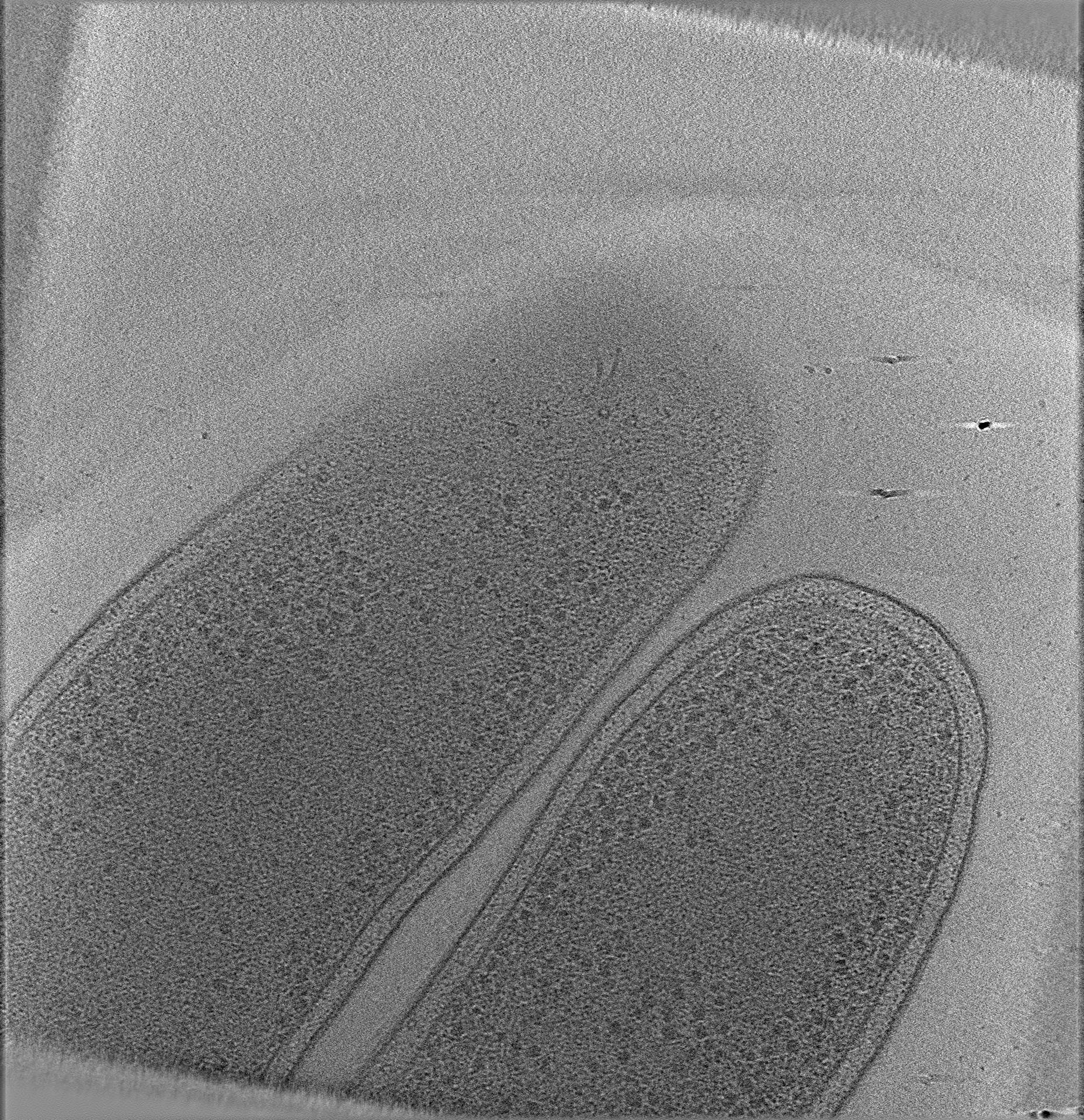
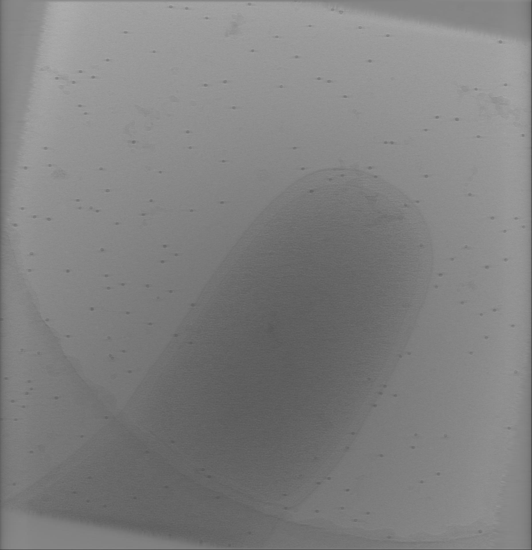
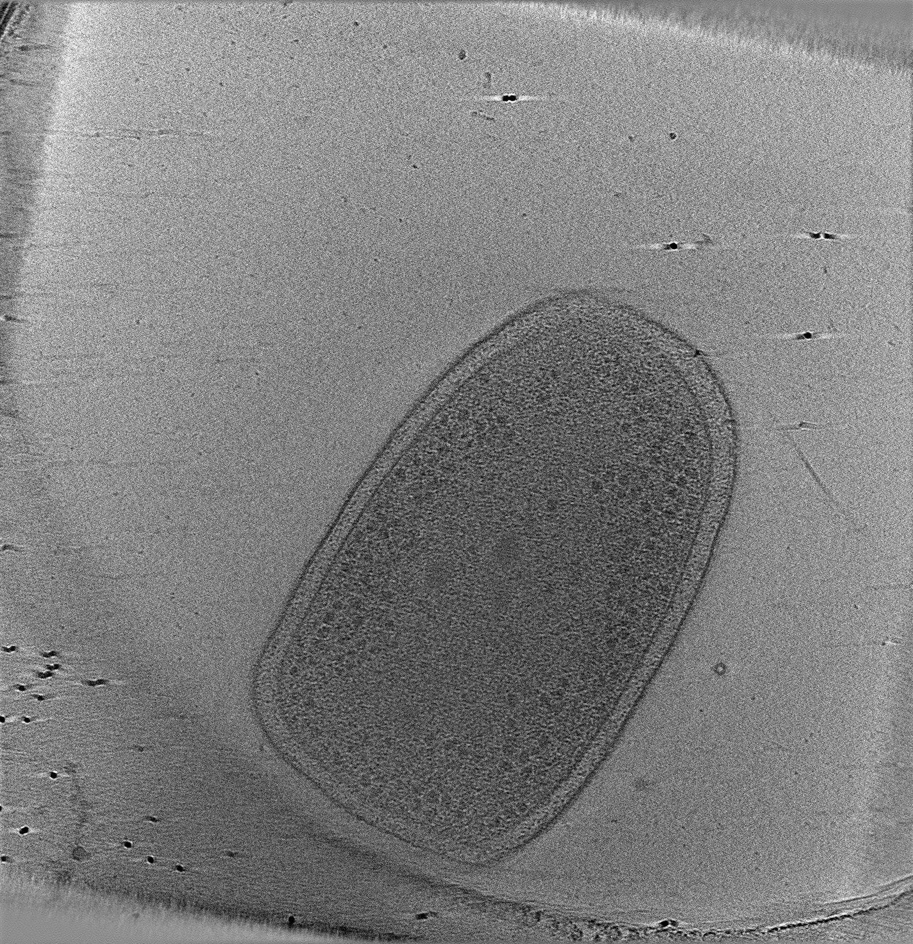
| Title | Electron cryotomogram of Vibrio cholerae O395 N1 | |||||||||
 Map data Map data | ||||||||||
 Sample Sample |
| |||||||||
| Biological species |   Vibrio cholerae (bacteria) Vibrio cholerae (bacteria) | |||||||||
| Method | electron tomography / cryo EM | |||||||||
 Authors Authors | Chang YW / Kjaer A / Ortega DR / Kovacikova G / Sutherland JA / Rettberg LA / Taylor RK / Jensen GJ | |||||||||
 Citation Citation | Journal: Nat Microbiol / Year: 2017 Title: Architecture of the Vibrio cholerae toxin-coregulated pilus machine revealed by electron cryotomography. Authors: Yi-Wei Chang / Andreas Kjær / Davi R Ortega / Gabriela Kovacikova / John A Sutherland / Lee A Rettberg / Ronald K Taylor / Grant J Jensen /   Abstract: Type IV pili (T4P) are filamentous appendages found on many Bacteria and Archaea. They are helical fibres of pilin proteins assembled by a multi-component macromolecular machine we call the basal ...Type IV pili (T4P) are filamentous appendages found on many Bacteria and Archaea. They are helical fibres of pilin proteins assembled by a multi-component macromolecular machine we call the basal body. Based on pilin features, T4P are classified into type IVa pili (T4aP) and type IVb pili (T4bP). T4aP are more widespread and are involved in cell motility, DNA transfer, host predation and electron transfer. T4bP are less prevalent and are mainly found in enteropathogenic bacteria, where they play key roles in host colonization. Following similar work on T4aP machines, here we use electron cryotomography to reveal the three-dimensional in situ structure of a T4bP machine in its piliated and non-piliated states. The specific machine we analyse is the Vibrio cholerae toxin-coregulated pilus machine (TCPM). Although only about half of the components of the TCPM show sequence homology to components of the previously analysed Myxococcus xanthus T4aP machine (T4aPM), we find that their structures are nevertheless remarkably similar. Based on homologies with components of the M. xanthus T4aPM and additional reconstructions of TCPM mutants in which the non-homologous proteins are individually deleted, we propose locations for all eight TCPM components within the complex. Non-homologous proteins in the T4aPM and TCPM are found to form similar structures, suggesting new hypotheses for their functions and evolutionary histories. | |||||||||
| History |
|
- Structure visualization
| Movie |
 Movie viewer Movie viewer |
|---|---|
| Structure viewer | EM map: SurfViewMolmilJmol/JSmol |
| Supplemental images |
- Downloads & links
-EMDB archive
| Map data | emd_3792.map.gz | 4.3 GB | EMDB map data format | |
|---|---|---|---|---|
| Header (meta data) | emd-3792-v30.xmlemd-3792.xml | 15.3 KB 15.3 KB | Display Display | EMDB header |
| Images |  emd_3792.png emd_3792.png | 256.4 KB | ||
| Others | emd_3792_additional.map.gz | 4.3 GB | ||
| Archive directory |  http://ftp.pdbj.org/pub/emdb/structures/EMD-3792ftp://ftp.pdbj.org/pub/emdb/structures/EMD-3792 http://ftp.pdbj.org/pub/emdb/structures/EMD-3792ftp://ftp.pdbj.org/pub/emdb/structures/EMD-3792 | HTTPS FTP |
-Related structure data
| Related structure data | 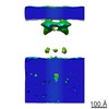 8492C 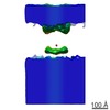 8493C 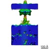 8494C  8495C 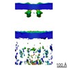 8496C 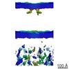 8497C  8498C 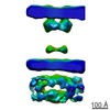 8499C 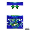 8500C  8501C 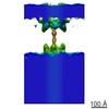 8502C 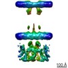 8503C C: citing same article ( |
|---|---|
| EM raw data | EMPIAR-10110 (Title: Tilt-series for the electron cryotomogram of Vibrio cholerae O395 N1 Data size: 51.3 Data #1: Different tilt series for the Vibrio cholerae toxin-coregulated pilus machine revealed by electron cryotomography [tilt series]) |
-Links
| EMDB pages | EMDB (EBI/PDBe) / EMDataResource |
|---|
-Map
| File | Download / File: emd_3792.map.gz / Format: CCP4 / Size: 1.3 GB / Type: IMAGE STORED AS SIGNED INTEGER (2 BYTES) | ||||||||||||||||||||||||||||||||||||||||||||||||||||||||||||
|---|---|---|---|---|---|---|---|---|---|---|---|---|---|---|---|---|---|---|---|---|---|---|---|---|---|---|---|---|---|---|---|---|---|---|---|---|---|---|---|---|---|---|---|---|---|---|---|---|---|---|---|---|---|---|---|---|---|---|---|---|---|
| Projections & slices | Image control
Images are generated by Spider. generated in cubic-lattice coordinate | ||||||||||||||||||||||||||||||||||||||||||||||||||||||||||||
| Voxel size | X=Y=Z: 7.8 Å | ||||||||||||||||||||||||||||||||||||||||||||||||||||||||||||
| Density |
| ||||||||||||||||||||||||||||||||||||||||||||||||||||||||||||
| Symmetry | Space group: 1 | ||||||||||||||||||||||||||||||||||||||||||||||||||||||||||||
| Details | EMDB XML:
CCP4 map header:
| ||||||||||||||||||||||||||||||||||||||||||||||||||||||||||||
 Z (Sec.)
 Y (Row.)
 X (Col.)


-Supplemental data
-Additional map: #1
| File | emd_3792_additional.map | ||||||||||||
|---|---|---|---|---|---|---|---|---|---|---|---|---|---|
| Projections & Slices |
| ||||||||||||
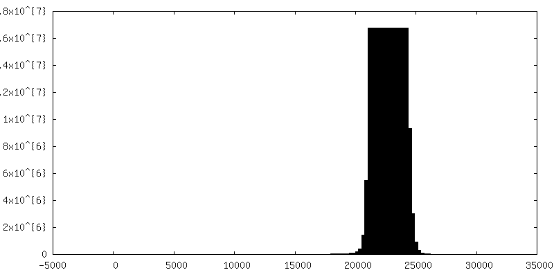
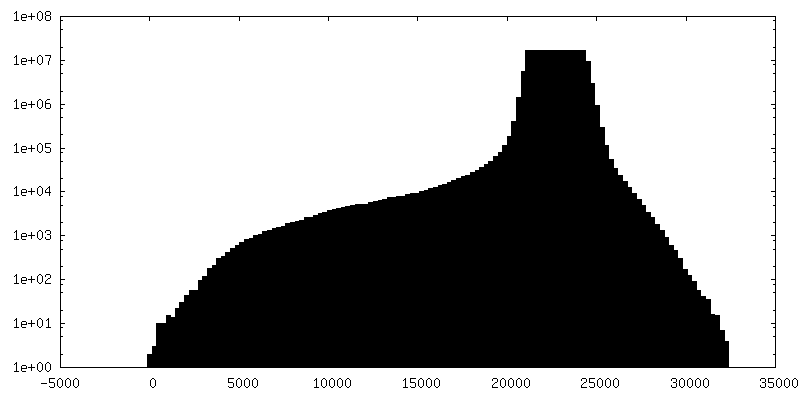
| Density Histograms |


- Sample components
-Entire : Vibrio Cholerae
| Entire | Name: Vibrio Cholerae (bacteria) |
|---|---|
| Components |
|
-Supramolecule #1: Vibrio Cholerae
| Supramolecule | Name: Vibrio Cholerae / type: cell / ID: 1 / Parent: 0 Details: Bacteria which is the causative agent of the diarrhoeal disease cholera. |
|---|
-Supramolecule #3: Vibrio Cholerae
| Supramolecule | Name: Vibrio Cholerae / type: cell / ID: 3 / Parent: 1 Details: Bacteria which is the causative agent of the diarrhoeal disease cholera. |
|---|---|
| Source (natural) | Organism: Vibrio cholerae (bacteria) / Strain: Wild-type |
-Supramolecule #2: Toxin-coregulated pilus machine
| Supramolecule | Name: Toxin-coregulated pilus machine / type: organelle_or_cellular_component / ID: 2 / Parent: 1 Details: Pilus - Bacteria which is the causative agent of the diarrhoeal disease cholera. |
|---|---|
| Source (natural) | Organism: Vibrio cholerae (bacteria) / Strain: Wild-type |
| Recombinant expression | Organism: Vibrio cholerae (bacteria) / Recombinant strain: Wild-type / Recombinant plasmid: pMT5-ToxT |
-Experimental details
-Structure determination
| Method | cryo EM |
|---|---|
 Processing Processing | electron tomography |
| Aggregation state | cell |
-Sample preparation
| Buffer | pH: 7 Component:
Details: V. cholerae O395 N1 strains (wild-type or TCPM component knockout mutants) harbouring the pMT5-ToxT plasmid were grown in 5 ml lysogeny broth (LB) medium. | |||||||||
|---|---|---|---|---|---|---|---|---|---|---|
| Grid | Model: Quantifoil R2/2 / Material: COPPER / Mesh: 200 / Pretreatment - Type: GLOW DISCHARGE | |||||||||
| Vitrification | Cryogen name: ETHANE-PROPANE | |||||||||
| Details | After 24 h of growth, the tube of V. cholerae culture was placed on a bench at room temperature for 1 min to sediment visible microcolonies. After the pellet was formed, the supernatant was gently removed by pipetting and the pellet was resuspended in fresh LB. The cells were incubated for 15 min prior to mixing with 10 nm colloidal gold. | |||||||||
| Sectioning | Other: NO SECTIONING | |||||||||
| Fiducial marker | Manufacturer: Sigma-Aldrich / Diameter: 10 nm |
- Electron microscopy
| Microscope | FEI POLARA 300 |
|---|---|
| Specialist optics | Energy filter - Name: GIF |
| Image recording | Film or detector model: GATAN K2 SUMMIT (4k x 4k) / Digitization - Dimensions - Width: 1856 pixel / Digitization - Dimensions - Height: 1918 pixel / Number grids imaged: 1 / Average electron dose: 1.5 e/Å2 |
| Electron beam | Acceleration voltage: 300 kV / Electron source:  FIELD EMISSION GUN FIELD EMISSION GUN |
| Electron optics | Illumination mode: FLOOD BEAM / Imaging mode: BRIGHT FIELD / Nominal defocus max: 6.0 µm / Nominal defocus min: 6.0 µm / Nominal magnification: 27500 |
| Sample stage | Specimen holder model: OTHER |
| Experimental equipment |  Model: Tecnai Polara / Image courtesy: FEI Company |
-Image processing
| Final reconstruction | Software - Name: TOMO3D / Software - details: SIRT reconstructions were then produced / Number images used: 121 |
|---|---|
| CTF correction | Software - Name: IMOD |
