 Movie
Movie Controller


+ Open data

- Basic information
| Entry |  | |||||||||
|---|---|---|---|---|---|---|---|---|---|---|
| Title | Structure of ADP-Form AsfvPrimPol Dodecamer | |||||||||
 Map data Map data | ||||||||||
 Sample Sample |
| |||||||||
 Keywords Keywords | VIRAL PROTEIN COMPLEX ADP / DNA BINDING PROTEIN-DNA COMPLEX | |||||||||
| Function / homology |  Function and homology information Function and homology informationhydrolase activity, acting on acid anhydrides / helicase activity / DNA replication / ATP binding Similarity search - Function | |||||||||
| Biological species |   African swine fever virus / African swine fever virus /  Homo sapiens (human) Homo sapiens (human) | |||||||||
| Method | single particle reconstruction / cryo EM / Resolution: 3.55 Å | |||||||||
 Authors Authors | Shi TH / Guo YY / Yan RH | |||||||||
| Funding support |  China, 1 items China, 1 items
| |||||||||
 Citation Citation | Journal: To Be Published Title: Structure of ADP-Form AsfvPrimPol Hexamer Authors: Shi TH / Guo YY / Yan RH | |||||||||
| History |
|
- Structure visualization
| Supplemental images |
|---|

- Downloads & links
-EMDB archive
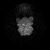
| Map data | emd_37885.map.gz | 107.3 MB | EMDB map data format | |
|---|---|---|---|---|
| Header (meta data) | emd-37885-v30.xmlemd-37885.xml | 16.5 KB 16.5 KB | Display Display | EMDB header |
| Images |  emd_37885.png emd_37885.png | 78.2 KB | ||
| Filedesc metadata | emd-37885.cif.gz | 6.1 KB | ||
| Others | emd_37885_half_map_1.map.gzemd_37885_half_map_2.map.gz | 200.7 MB 200.7 MB | ||
| Archive directory |  http://ftp.pdbj.org/pub/emdb/structures/EMD-37885ftp://ftp.pdbj.org/pub/emdb/structures/EMD-37885 http://ftp.pdbj.org/pub/emdb/structures/EMD-37885ftp://ftp.pdbj.org/pub/emdb/structures/EMD-37885 | HTTPS FTP |
-Related structure data
| Related structure data |  8ww9MC  8wvzC  8ww6C 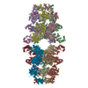 8ww7C  8ww8C  8wwaC M: atomic model generated by this map C: citing same article ( |
|---|---|
| Similar structure data |


-Links
| EMDB pages | EMDB (EBI/PDBe) / EMDataResource |
|---|---|
| Related items in Molecule of the Month |


-Map
| File | Download / File: emd_37885.map.gz / Format: CCP4 / Size: 216 MB / Type: IMAGE STORED AS FLOATING POINT NUMBER (4 BYTES) | ||||||||||||||||||||||||||||||||||||
|---|---|---|---|---|---|---|---|---|---|---|---|---|---|---|---|---|---|---|---|---|---|---|---|---|---|---|---|---|---|---|---|---|---|---|---|---|---|
| Projections & slices | Image control
Images are generated by Spider. | ||||||||||||||||||||||||||||||||||||
| Voxel size | X=Y=Z: 1.095 Å | ||||||||||||||||||||||||||||||||||||
| Density |
| ||||||||||||||||||||||||||||||||||||
| Symmetry | Space group: 1 | ||||||||||||||||||||||||||||||||||||
| Details | EMDB XML:
|
 Z (Sec.)
 Y (Row.)
 X (Col.)


-Supplemental data
-Half map: #1
| File | emd_37885_half_map_1.map | ||||||||||||
|---|---|---|---|---|---|---|---|---|---|---|---|---|---|
| Projections & Slices |
| ||||||||||||
| Density Histograms |


-Half map: #2
| File | emd_37885_half_map_2.map | ||||||||||||
|---|---|---|---|---|---|---|---|---|---|---|---|---|---|
| Projections & Slices |
| ||||||||||||
| Density Histograms |


- Sample components
-Entire : C962R ADP-DNA dodecamer
| Entire | Name: C962R ADP-DNA dodecamer |
|---|---|
| Components |
|
-Supramolecule #1: C962R ADP-DNA dodecamer
| Supramolecule | Name: C962R ADP-DNA dodecamer / type: complex / ID: 1 / Parent: 0 / Macromolecule list: #1-#3 |
|---|---|
| Source (natural) | Organism: African swine fever virus |
-Macromolecule #1: DNA (29-MER)
| Macromolecule | Name: DNA (29-MER) / type: dna / ID: 1 / Number of copies: 2 / Classification: DNA |
|---|---|
| Source (natural) | Organism: Homo sapiens (human) |
| Molecular weight | Theoretical: 8.776634 KDa |
| Sequence | String: (DT)(DT)(DT)(DT)(DT)(DT)(DT)(DT)(DT)(DT) (DT)(DT)(DT)(DT)(DT)(DT)(DT)(DT)(DT)(DT) (DT)(DT)(DT)(DT)(DT)(DT)(DT)(DT)(DT) |
-Macromolecule #2: DNA (5'-D(P*AP*AP*AP*A)-3')
| Macromolecule | Name: DNA (5'-D(P*AP*AP*AP*A)-3') / type: dna / ID: 2 / Number of copies: 2 / Classification: DNA |
|---|---|
| Source (natural) | Organism: Homo sapiens (human) |
| Molecular weight | Theoretical: 1.20787 KDa |
| Sequence | String: (DA)(DA)(DA)(DA) |
-Macromolecule #3: Putative primase C962R
| Macromolecule | Name: Putative primase C962R / type: protein_or_peptide / ID: 3 / Number of copies: 12 / Enantiomer: LEVO |
|---|---|
| Source (natural) | Organism: African swine fever virus |
| Molecular weight | Theoretical: 112.721516 KDa |
| Recombinant expression | Organism: Homo sapiens (human) |
| Sequence | String: MREESWEDHD TIQLTAQRKY LAEVQALETL LTRELSVFLT EPGSKKTNII NRITGKTYAL PSTELLRLYE HLEQCRKQGA LMYFLERQG TYSGLMLDYD LKLNTNAVPP LEPPALSRLC HRIFVHIKNS SVLPEGSHKI HFFFTLKPEV VQGKYGFHVL I PGLKLAAS ...String: MREESWEDHD TIQLTAQRKY LAEVQALETL LTRELSVFLT EPGSKKTNII NRITGKTYAL PSTELLRLYE HLEQCRKQGA LMYFLERQG TYSGLMLDYD LKLNTNAVPP LEPPALSRLC HRIFVHIKNS SVLPEGSHKI HFFFTLKPEV VQGKYGFHVL I PGLKLAAS TKKSIIGSLQ HDATVQKILH EQGVTNPESC LDPHSASVPS LLYGSSKLNH KPYQLKTGFE LVFDSSDPDY IP IHQIKNL ESYNLVSELS LTNEQGSLVR PVYCAADIAA EKEEEIPTED HSLSILMLHD PEARYLHKIL NLLPPEYYVE YPL WSNVVF ALANTSANYR PLAEWFSQKC PEKWNTGGKE KLEKLWNDAS HHTEKKITKR SIMYWAHKHA PQQYKEIVEQ GYFS ILAEY VYSYNGMLEH YMIAKVIYAM MGNKFVVDVD SNGKYVWFEF VLPGQPMNQG EIWKWRKEVN PDELHIYISE NFSRV MDRI TEHIKYHLSQ PHESNILNYY KKLLKAFERS KSKIFNDSFK KGVIRQAEFL FRQRSFIQTL DTNPHLLGVG NGVLSI ETI PAKLINHFHE HPIHQYTHIC YVPFNPENPW TKLLLNALQD IIPELDARLW IMFYLSTAIF RGLKEALMLL WLGGGCN GK TFLMRLVAMV LGDHYASKLN ISLLTSCRET AEKPNSAFMR LKGRGYGYFE ETNKSEVLNT SRLKEMVNPG DVTARELN Q KQESFQMTAT MVAASNYNFI IDTTDHGTWR RLRHYRSKVK FCHNPDPSNP YEKKEDPRFI HEYIMDPDCQ NAFFSILVY FWEKLQKEYN GQIKKVFCPT IESETEAYRK SQDTLHRFIT ERVVESPSAE TVYNLSEVVT AYAEWYNTNI NVKRHIALEL SQELENSVL EKYLQWSPNK TRILKGCRIL HKFETLQPGE SYIGVSTAGT LLNTPICEPK NKWWEWSPNP SAPPEKEASA P TPLEDYKD DDDK UniProtKB: Putative primase C962R |
-Macromolecule #4: ADENOSINE-5'-DIPHOSPHATE
| Macromolecule | Name: ADENOSINE-5'-DIPHOSPHATE / type: ligand / ID: 4 / Number of copies: 12 / Formula: ADP |
|---|---|
| Molecular weight | Theoretical: 427.201 Da |
| Chemical component information |  ChemComp-ADP: |
-Experimental details
-Structure determination
| Method | cryo EM |
|---|---|
 Processing Processing | single particle reconstruction |
| Aggregation state | particle |
-Sample preparation
| Buffer | pH: 7.5 |
|---|---|
| Vitrification | Cryogen name: ETHANE |
- Electron microscopy
| Microscope | FEI TITAN KRIOS |
|---|---|
| Image recording | Film or detector model: GATAN K3 BIOQUANTUM (6k x 4k) / Average electron dose: 50.0 e/Å2 |
| Electron beam | Acceleration voltage: 300 kV / Electron source:  FIELD EMISSION GUN FIELD EMISSION GUN |
| Electron optics | Illumination mode: FLOOD BEAM / Imaging mode: BRIGHT FIELD / Nominal defocus max: 2.2 µm / Nominal defocus min: 1.2 µm |
| Sample stage | Specimen holder model: FEI TITAN KRIOS AUTOGRID HOLDER / Cooling holder cryogen: NITROGEN |
| Experimental equipment |  Model: Titan Krios / Image courtesy: FEI Company |
-Image processing
| Startup model | Type of model: OTHER |
|---|---|
| Final reconstruction | Resolution.type: BY AUTHOR / Resolution: 3.55 Å / Resolution method: FSC 0.143 CUT-OFF / Number images used: 200521 |
| Initial angle assignment | Type: ANGULAR RECONSTITUTION |
| Final angle assignment | Type: MAXIMUM LIKELIHOOD |
