 Movie
Movie Controller


+ Open data

- Basic information
| Entry |  | |||||||||
|---|---|---|---|---|---|---|---|---|---|---|
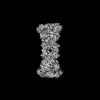
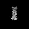
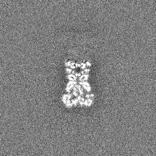
| Title | Cryo-EM structure of DSR2 | |||||||||
 Map data Map data | ||||||||||
 Sample Sample |
| |||||||||
 Keywords Keywords | Anti-phage immune / NADase / Defense-associated sirtuin 2 / DSR2 / Phage tail tube protein / DSR Anti Defense 1 / DSAD1 / Bacteria-phage interaction / SURFACTANT PROTEIN | |||||||||
| Function / homology | SIR2-like domain / SIR2-like domain / DHS-like NAD/FAD-binding domain superfamily / SIR2-like domain-containing protein Function and homology information Function and homology information | |||||||||
| Biological species |  | |||||||||
| Method | single particle reconstruction / cryo EM / Resolution: 4.15 Å | |||||||||
 Authors Authors | Gao A / Huang J / Zhu K | |||||||||
| Funding support | 1 items
| |||||||||
 Citation Citation | Journal: Nat Commun / Year: 2024 Title: Molecular basis of bacterial DSR2 anti-phage defense and viral immune evasion. Authors: Jiafeng Huang / Keli Zhu / Yina Gao / Feng Ye / Zhaolong Li / Yao Ge / Songqing Liu / Jing Yang / Ang Gao /  Abstract: Defense-associated sirtuin 2 (DSR2) systems are widely distributed across prokaryotic genomes, providing robust protection against phage infection. DSR2 recognizes phage tail tube proteins and ...Defense-associated sirtuin 2 (DSR2) systems are widely distributed across prokaryotic genomes, providing robust protection against phage infection. DSR2 recognizes phage tail tube proteins and induces abortive infection by depleting intracellular NAD, a process that is counteracted by another phage-encoded protein, DSR Anti Defense 1 (DSAD1). Here, we present cryo-EM structures of Bacillus subtilis DSR2 in its apo, Tube-bound, and DSAD1-bound states. DSR2 assembles into an elongated tetramer, with four NADase catalytic modules clustered in the center and the regulatory-sensing modules distributed at four distal corners. Interestingly, monomeric Tube protein, rather than its oligomeric states, docks at each corner of the DSR2 tetramer to form a 4:4 DSR2-Tube assembly, which is essential for DSR2 NADase activity. DSAD1 competes with Tube for binding to DSR2 by occupying an overlapping region, thereby inhibiting DSR2 immunity. Thus, our results provide important insights into the assembly, activation and inhibition of the DSR2 anti-phage defense system. | |||||||||
| History |
|
- Structure visualization
| Supplemental images |
|---|

- Downloads & links
-EMDB archive
| Map data | emd_37610.map.gz | 506.6 MB | EMDB map data format | |
|---|---|---|---|---|
| Header (meta data) | emd-37610-v30.xmlemd-37610.xml | 13.9 KB 13.9 KB | Display Display | EMDB header |
| Images | 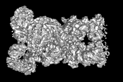 emd_37610.png emd_37610.png | 83.7 KB | ||
| Filedesc metadata | emd-37610.cif.gz | 5.7 KB | ||
| Others | emd_37610_half_map_1.map.gzemd_37610_half_map_2.map.gz | 498.3 MB 498.3 MB | ||
| Archive directory |  http://ftp.pdbj.org/pub/emdb/structures/EMD-37610ftp://ftp.pdbj.org/pub/emdb/structures/EMD-37610 http://ftp.pdbj.org/pub/emdb/structures/EMD-37610ftp://ftp.pdbj.org/pub/emdb/structures/EMD-37610 | HTTPS FTP |
-Validation report
| Summary document | emd_37610_validation.pdf.gz | 1.1 MB | Display | EMDB validaton report |
|---|---|---|---|---|
| Full document | emd_37610_full_validation.pdf.gz | 1.1 MB | Display | |
| Data in XML | emd_37610_validation.xml.gz | 19.1 KB | Display | |
| Data in CIF | emd_37610_validation.cif.gz | 22.7 KB | Display | |
| Arichive directory | https://ftp.pdbj.org/pub/emdb/validation_reports/EMD-37610ftp://ftp.pdbj.org/pub/emdb/validation_reports/EMD-37610 | HTTPS FTP |
-Related structure data
| Related structure data | 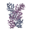 8wkxMC  8wksC 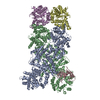 8wktC M: atomic model generated by this map C: citing same article ( |
|---|---|
| Similar structure data |
-Links
| EMDB pages | EMDB (EBI/PDBe) / EMDataResource |
|---|---|
| Related items in Molecule of the Month |


-Map
| File | Download / File: emd_37610.map.gz / Format: CCP4 / Size: 536.4 MB / Type: IMAGE STORED AS FLOATING POINT NUMBER (4 BYTES) | ||||||||||||||||||||||||||||||||||||
|---|---|---|---|---|---|---|---|---|---|---|---|---|---|---|---|---|---|---|---|---|---|---|---|---|---|---|---|---|---|---|---|---|---|---|---|---|---|
| Projections & slices | Image control
Images are generated by Spider. | ||||||||||||||||||||||||||||||||||||
| Voxel size | X=Y=Z: 1.07 Å | ||||||||||||||||||||||||||||||||||||
| Density |
| ||||||||||||||||||||||||||||||||||||
| Symmetry | Space group: 1 | ||||||||||||||||||||||||||||||||||||
| Details | EMDB XML:
|
 Z (Sec.)
 Y (Row.)
 X (Col.)


-Supplemental data
-Half map: #2
| File | emd_37610_half_map_1.map | ||||||||||||
|---|---|---|---|---|---|---|---|---|---|---|---|---|---|
| Projections & Slices |
| ||||||||||||
| Density Histograms |


-Half map: #1
| File | emd_37610_half_map_2.map | ||||||||||||
|---|---|---|---|---|---|---|---|---|---|---|---|---|---|
| Projections & Slices |
| ||||||||||||
| Density Histograms |


- Sample components
-Entire : DSR2
| Entire | Name: DSR2 |
|---|---|
| Components |
|
-Supramolecule #1: DSR2
| Supramolecule | Name: DSR2 / type: complex / ID: 1 / Parent: 0 / Macromolecule list: all |
|---|---|
| Source (natural) | Organism: |
-Macromolecule #1: SIR2-like domain-containing protein
| Macromolecule | Name: SIR2-like domain-containing protein / type: protein_or_peptide / ID: 1 / Number of copies: 2 / Enantiomer: LEVO |
|---|---|
| Source (natural) | Organism: |
| Molecular weight | Theoretical: 118.635789 KDa |
| Recombinant expression | Organism: |
| Sequence | String: MVKVDLESKR YGEKLKEVFL MLDNNVVECI KEITESSRNG KLVFFVGAGV STLSDYPQWW RLVDKYHEEL YGSPKKGNYS SDEYLRIPQ IFYNVKGEMA FDGILKDFFQ VDKPTNPIHD KILAMNPAHV ITTNYDNLID TACWKRGKYF SVISAEEDVA N ATSSRYLL ...String: MVKVDLESKR YGEKLKEVFL MLDNNVVECI KEITESSRNG KLVFFVGAGV STLSDYPQWW RLVDKYHEEL YGSPKKGNYS SDEYLRIPQ IFYNVKGEMA FDGILKDFFQ VDKPTNPIHD KILAMNPAHV ITTNYDNLID TACWKRGKYF SVISAEEDVA N ATSSRYLL KVHGDFRKGF KGENVVLKED DYLNYDQNYP LISNLMKTII ATHTIVFIGY GLGDYNINML LNWVRKLQKD SF HKPFFIR TDPSPIENET LIYYENKGLR IIDAASLIDS NEYDYLERYS AVMDLLIESQ ENKFITKDDE VIDYIYGKIS PLF ALQYIR KIDLKHVFEY DYHFEVNGTV VRHKNKGFGY MERFFELKES CDERSKLSKK QYERFNALFN FFEKNGVICM AKDA GTLNT SIEINSLAYH GKYDVMKKFI EEQSVSIEDD YKKAFFLACL GRWEESYDLY SNIILNSIDE SNGCVYYLSQ INRYR IYQS ITQAVTQFNG LGLLTFGRHY KPFTDEFLAR IEREMTNFNI DDLFNGMPFE FQKKYKILEF LSDNQFLYDD TVKLFE LTN KVRSEMSEGS YSFGMSSDIV VLLRLYDNLR FLYENCLWSV SFHEFHQYIR NSMSLLIEKA EYERTRDIDE LGFSFFG KK SGFFMEYYDF VNISRHFKID DIKNLERSCS IDKIRFGEQE KIEEYLVGIA EEITKQFSAN GMNVVFYTQF ISEAKAAL Y FAKYVKLSEE GLGKIVKALL FYFPERDLDI GKRYVWLERL TKCNELPKSI ISIIDDFLVL QAEKHIDQNY SEVSSNGLY SRDYGALIKH FEKNFISKRL SEITLCLTQD KQKQIDFLFK LLPLLSTNAK SHLLSFKSVE NINDLMNGIR IGLIDEFTPE HEELIIEYL ETRKVNYIVE KEKGIQTFSS NDYMSTFGIW YFLEEINNSK MEEFIGMDDQ YDFFVDPENF DYKKFIPSWL K NYNDKLLG KIAGNKHMKH HVIEVLKERV KNSNDKRYLE ILMNYFI UniProtKB: SIR2-like domain-containing protein |
-Experimental details
-Structure determination
| Method | cryo EM |
|---|---|
 Processing Processing | single particle reconstruction |
| Aggregation state | particle |
-Sample preparation
| Buffer | pH: 7.5 |
|---|---|
| Vitrification | Cryogen name: ETHANE |
- Electron microscopy
| Microscope | FEI TITAN |
|---|---|
| Image recording | Film or detector model: GATAN K3 (6k x 4k) / Average electron dose: 60.0 e/Å2 |
| Electron beam | Acceleration voltage: 300 kV / Electron source:  FIELD EMISSION GUN FIELD EMISSION GUN |
| Electron optics | Illumination mode: FLOOD BEAM / Imaging mode: BRIGHT FIELD / Nominal defocus max: 1.8 µm / Nominal defocus min: 1.2 µm |
-Image processing
| Startup model | Type of model: NONE |
|---|---|
| Final reconstruction | Resolution.type: BY AUTHOR / Resolution: 4.15 Å / Resolution method: FSC 0.143 CUT-OFF / Number images used: 80223 |
| Initial angle assignment | Type: PROJECTION MATCHING |
| Final angle assignment | Type: PROJECTION MATCHING |
