Movie
Movie Controller


+ Open data

- Basic information
| Entry |  | |||||||||
|---|---|---|---|---|---|---|---|---|---|---|
| Title | ADP-bound purinergic receptor 1 in complex with miniGs/q | |||||||||
 Map data Map data | ||||||||||
 Sample Sample |
| |||||||||
 Keywords Keywords | GPCR / MEMBRANE PROTEIN | |||||||||
| Function / homology |  Function and homology information Function and homology informationG protein-coupled ATP receptor activity / cellular response to purine-containing compound / relaxation of muscle / G protein-coupled ADP receptor activity / A1 adenosine receptor binding / G protein-coupled purinergic nucleotide receptor signaling pathway / positive regulation of inositol trisphosphate biosynthetic process / P2Y receptors / positive regulation of penile erection / G protein-coupled purinergic nucleotide receptor activity ...G protein-coupled ATP receptor activity / cellular response to purine-containing compound / relaxation of muscle / G protein-coupled ADP receptor activity / A1 adenosine receptor binding / G protein-coupled purinergic nucleotide receptor signaling pathway / positive regulation of inositol trisphosphate biosynthetic process / P2Y receptors / positive regulation of penile erection / G protein-coupled purinergic nucleotide receptor activity / negative regulation of norepinephrine secretion / positive regulation of monoatomic ion transport / glial cell migration / signaling receptor regulator activity / positive regulation of hormone secretion / regulation of presynaptic cytosolic calcium ion concentration / response to growth factor / signal transduction involved in regulation of gene expression / G protein-coupled adenosine receptor signaling pathway / eating behavior / regulation of synaptic vesicle exocytosis / cellular response to ATP / response to mechanical stimulus / establishment of localization in cell / monoatomic ion transport / presynaptic active zone membrane / blood vessel diameter maintenance / protein localization to plasma membrane / platelet activation / ADP binding / electron transport chain / adenylate cyclase-inhibiting G protein-coupled receptor signaling pathway / Olfactory Signaling Pathway / Activation of the phototransduction cascade / G protein-coupled acetylcholine receptor signaling pathway / G beta:gamma signalling through PLC beta / Presynaptic function of Kainate receptors / Thromboxane signalling through TP receptor / Activation of G protein gated Potassium channels / Inhibition of voltage gated Ca2+ channels via Gbeta/gamma subunits / G-protein activation / Glucagon signaling in metabolic regulation / G beta:gamma signalling through CDC42 / Prostacyclin signalling through prostacyclin receptor / Synthesis, secretion, and inactivation of Glucagon-like Peptide-1 (GLP-1) / G beta:gamma signalling through BTK / photoreceptor disc membrane / ADP signalling through P2Y purinoceptor 12 / Glucagon-type ligand receptors / Sensory perception of sweet, bitter, and umami (glutamate) taste / Adrenaline,noradrenaline inhibits insulin secretion / Vasopressin regulates renal water homeostasis via Aquaporins / regulation of cell shape / Glucagon-like Peptide-1 (GLP1) regulates insulin secretion / G alpha (z) signalling events / cellular response to catecholamine stimulus / ADP signalling through P2Y purinoceptor 1 / G beta:gamma signalling through PI3Kgamma / ADORA2B mediated anti-inflammatory cytokines production / adenylate cyclase-activating dopamine receptor signaling pathway / Cooperation of PDCL (PhLP1) and TRiC/CCT in G-protein beta folding / GPER1 signaling / cellular response to prostaglandin E stimulus / heterotrimeric G-protein complex / Inactivation, recovery and regulation of the phototransduction cascade / G alpha (12/13) signalling events / G-protein beta-subunit binding / extracellular vesicle / positive regulation of cytosolic calcium ion concentration / sensory perception of taste / Thrombin signalling through proteinase activated receptors (PARs) / signaling receptor complex adaptor activity / signaling receptor activity / retina development in camera-type eye / fibroblast proliferation / cell body / GTPase binding / scaffold protein binding / Ca2+ pathway / High laminar flow shear stress activates signaling by PIEZO1 and PECAM1:CDH5:KDR in endothelial cells / G alpha (i) signalling events / basolateral plasma membrane / G alpha (s) signalling events / G alpha (q) signalling events / phospholipase C-activating G protein-coupled receptor signaling pathway / Ras protein signal transduction / positive regulation of ERK1 and ERK2 cascade / periplasmic space / electron transfer activity / cell population proliferation / postsynaptic membrane / cell surface receptor signaling pathway / cilium / Extra-nuclear estrogen signaling / apical plasma membrane / postsynaptic density / iron ion binding / G protein-coupled receptor signaling pathway / protein heterodimerization activity / lysosomal membrane Similarity search - Function | |||||||||
| Biological species |  Homo sapiens (human) Homo sapiens (human) | |||||||||
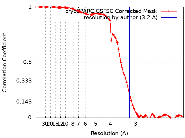
| Method | single particle reconstruction / cryo EM / Resolution: 3.2 Å | |||||||||
 Authors Authors | Gu QC / Tang WQ | |||||||||
| Funding support |  China, 1 items China, 1 items
| |||||||||
 Citation Citation | Journal: To Be Published Title: ADP-bound purinergic receptor 1 in complex with miniGs/q Authors: Gu QC / Tang WQ #1: Journal: To Be PublishedTitle: Real-space refinement in PHENIX for cryo-EM and crystallography Authors: Gu QC / Tang WQ | |||||||||
| History |
|
- Structure visualization
| Supplemental images |
|---|

- Downloads & links
-EMDB archive
| Map data | emd_37591.map.gz | 59.7 MB | EMDB map data format | |
|---|---|---|---|---|
| Header (meta data) | emd-37591-v30.xmlemd-37591.xml | 26.6 KB 26.6 KB | Display Display | EMDB header |
| FSC (resolution estimation) | emd_37591_fsc.xml | 7.6 KB | Display | FSC data file |
| Images |  emd_37591.png emd_37591.png | 31.4 KB | ||
| Masks | emd_37591_msk_1.map | 64 MB | Mask map | |
| Filedesc metadata | emd-37591.cif.gz | 7.9 KB | ||
| Others | emd_37591_half_map_1.map.gzemd_37591_half_map_2.map.gz | 59.5 MB 59.5 MB | ||
| Archive directory |  http://ftp.pdbj.org/pub/emdb/structures/EMD-37591ftp://ftp.pdbj.org/pub/emdb/structures/EMD-37591 http://ftp.pdbj.org/pub/emdb/structures/EMD-37591ftp://ftp.pdbj.org/pub/emdb/structures/EMD-37591 | HTTPS FTP |
-Related structure data
| Related structure data |  8wjxMC M: atomic model generated by this map C: citing same article ( |
|---|---|
| Similar structure data |
-Links
| EMDB pages | EMDB (EBI/PDBe) / EMDataResource |
|---|---|
| Related items in Molecule of the Month |


-Map
| File | Download / File: emd_37591.map.gz / Format: CCP4 / Size: 64 MB / Type: IMAGE STORED AS FLOATING POINT NUMBER (4 BYTES) | ||||||||||||||||||||||||||||||||||||
|---|---|---|---|---|---|---|---|---|---|---|---|---|---|---|---|---|---|---|---|---|---|---|---|---|---|---|---|---|---|---|---|---|---|---|---|---|---|
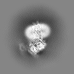
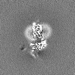
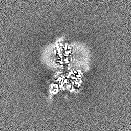
| Projections & slices | Image control
Images are generated by Spider. | ||||||||||||||||||||||||||||||||||||
| Voxel size | X=Y=Z: 1 Å | ||||||||||||||||||||||||||||||||||||
| Density |
| ||||||||||||||||||||||||||||||||||||
| Symmetry | Space group: 1 | ||||||||||||||||||||||||||||||||||||
| Details | EMDB XML:
|
 Z (Sec.)
 Y (Row.)
 X (Col.)


-Supplemental data
-Mask #1
| File | emd_37591_msk_1.map | ||||||||||||
|---|---|---|---|---|---|---|---|---|---|---|---|---|---|
| Projections & Slices |
| ||||||||||||
| Density Histograms |


-Half map: #2
| File | emd_37591_half_map_1.map | ||||||||||||
|---|---|---|---|---|---|---|---|---|---|---|---|---|---|
| Projections & Slices |
| ||||||||||||
| Density Histograms |


-Half map: #1
| File | emd_37591_half_map_2.map | ||||||||||||
|---|---|---|---|---|---|---|---|---|---|---|---|---|---|
| Projections & Slices |
| ||||||||||||
| Density Histograms |


- Sample components
-Entire : ADP-bound purinergic receptor 1 in complex with miniGs/q
| Entire | Name: ADP-bound purinergic receptor 1 in complex with miniGs/q |
|---|---|
| Components |
|
-Supramolecule #1: ADP-bound purinergic receptor 1 in complex with miniGs/q
| Supramolecule | Name: ADP-bound purinergic receptor 1 in complex with miniGs/q type: complex / ID: 1 / Parent: 0 / Macromolecule list: #1-#5 |
|---|---|
| Source (natural) | Organism: Homo sapiens (human) |
-Macromolecule #1: GNAS complex locus
| Macromolecule | Name: GNAS complex locus / type: protein_or_peptide / ID: 1 / Number of copies: 1 / Enantiomer: LEVO |
|---|---|
| Source (natural) | Organism: Homo sapiens (human) |
| Molecular weight | Theoretical: 30.840807 KDa |
| Recombinant expression | Organism:  |
| Sequence | String: MGMGHHHHHH ENLYFQGNSK TEDQRNEEKA QREANKKIEK QLQKDKQVYR ATHRLLLLGA DNSGKSTIVK QMRILHGGSG GSGGTSGIF ETKFQVDKVN FHMFDVGGQR DERRKWIQCF NDVTAIIFVV DSSDYNRLQE ALNLFKSIWN NRWLRTISVI L FLNKQDLL ...String: MGMGHHHHHH ENLYFQGNSK TEDQRNEEKA QREANKKIEK QLQKDKQVYR ATHRLLLLGA DNSGKSTIVK QMRILHGGSG GSGGTSGIF ETKFQVDKVN FHMFDVGGQR DERRKWIQCF NDVTAIIFVV DSSDYNRLQE ALNLFKSIWN NRWLRTISVI L FLNKQDLL AEKVLAGKSK IEDYFPEFAR YTTPEDATPE PGEDPRVTRA KYFIRDEFLR ISTASGDGRH YCYPHFTCAV DT ENARRIF NDCKDIILQM NLREYNLV |
-Macromolecule #2: Guanine nucleotide-binding protein G(I)/G(S)/G(T) subunit beta-1
| Macromolecule | Name: Guanine nucleotide-binding protein G(I)/G(S)/G(T) subunit beta-1 type: protein_or_peptide / ID: 2 / Number of copies: 1 / Enantiomer: LEVO |
|---|---|
| Source (natural) | Organism: Homo sapiens (human) |
| Molecular weight | Theoretical: 41.055867 KDa |
| Recombinant expression | Organism:   Spodoptera frugiperda (fall armyworm) Spodoptera frugiperda (fall armyworm) |
| Sequence | String: MHHHHHHGSL LQSELDQLRQ EAEQLKNQIR DARKACADAT LSQITNNIDP VGRIQMRTRR TLRGHLAKIY AMHWGTDSRL LVSASQDGK LIIWDSYTTN KVHAIPLRSS WVMTCAYAPS GNYVACGGLD NICSIYNLKT REGNVRVSRE LAGHTGYLSC C RFLDDNQI ...String: MHHHHHHGSL LQSELDQLRQ EAEQLKNQIR DARKACADAT LSQITNNIDP VGRIQMRTRR TLRGHLAKIY AMHWGTDSRL LVSASQDGK LIIWDSYTTN KVHAIPLRSS WVMTCAYAPS GNYVACGGLD NICSIYNLKT REGNVRVSRE LAGHTGYLSC C RFLDDNQI VTSSGDTTCA LWDIETGQQT TTFTGHTGDV MSLSLAPDTR LFVSGACDAS AKLWDVREGM CRQTFTGHES DI NAICFFP NGNAFATGSD DATCRLFDLR ADQELMTYSH DNIICGITSV SFSKSGRLLL AGYDDFNCNV WDALKADRAG VLA GHDNRV SCLGVTDDGM AVATGSWDSF LKIWNGSSGG GGSGGGGSSG VSGWRLFKKI S UniProtKB: Guanine nucleotide-binding protein G(I)/G(S)/G(T) subunit beta-1 |
-Macromolecule #3: Guanine nucleotide-binding protein G(I)/G(S)/G(O) subunit gamma-2
| Macromolecule | Name: Guanine nucleotide-binding protein G(I)/G(S)/G(O) subunit gamma-2 type: protein_or_peptide / ID: 3 / Number of copies: 1 / Enantiomer: LEVO |
|---|---|
| Source (natural) | Organism: Homo sapiens (human) |
| Molecular weight | Theoretical: 7.861143 KDa |
| Recombinant expression | Organism: Spodoptera frugiperda (fall armyworm) |
| Sequence | String: MASNNTASIA QARKLVEQLK MEANIDRIKV SKAAADLMAY CEAHAKEDPL LTPVPASENP FREKKFFCAI L UniProtKB: Guanine nucleotide-binding protein G(I)/G(S)/G(O) subunit gamma-2 |
-Macromolecule #4: Nanobody 35
| Macromolecule | Name: Nanobody 35 / type: protein_or_peptide / ID: 4 / Number of copies: 1 / Enantiomer: LEVO |
|---|---|
| Source (natural) | Organism: Homo sapiens (human) |
| Molecular weight | Theoretical: 17.057271 KDa |
| Recombinant expression | Organism: |
| Sequence | String: MKYLLPTAAA GLLLLAAQPA MAMQVQLQES GGGLVQPGGS LRLSCAASGF TFSNYKMNWV RQAPGKGLEW VSDISQSGAS ISYTGSVKG RFTISRDNAK NTLYLQMNSL KPEDTAVYYC ARCPAPFTRD CFDVTSTTYA YRGQGTQVTV SSHHHHHH |
-Macromolecule #5: Soluble cytochrome b562,P2Y purinoceptor 1
| Macromolecule | Name: Soluble cytochrome b562,P2Y purinoceptor 1 / type: protein_or_peptide / ID: 5 / Number of copies: 1 / Enantiomer: LEVO |
|---|---|
| Source (natural) | Organism: Homo sapiens (human) |
| Molecular weight | Theoretical: 76.586242 KDa |
| Recombinant expression | Organism: Spodoptera frugiperda (fall armyworm) |
| Sequence | String: MKTIIALSYI FCLVFADYKD DDDAGRAHHH HHHHHHHENL YFQSGAPADL EDNWETLNDN LKVIEKADNA AQVKDALTKM RAAALDAQK ATPPKLEDKS PDSPEMKDFR HGFDILVGQI DDALKLANEG KVKEAQAAAE QLKTTRNAYI QKYLEFLEVL F QGPTEVLW ...String: MKTIIALSYI FCLVFADYKD DDDAGRAHHH HHHHHHHENL YFQSGAPADL EDNWETLNDN LKVIEKADNA AQVKDALTKM RAAALDAQK ATPPKLEDKS PDSPEMKDFR HGFDILVGQI DDALKLANEG KVKEAQAAAE QLKTTRNAYI QKYLEFLEVL F QGPTEVLW PAVPNGTDAA FLAGPGSSWG NSTVASTAAV SSSFKCALTK TGFQFYYLPA VYILVFIIGF LGNSVAIWMF VF HMKPWSG ISVYMFNLAL ADFLYVLTLP ALIFYYFNKT DWIFGDAMCK LQRFIFHVNL YGSILFLTCI SAHRYSGVVY PLK SLGRLK KKNAICISVL VWLIVVVAIS PILFYSGTGV RKNKTITCYD TTSDEYLRSY FIYSMCTTVA MFCVPLVLIL GCYG LIVRA LIYKDLDNSP LRRKSIYLVI IVLTVFAVSY IPFHVMKTMN LRARLDFQTP AMCAFNDRVY ATYQVTRGLA SLNSC VDPI LYFLAGDTFR RRLSRATRKA SRRSEANLQS KSEDMTLNVF TLEDFVGDWE QTAAYNLDQV LEQGGVSSLL QNLAVS VTP IQRIVRSGEN ALKIDIHVII PYEGLSADQM AQIEEVFKVV YPVDDHHFKV ILPYGTLVID GVTPNMLNYF GRPYEGI AV FDGKKITVTG TLWNGNKIID ERLITPDGSM LFRVTINS UniProtKB: Soluble cytochrome b562, P2Y purinoceptor 1 |
-Macromolecule #6: ADENOSINE-5'-DIPHOSPHATE
| Macromolecule | Name: ADENOSINE-5'-DIPHOSPHATE / type: ligand / ID: 6 / Number of copies: 1 / Formula: ADP |
|---|---|
| Molecular weight | Theoretical: 427.201 Da |
| Chemical component information |  ChemComp-ADP: |
-Macromolecule #7: CHOLESTEROL
| Macromolecule | Name: CHOLESTEROL / type: ligand / ID: 7 / Number of copies: 1 / Formula: CLR |
|---|---|
| Molecular weight | Theoretical: 386.654 Da |
| Chemical component information |  ChemComp-CLR: |
-Experimental details
-Structure determination
| Method | cryo EM |
|---|---|
 Processing Processing | single particle reconstruction |
| Aggregation state | particle |
-Sample preparation
| Buffer | pH: 7.5 |
|---|---|
| Vitrification | Cryogen name: ETHANE |
- Electron microscopy
| Microscope | FEI TALOS ARCTICA |
|---|---|
| Image recording | Film or detector model: GATAN K2 SUMMIT (4k x 4k) / Average electron dose: 60.0 e/Å2 |
| Electron beam | Acceleration voltage: 200 kV / Electron source:  FIELD EMISSION GUN FIELD EMISSION GUN |
| Electron optics | Illumination mode: FLOOD BEAM / Imaging mode: BRIGHT FIELD / Nominal defocus max: 2.0 µm / Nominal defocus min: 1.2 µm |
| Experimental equipment |  Model: Talos Arctica / Image courtesy: FEI Company |