 Movie
Movie Controller

[English] 日本語
 Yorodumi
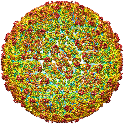
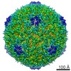
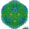
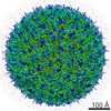
- EMDB-3752: The cryo-EM structure of Tick-borne encephalitis virus mature particle -
+ Open data

- Basic information
| Entry | Database: EMDB / ID: EMD-3752 | ||||||||||||
|---|---|---|---|---|---|---|---|---|---|---|---|---|---|
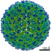
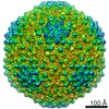
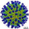
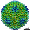
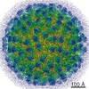
| Title | The cryo-EM structure of Tick-borne encephalitis virus mature particle | ||||||||||||
 Map data Map data | Mature tick-borne encephalitis virus particle | ||||||||||||
 Sample Sample |
| ||||||||||||
 Keywords Keywords | tick-borne encephalitis virus / mature particle / cryo-EM / VIRUS | ||||||||||||
| Function / homology |  Function and homology information Function and homology informationflavivirin / symbiont-mediated suppression of host JAK-STAT cascade via inhibition of STAT2 activity / symbiont-mediated suppression of host JAK-STAT cascade via inhibition of STAT1 activity / viral capsid / nucleoside-triphosphate phosphatase / double-stranded RNA binding / clathrin-dependent endocytosis of virus by host cell / mRNA (guanine-N7)-methyltransferase / methyltransferase cap1 / methyltransferase cap1 activity ...flavivirin / symbiont-mediated suppression of host JAK-STAT cascade via inhibition of STAT2 activity / symbiont-mediated suppression of host JAK-STAT cascade via inhibition of STAT1 activity / viral capsid / nucleoside-triphosphate phosphatase / double-stranded RNA binding / clathrin-dependent endocytosis of virus by host cell / mRNA (guanine-N7)-methyltransferase / methyltransferase cap1 / methyltransferase cap1 activity / mRNA 5'-cap (guanine-N7-)-methyltransferase activity / RNA helicase activity / protein dimerization activity / symbiont-mediated suppression of host innate immune response / host cell perinuclear region of cytoplasm / host cell endoplasmic reticulum membrane / RNA helicase / symbiont-mediated suppression of host type I interferon-mediated signaling pathway / serine-type endopeptidase activity / symbiont-mediated activation of host autophagy / RNA-directed RNA polymerase / viral RNA genome replication / RNA-directed RNA polymerase activity / fusion of virus membrane with host endosome membrane / viral envelope / virion attachment to host cell / host cell nucleus / virion membrane / structural molecule activity / ATP hydrolysis activity / proteolysis / extracellular region / ATP binding / metal ion binding Similarity search - Function | ||||||||||||
| Biological species |  Tick-borne encephalitis virus (strain Hypr) / Tick-borne encephalitis virus (strain HYPR) Tick-borne encephalitis virus (strain Hypr) / Tick-borne encephalitis virus (strain HYPR) | ||||||||||||
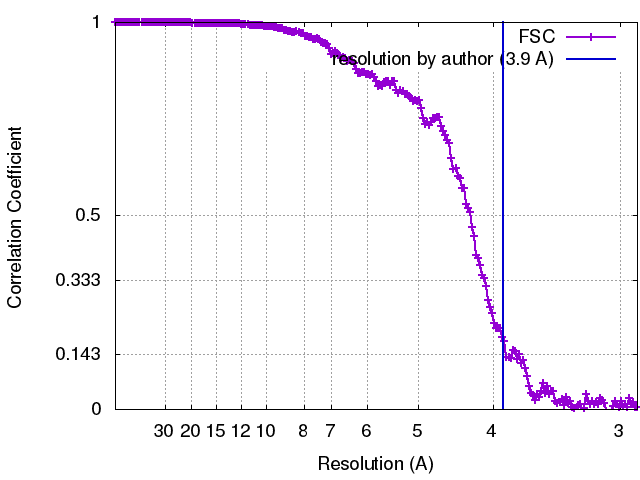
| Method | single particle reconstruction / cryo EM / Resolution: 3.9 Å | ||||||||||||
 Authors Authors | Fuzik T / Plevka P | ||||||||||||
| Funding support |  Germany, Germany,  Czech Republic, 3 items Czech Republic, 3 items
| ||||||||||||
 Citation Citation | Journal: Nat Commun / Year: 2018 Title: Structure of tick-borne encephalitis virus and its neutralization by a monoclonal antibody. Authors: Tibor Füzik / Petra Formanová / Daniel Růžek / Kentaro Yoshii / Matthias Niedrig / Pavel Plevka /  Abstract: Tick-borne encephalitis virus (TBEV) causes 13,000 cases of human meningitis and encephalitis annually. However, the structure of the TBEV virion and its interactions with antibodies are unknown. ...Tick-borne encephalitis virus (TBEV) causes 13,000 cases of human meningitis and encephalitis annually. However, the structure of the TBEV virion and its interactions with antibodies are unknown. Here, we present cryo-EM structures of the native TBEV virion and its complex with Fab fragments of neutralizing antibody 19/1786. Flavivirus genome delivery depends on membrane fusion that is triggered at low pH. The virion structure indicates that the repulsive interactions of histidine side chains, which become protonated at low pH, may contribute to the disruption of heterotetramers of the TBEV envelope and membrane proteins and induce detachment of the envelope protein ectodomains from the virus membrane. The Fab fragments bind to 120 out of the 180 envelope glycoproteins of the TBEV virion. Unlike most of the previously studied flavivirus-neutralizing antibodies, the Fab fragments do not lock the E-proteins in the native-like arrangement, but interfere with the process of virus-induced membrane fusion. | ||||||||||||
| History |
|
- Structure visualization
| Movie |
Movie viewer |
|---|---|
| Structure viewer | EM map: SurfViewMolmilJmol/JSmol |
| Supplemental images |
- Downloads & links
-EMDB archive
| Map data | emd_3752.map.gz | 49.2 MB | EMDB map data format | |
|---|---|---|---|---|
| Header (meta data) | emd-3752-v30.xmlemd-3752.xml | 17.8 KB 17.8 KB | Display Display | EMDB header |
| FSC (resolution estimation) | emd_3752_fsc.xml | 21 KB | Display | FSC data file |
| Images |  emd_3752.png emd_3752.png | 336.9 KB | ||
| Filedesc metadata | emd-3752.cif.gz | 6.6 KB | ||
| Archive directory |  http://ftp.pdbj.org/pub/emdb/structures/EMD-3752ftp://ftp.pdbj.org/pub/emdb/structures/EMD-3752 http://ftp.pdbj.org/pub/emdb/structures/EMD-3752ftp://ftp.pdbj.org/pub/emdb/structures/EMD-3752 | HTTPS FTP |
-Related structure data
| Related structure data | 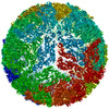 5o6aMC 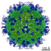 3754C 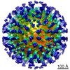 3755C  5o6vC M: atomic model generated by this map C: citing same article ( |
|---|---|
| Similar structure data |

-Links
| EMDB pages | EMDB (EBI/PDBe) / EMDataResource |
|---|---|
| Related items in Molecule of the Month |


-Map
| File | Download / File: emd_3752.map.gz / Format: CCP4 / Size: 178 MB / Type: IMAGE STORED AS FLOATING POINT NUMBER (4 BYTES) | ||||||||||||||||||||||||||||||||||||||||||||||||||||||||||||||||||||
|---|---|---|---|---|---|---|---|---|---|---|---|---|---|---|---|---|---|---|---|---|---|---|---|---|---|---|---|---|---|---|---|---|---|---|---|---|---|---|---|---|---|---|---|---|---|---|---|---|---|---|---|---|---|---|---|---|---|---|---|---|---|---|---|---|---|---|---|---|---|
| Annotation | Mature tick-borne encephalitis virus particle | ||||||||||||||||||||||||||||||||||||||||||||||||||||||||||||||||||||
| Projections & slices | Image control
Images are generated by Spider. | ||||||||||||||||||||||||||||||||||||||||||||||||||||||||||||||||||||
| Voxel size | X=Y=Z: 1.45 Å | ||||||||||||||||||||||||||||||||||||||||||||||||||||||||||||||||||||
| Density |
| ||||||||||||||||||||||||||||||||||||||||||||||||||||||||||||||||||||
| Symmetry | Space group: 1 | ||||||||||||||||||||||||||||||||||||||||||||||||||||||||||||||||||||
| Details | EMDB XML:
CCP4 map header:
| ||||||||||||||||||||||||||||||||||||||||||||||||||||||||||||||||||||
 Y (Sec.)
 X (Row.)
 Z (Col.)


-Supplemental data
- Sample components
-Entire : Tick-borne encephalitis virus (strain HYPR)
| Entire | Name: Tick-borne encephalitis virus (strain HYPR) |
|---|---|
| Components |
|
-Supramolecule #1: Tick-borne encephalitis virus (strain HYPR)
| Supramolecule | Name: Tick-borne encephalitis virus (strain HYPR) / type: virus / ID: 1 / Parent: 0 / Macromolecule list: #1-#2 / NCBI-ID: 70733 Sci species name: Tick-borne encephalitis virus (strain HYPR) Virus type: VIRION / Virus isolate: SEROTYPE / Virus enveloped: Yes / Virus empty: No |
|---|---|
| Molecular weight | Theoretical: 22 MDa |
| Virus shell | Shell ID: 1 / Name: Mature particle / Diameter: 500.0 Å / T number (triangulation number): 3 |
-Macromolecule #1: Envelope protein
| Macromolecule | Name: Envelope protein / type: protein_or_peptide / ID: 1 / Number of copies: 3 / Enantiomer: LEVO / EC number: flavivirin |
|---|---|
| Source (natural) | Organism: Tick-borne encephalitis virus (strain Hypr) / Strain: Hypr |
| Molecular weight | Theoretical: 53.667418 KDa |
| Sequence | String: SRCTHLENRD FVTGTQGTTR VTLVLELGGC VTITAEGKPS MDVWLDAIYQ ENPAQTREYC LHAKLSDTKV AARCPTMGPA TLAEEHQGG TVCKRDQSDR GWGNHCGLFG KGSIVACVKA ACEAKKKATG HVYDANKIVY TVKVEPHTGD YVAANETHSG R KTASFTVS ...String: SRCTHLENRD FVTGTQGTTR VTLVLELGGC VTITAEGKPS MDVWLDAIYQ ENPAQTREYC LHAKLSDTKV AARCPTMGPA TLAEEHQGG TVCKRDQSDR GWGNHCGLFG KGSIVACVKA ACEAKKKATG HVYDANKIVY TVKVEPHTGD YVAANETHSG R KTASFTVS SEKTILTMGE YGDVSLLCRV ASGVDLAQTV ILELDKTVEH LPTAWQVHRD WFNDLALPWK HEGARNWNNA ER LVEFGAP HAVKMDVYNL GDQTGVLLKA LAGVPVAHIE GTKYHLKSGH VTCEVGLEKL KMKGLTYTMC DKTKFTWKRA PTD SGHDTV VMEVTFSGTK PCRIPVRAVA HGSPDVNVAM LITPNPTIEN NGGGFIEMQL PPGDNIIYVG ELSYQWFQKG SSIG RVFQK TKKGIERLTV IGEHAWDFGS AGGFLSSIGK ALHTVLGGAF NSIFGGVGFL PKLLLGVALA WLGLNMRNPT MSMSF LLAG VLVLAMTLGV GA UniProtKB: Genome polyprotein |
-Macromolecule #2: Small envelope protein M
| Macromolecule | Name: Small envelope protein M / type: protein_or_peptide / ID: 2 / Number of copies: 3 / Enantiomer: LEVO / EC number: flavivirin |
|---|---|
| Source (natural) | Organism: Tick-borne encephalitis virus (strain Hypr) / Strain: Hypr |
| Molecular weight | Theoretical: 8.339867 KDa |
| Sequence | String: SVLIPSHAQG ELTGRGHKWL EGDSLRTHLT RVEGWVWKNR LLALAMVTVV WLTLESVVTR VAVLVVLLCL APVYA UniProtKB: Genome polyprotein |
-Macromolecule #3: 2-acetamido-2-deoxy-beta-D-glucopyranose
| Macromolecule | Name: 2-acetamido-2-deoxy-beta-D-glucopyranose / type: ligand / ID: 3 / Number of copies: 3 / Formula: NAG |
|---|---|
| Molecular weight | Theoretical: 221.208 Da |
| Chemical component information |  ChemComp-NAG: |
-Experimental details
-Structure determination
| Method | cryo EM |
|---|---|
 Processing Processing | single particle reconstruction |
| Aggregation state | particle |
-Sample preparation
| Buffer | pH: 8.5 Component:
| ||||||||||||
|---|---|---|---|---|---|---|---|---|---|---|---|---|---|
| Grid | Model: Quantifoil R2/1 / Material: COPPER / Mesh: 200 / Support film - Material: CARBON / Support film - topology: HOLEY ARRAY / Pretreatment - Type: GLOW DISCHARGE / Pretreatment - Time: 25 sec. / Pretreatment - Atmosphere: NITROGEN / Pretreatment - Pressure: 0.007 kPa | ||||||||||||
| Vitrification | Cryogen name: ETHANE / Chamber humidity: 100 % / Chamber temperature: 298 K / Instrument: FEI VITROBOT MARK IV Details: Wait time: 10 s Blot time: 2 s Blot force: -2 Drain time: 0 s. |
- Electron microscopy
| Microscope | FEI TITAN KRIOS |
|---|---|
| Image recording | Film or detector model: FEI FALCON II (4k x 4k) / Detector mode: INTEGRATING / Digitization - Dimensions - Width: 4096 pixel / Digitization - Dimensions - Height: 4096 pixel / Digitization - Frames/image: 1-7 / Number grids imaged: 1 / Number real images: 5426 / Average exposure time: 0.5 sec. / Average electron dose: 22.0 e/Å2 |
| Electron beam | Acceleration voltage: 300 kV / Electron source:  FIELD EMISSION GUN FIELD EMISSION GUN |
| Electron optics | C2 aperture diameter: 100.0 µm / Calibrated defocus max: 3.0 µm / Calibrated defocus min: 1.0 µm / Calibrated magnification: 75000 / Illumination mode: FLOOD BEAM / Imaging mode: BRIGHT FIELD / Cs: 2.7 mm / Nominal defocus max: 3.0 µm / Nominal defocus min: 1.5 µm / Nominal magnification: 75000 |
| Sample stage | Specimen holder model: FEI TITAN KRIOS AUTOGRID HOLDER / Cooling holder cryogen: NITROGEN |
| Experimental equipment |  Model: Titan Krios / Image courtesy: FEI Company |
+Image processing
-Atomic model buiding 1
| Initial model | PDB ID: Chain - Chain ID: A / Chain - Residue range: 1-395 / Chain - Source name: PDB / Chain - Initial model type: experimental model |
|---|---|
| Details | An initial model was generated based on the known crystal structure of the ecto-domain (PDB:1SVB), Dengue virus type 2 and the Zika virus (PDB:5IRE,3J27) using the program Modeller. The model was rigid-body fitted to the electron density map of the TBEV particle using the program Chimera. Subsequently, the structure was manually corrected using the program Coot, followed by real-space refinement in Phenix and reciprocal space refinement in Refmac5. |
| Refinement | Space: RECIPROCAL / Protocol: OTHER |
| Output model | PDB-5o6a: |

