+ Open data
- Basic information
| Entry |  | |||||||||
|---|---|---|---|---|---|---|---|---|---|---|
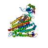
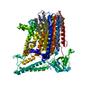
| Title | mouse TMEM63b in LMNG-CHS micelle | |||||||||
 Map data Map data | sharpened map | |||||||||
 Sample Sample |
| |||||||||
 Keywords Keywords | Scramblase / LIPID TRANSPORT | |||||||||
| Function / homology |  Function and homology information Function and homology informationalveolar lamellar body membrane / sphingomyelin transfer activity / surfactant secretion / phosphatidylcholine transfer activity / mechanosensitive monoatomic cation channel activity / osmolarity-sensing monoatomic cation channel activity / intestinal stem cell homeostasis / phospholipid scramblase activity / drinking behavior / mechanosensitive monoatomic ion channel activity ...alveolar lamellar body membrane / sphingomyelin transfer activity / surfactant secretion / phosphatidylcholine transfer activity / mechanosensitive monoatomic cation channel activity / osmolarity-sensing monoatomic cation channel activity / intestinal stem cell homeostasis / phospholipid scramblase activity / drinking behavior / mechanosensitive monoatomic ion channel activity / calcium-activated cation channel activity / exocytosis / bioluminescence / generation of precursor metabolites and energy / sensory perception of sound / actin cytoskeleton / early endosome membrane / lysosomal membrane / endoplasmic reticulum membrane / plasma membrane Similarity search - Function | |||||||||
| Biological species |   | |||||||||
| Method | single particle reconstruction / cryo EM / Resolution: 3.4 Å | |||||||||
 Authors Authors | Miyata Y / Takahashi K / Lee Y / Sultan CS / Kuribayashi R / Takahashi M / Hata K / Bamba T / Izumi Y / Liu K ...Miyata Y / Takahashi K / Lee Y / Sultan CS / Kuribayashi R / Takahashi M / Hata K / Bamba T / Izumi Y / Liu K / Uemura T / Nomura N / Iwata S / Nagata S / Nishizawa T / Segawa K | |||||||||
| Funding support |  Japan, 1 items Japan, 1 items
| |||||||||
 Citation Citation |  Journal: Nat Struct Mol Biol / Year: 2025 Journal: Nat Struct Mol Biol / Year: 2025Title: Membrane structure-responsive lipid scrambling by TMEM63B to control plasma membrane lipid distribution. Authors: Yugo Miyata / Katsuya Takahashi / Yongchan Lee / Cheryl S Sultan / Risa Kuribayashi / Masatomo Takahashi / Kosuke Hata / Takeshi Bamba / Yoshihiro Izumi / Kehong Liu / Tomoko Uemura / ...Authors: Yugo Miyata / Katsuya Takahashi / Yongchan Lee / Cheryl S Sultan / Risa Kuribayashi / Masatomo Takahashi / Kosuke Hata / Takeshi Bamba / Yoshihiro Izumi / Kehong Liu / Tomoko Uemura / Norimichi Nomura / So Iwata / Shigekazu Nagata / Tomohiro Nishizawa / Katsumori Segawa /  Abstract: Phospholipids are asymmetrically distributed in the plasma membrane (PM), with phosphatidylcholine and sphingomyelin abundant in the outer leaflet. However, the mechanisms by which their distribution ...Phospholipids are asymmetrically distributed in the plasma membrane (PM), with phosphatidylcholine and sphingomyelin abundant in the outer leaflet. However, the mechanisms by which their distribution is regulated remain unclear. Here, we show that transmembrane protein 63B (TMEM63B) functions as a membrane structure-responsive lipid scramblase localized at the PM and lysosomes, activating bidirectional lipid translocation upon changes in membrane curvature and thickness. TMEM63B contains two intracellular loops with palmitoylated cysteine residue clusters essential for its scrambling function. TMEM63B deficiency alters phosphatidylcholine and sphingomyelin distributions in the PM. Persons with heterozygous mutations in TMEM63B are known to develop neurodevelopmental disorders. We show that V44M, the most frequent substitution, confers constitutive scramblase activity on TMEM63B, disrupting PM phospholipid asymmetry. We determined the cryo-electron microscopy structures of TMEM63B in its open and closed conformations, uncovering a lipid translocation pathway formed in response to changes in the membrane environment. Together, our results identify TMEM63B as a membrane structure-responsive scramblase that controls PM lipid distribution and we reveal the molecular basis for lipid scrambling and its biological importance. | |||||||||
| History |
|
- Structure visualization
| Supplemental images |
|---|
- Downloads & links
-EMDB archive
| Map data |  emd_37501.map.gz emd_37501.map.gz | 14.7 MB |  EMDB map data format EMDB map data format | |
|---|---|---|---|---|
| Header (meta data) |  emd-37501-v30.xml emd-37501-v30.xml emd-37501.xml emd-37501.xml | 24.3 KB 24.3 KB | Display Display |  EMDB header EMDB header |
| FSC (resolution estimation) |  emd_37501_fsc.xml emd_37501_fsc.xml | 5.2 KB | Display |  FSC data file FSC data file |
| Images |  emd_37501.png emd_37501.png | 139.9 KB | ||
| Masks |  emd_37501_msk_1.map emd_37501_msk_1.map emd_37501_msk_2.map emd_37501_msk_2.map | 15.6 MB 15.6 MB |  Mask map Mask map | |
| Filedesc metadata |  emd-37501.cif.gz emd-37501.cif.gz | 7.8 KB | ||
| Others |  emd_37501_half_map_1.map.gz emd_37501_half_map_1.map.gz emd_37501_half_map_2.map.gz emd_37501_half_map_2.map.gz | 14.5 MB 14.5 MB | ||
| Archive directory |  http://ftp.pdbj.org/pub/emdb/structures/EMD-37501 http://ftp.pdbj.org/pub/emdb/structures/EMD-37501 ftp://ftp.pdbj.org/pub/emdb/structures/EMD-37501 ftp://ftp.pdbj.org/pub/emdb/structures/EMD-37501 | HTTPS FTP |
-Validation report
| Summary document |  emd_37501_validation.pdf.gz emd_37501_validation.pdf.gz | 809.2 KB | Display |  EMDB validaton report EMDB validaton report |
|---|---|---|---|---|
| Full document |  emd_37501_full_validation.pdf.gz emd_37501_full_validation.pdf.gz | 808.7 KB | Display | |
| Data in XML |  emd_37501_validation.xml.gz emd_37501_validation.xml.gz | 11.8 KB | Display | |
| Data in CIF |  emd_37501_validation.cif.gz emd_37501_validation.cif.gz | 15 KB | Display | |
| Arichive directory |  https://ftp.pdbj.org/pub/emdb/validation_reports/EMD-37501 https://ftp.pdbj.org/pub/emdb/validation_reports/EMD-37501 ftp://ftp.pdbj.org/pub/emdb/validation_reports/EMD-37501 ftp://ftp.pdbj.org/pub/emdb/validation_reports/EMD-37501 | HTTPS FTP |
-Related structure data
| Related structure data |  8wg3MC  8wg4C M: atomic model generated by this map C: citing same article ( |
|---|---|
| Similar structure data | Similarity search - Function & homology  F&H Search F&H Search |
- Links
Links
| EMDB pages |  EMDB (EBI/PDBe) / EMDB (EBI/PDBe) /  EMDataResource EMDataResource |
|---|---|
| Related items in Molecule of the Month |
- Map
Map
| File |  Download / File: emd_37501.map.gz / Format: CCP4 / Size: 15.6 MB / Type: IMAGE STORED AS FLOATING POINT NUMBER (4 BYTES) Download / File: emd_37501.map.gz / Format: CCP4 / Size: 15.6 MB / Type: IMAGE STORED AS FLOATING POINT NUMBER (4 BYTES) | ||||||||||||||||||||||||||||||||||||
|---|---|---|---|---|---|---|---|---|---|---|---|---|---|---|---|---|---|---|---|---|---|---|---|---|---|---|---|---|---|---|---|---|---|---|---|---|---|
| Annotation | sharpened map | ||||||||||||||||||||||||||||||||||||
| Projections & slices | Image control
Images are generated by Spider. | ||||||||||||||||||||||||||||||||||||
| Voxel size | X=Y=Z: 1.55625 Å | ||||||||||||||||||||||||||||||||||||
| Density |
| ||||||||||||||||||||||||||||||||||||
| Symmetry | Space group: 1 | ||||||||||||||||||||||||||||||||||||
| Details | EMDB XML:
|
-Supplemental data
-Mask #1
| File |  emd_37501_msk_1.map emd_37501_msk_1.map | ||||||||||||
|---|---|---|---|---|---|---|---|---|---|---|---|---|---|
| Projections & Slices |
| ||||||||||||
| Density Histograms |
-Mask #2
| File |  emd_37501_msk_2.map emd_37501_msk_2.map | ||||||||||||
|---|---|---|---|---|---|---|---|---|---|---|---|---|---|
| Projections & Slices |
| ||||||||||||
| Density Histograms |
-Half map: halfmapA
| File | emd_37501_half_map_1.map | ||||||||||||
|---|---|---|---|---|---|---|---|---|---|---|---|---|---|
| Annotation | halfmapA | ||||||||||||
| Projections & Slices |
| ||||||||||||
| Density Histograms |
-Half map: halfmapB
| File | emd_37501_half_map_2.map | ||||||||||||
|---|---|---|---|---|---|---|---|---|---|---|---|---|---|
| Annotation | halfmapB | ||||||||||||
| Projections & Slices |
| ||||||||||||
| Density Histograms |
- Sample components
-Entire : mouse TMEM63B
| Entire | Name: mouse TMEM63B |
|---|---|
| Components |
|
-Supramolecule #1: mouse TMEM63B
| Supramolecule | Name: mouse TMEM63B / type: complex / ID: 1 / Parent: 0 / Macromolecule list: #1 |
|---|---|
| Source (natural) | Organism:  |
| Molecular weight | Theoretical: 100 KDa |
-Macromolecule #1: CSC1-like protein 2,Green fluorescent protein
| Macromolecule | Name: CSC1-like protein 2,Green fluorescent protein / type: protein_or_peptide / ID: 1 / Number of copies: 1 / Enantiomer: LEVO |
|---|---|
| Source (natural) | Organism:  |
| Molecular weight | Theoretical: 126.759461 KDa |
| Recombinant expression | Organism:  Homo sapiens (human) Homo sapiens (human) |
| Sequence | String: MLPFLLATLG TAALNSSNPK DYCYSARIRS TVLQGLPFGG VPTVLALDFM CFLALLFLFS ILRKVAWDYG RLALVTDADR LRRQERERV EQEYVASAMH GDSHDRYERL TSVSSSVDFD QRDNGF(P1L)SWL TAIFRIKDDE IRDKCGGDAV HYLSFQR HI IGLLVVVGVL ...String: MLPFLLATLG TAALNSSNPK DYCYSARIRS TVLQGLPFGG VPTVLALDFM CFLALLFLFS ILRKVAWDYG RLALVTDADR LRRQERERV EQEYVASAMH GDSHDRYERL TSVSSSVDFD QRDNGF(P1L)SWL TAIFRIKDDE IRDKCGGDAV HYLSFQR HI IGLLVVVGVL SVGIVLPVNF SGDLLENNAY SFGRTTIANL KSGNNLLWLH TSFAFLYLLL TVYSMRRHTS KMRYKEDD L VKRTLFINGI SKYAESEKIK KHFEEAYPNC TVLEARPCYN VARLMFLDAE RKKAERGKLY FTNLQSKENV PAMINPKPC GHLCCCVVRG CEQVEAIEYY TKLEQRLKED YRREKEKVNE KPLGMAFVTF HNETITAIIL KDFNVCKCQG CTCRGEPRAS SCSEALHIS NWTVTYAPDP QNIYWEHLSI RGFIWWLRCL VINVVLFILL FFLTTPAIII TTMDKFNVTK PVEYLNNPII T QFFPTLLL WCFSALLPTI VYYSAFFEAH WTRSGENRTT MHKCYTFLIF MVLLLPSLGL SSLDLFFRWL FDKKFLAEAA IR FECVFLP DNGAFFVNYV IASAFIGNAM DLLRIPGLLM YMIRLCLARS AAERRNVKRH QAYEFQFGAA YAWMMCVFTV VMT YSITCP IIVPFGLMYM LLKHLVDRYN LYYAYLPAKL DKKIHSGAVN QVVAAPILCL FWLLFFSTMR TGFLAPTSMF TFVV LVITI VICLCHVCFG HFKYLSAHNY KIEHTETDAV SSRSNGRPPT AGAVPKSAKY IAQVLQDSEG DGDGDGAPGS SGDEP PSSS SQDEELLMPP DGLTDTDFQS CEDSLIENEI HQGTENLYFQ GSAAAAVSKG EELFTGVVPI LVELDGDVNG HKFSVS GEG EGDATYGKLT LKFICTTGKL PVPWPTLVTT LTYGVQCFSR YPDHMKQHDF FKSAMPEGYV QERTIFFKDD GNYKTRA EV KFEGDTLVNR IELKGIDFKE DGNILGHKLE YNYNSHNVYI MADKQKNGIK VNFKIRHNIE DGSVQLADHY QQNTPIGD G PVLLPDNHYL STQSKLSKDP NEKRDHMVLL EFVTAAGITL GMDELYKSGL RSWSHPQFEK GGGSGGGSGG SAWSHPQFE K UniProtKB: Mechanosensitive cation channel TMEM63B, Green fluorescent protein |
-Macromolecule #2: CHOLESTEROL HEMISUCCINATE
| Macromolecule | Name: CHOLESTEROL HEMISUCCINATE / type: ligand / ID: 2 / Number of copies: 3 / Formula: Y01 |
|---|---|
| Molecular weight | Theoretical: 486.726 Da |
| Chemical component information |  ChemComp-Y01: |
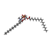
-Macromolecule #3: 1-palmitoyl-2-oleoyl-sn-glycero-3-phosphocholine
| Macromolecule | Name: 1-palmitoyl-2-oleoyl-sn-glycero-3-phosphocholine / type: ligand / ID: 3 / Number of copies: 1 / Formula: LBN |
|---|---|
| Molecular weight | Theoretical: 760.076 Da |
| Chemical component information |  ChemComp-LBN: |
-Experimental details
-Structure determination
| Method | cryo EM |
|---|---|
 Processing Processing | single particle reconstruction |
| Aggregation state | 2D array |
- Sample preparation
| Concentration | 5 mg/mL |
|---|---|
| Buffer | pH: 8 |
| Grid | Model: Quantifoil R0.6/1 / Material: COPPER / Mesh: 300 / Support film - Material: CARBON / Support film - topology: HOLEY |
| Vitrification | Cryogen name: ETHANE / Chamber humidity: 100 % / Chamber temperature: 279 K / Instrument: FEI VITROBOT MARK IV |
- Electron microscopy
| Microscope | FEI TITAN KRIOS |
|---|---|
| Image recording | Film or detector model: GATAN K3 BIOQUANTUM (6k x 4k) / Average electron dose: 50.0 e/Å2 |
| Electron beam | Acceleration voltage: 300 kV / Electron source:  FIELD EMISSION GUN FIELD EMISSION GUN |
| Electron optics | Illumination mode: FLOOD BEAM / Imaging mode: BRIGHT FIELD / Nominal defocus max: 1.6 µm / Nominal defocus min: 0.8 µm / Nominal magnification: 105000 |
| Sample stage | Cooling holder cryogen: NITROGEN |
| Experimental equipment |  Model: Titan Krios / Image courtesy: FEI Company |
 Movie
Movie Controller


 Z (Sec.)
 Y (Row.)
 X (Col.)


