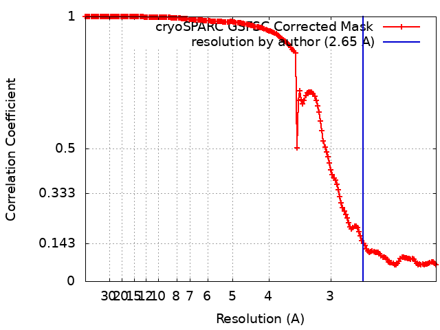
Journal: Plant Commun / Year: 2024 Title: Structural and spectroscopic insights into fucoxanthin chlorophyll a/c-binding proteins of diatoms in diverse oligomeric states. Authors: Cuicui Zhou / Yue Feng / Zhenhua Li / Lili Shen / Xiaoyi Li / Yumei Wang / Guangye Han / Tingyun Kuang / Cheng Liu / Jian-Ren Shen / Wenda Wang / Abstract: Diatoms, a group of prevalent marine algae, significantly contribute to global primary productivity. Their substantial biomass is linked to enhanced absorption of blue-green light underwater, ...Diatoms, a group of prevalent marine algae, significantly contribute to global primary productivity. Their substantial biomass is linked to enhanced absorption of blue-green light underwater, facilitated by fucoxanthin chlorophyll a/c-binding proteins (FCPs), exhibiting oligomeric diversity across diatom species. Utilizing mild CN-PAGE analysis on solubilized thylakoid membranes, we displayed monomeric, dimeric, trimeric, tetrameric and pentameric FCPs in diatoms. Mass spectrometry analysis revealed each oligomeric FCP has specific protein compositions, constituting a large Lhcf family of FCP antennas. In addition, we resolved the structures of Thalassiosira pseudonana FCP (Tp-FCP) homotrimer and Chaetoceros gracilis FCP (Cg-FCP) pentamer by cryo-electron microscopy at 2.73 Å and 2.65 Å resolutions, respectively. The distinct pigment composition and organization in various oligomeric FCPs change their blue-green light-harvesting, excitation energy transfer pathways. In comparison to dimeric and trimeric FCPs, Cg-FCP tetramer and Cg-FCP pentamer exhibit stronger absorption by Chls c, red-shifted and broader Chl a fluorescence emission, as well as more robust circular dichroism signals originating from Chl a-carotenoid dimers. These spectroscopic characteristics indicate that Chl a molecules in Cg-FCP tetramer and Cg-FCP pentamer are more heterogeneous than in both dimers and Tp-FCP trimer. The structural and spectroscopic insights provided by this study contribute to a better understanding of the mechanisms that empower diatoms to adapt to fluctuating light environments.
In the structure databanks used in Yorodumi, some data are registered as the other names, "COVID-19 virus" and "2019-nCoV". Here are the details of the virus and the list of structure data.
Jan 31, 2019. EMDB accession codes are about to change! (news from PDBe EMDB page)
EMDB accession codes are about to change! (news from PDBe EMDB page)
The allocation of 4 digits for EMDB accession codes will soon come to an end. Whilst these codes will remain in use, new EMDB accession codes will include an additional digit and will expand incrementally as the available range of codes is exhausted. The current 4-digit format prefixed with “EMD-” (i.e. EMD-XXXX) will advance to a 5-digit format (i.e. EMD-XXXXX), and so on. It is currently estimated that the 4-digit codes will be depleted around Spring 2019, at which point the 5-digit format will come into force.
The EM Navigator/Yorodumi systems omit the EMD- prefix.
Related info.:Q: What is EMD? / ID/Accession-code notation in Yorodumi/EM Navigator
Yorodumi is a browser for structure data from EMDB, PDB, SASBDB, etc.
This page is also the successor to EM Navigator detail page, and also detail information page/front-end page for Omokage search.
The word "yorodu" (or yorozu) is an old Japanese word meaning "ten thousand". "mi" (miru) is to see.
Related info.:EMDB / PDB / SASBDB / Comparison of 3 databanks / Yorodumi Search / Aug 31, 2016. New EM Navigator & Yorodumi / Yorodumi Papers / Jmol/JSmol / Function and homology information / Changes in new EM Navigator and Yorodumi
 Movie
Movie Controller


 Open data
 Basic information
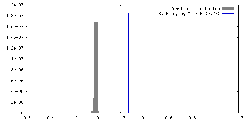
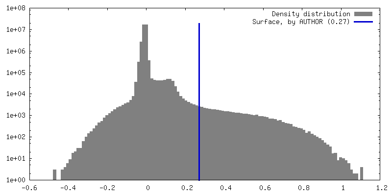
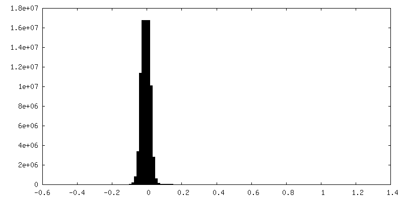
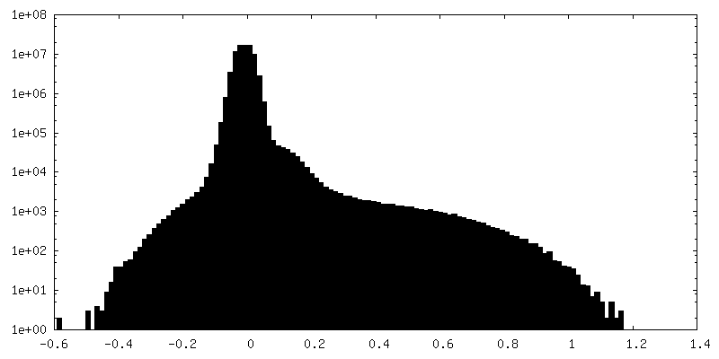
 Map data
 Sample
 Keywords
 Chaetoceros neogracilis (Diatom)
 Authors
 China, 1 items
  Citation
 Structure visualization
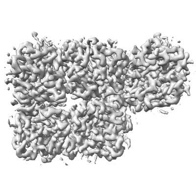
 Downloads & links
 EMDB map data format
 emd_37442.png
 http://ftp.pdbj.org/pub/emdb/structures/EMD-37442
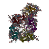


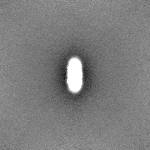
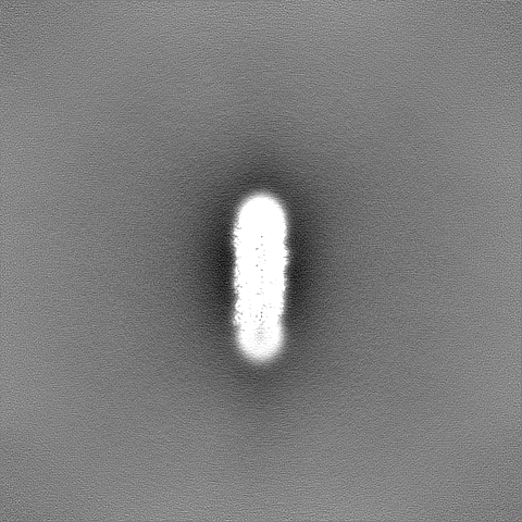
 Z (Sec.)
 Y (Row.)
 X (Col.)


 Sample components


 Processing
 Electron microscopy
 FIELD EMISSION GUN