Movie
Movie Controller


+ Open data

- Basic information
| Entry |  | |||||||||
|---|---|---|---|---|---|---|---|---|---|---|
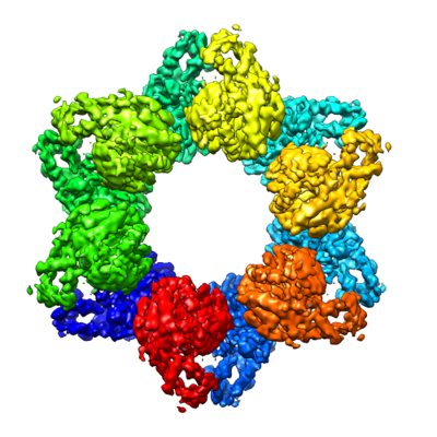
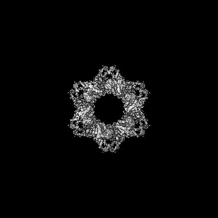
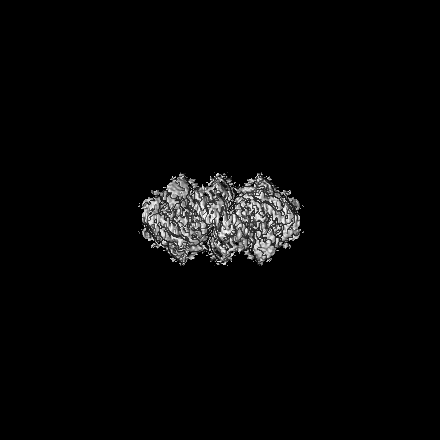
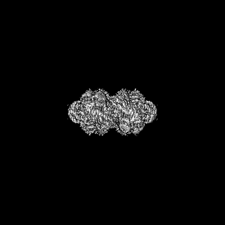
| Title | Structure of a bacterial protein | |||||||||
 Map data Map data | main map | |||||||||
 Sample Sample |
| |||||||||
 Keywords Keywords | immune system / cell growth inhibition | |||||||||
| Function / homology | SIR2-like domain / DHS-like NAD/FAD-binding domain superfamily / SIR2-like domain-containing protein Function and homology information Function and homology information | |||||||||
| Biological species |  Paenibacillus sp. 453mf (bacteria) Paenibacillus sp. 453mf (bacteria) | |||||||||
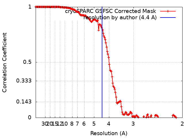
| Method | single particle reconstruction / cryo EM / Resolution: 4.4 Å | |||||||||
 Authors Authors | Yu G / Liao F / Li X / Li Z / Zhang H | |||||||||
| Funding support |  China, 1 items China, 1 items
| |||||||||
 Citation Citation | Journal: To Be Published Title: structure of a bacterial protein Authors: Yu G / Liao F / Li X / Li Z / Zhang H | |||||||||
| History |
|
- Structure visualization
| Supplemental images |
|---|
- Downloads & links
-EMDB archive
| Map data | emd_37384.map.gz | 306.4 MB | EMDB map data format | |
|---|---|---|---|---|
| Header (meta data) | emd-37384-v30.xmlemd-37384.xml | 15.5 KB 15.5 KB | Display Display | EMDB header |
| FSC (resolution estimation) | emd_37384_fsc.xml | 14.6 KB | Display | FSC data file |
| Images |  emd_37384.png emd_37384.png | 157.6 KB | ||
| Filedesc metadata | emd-37384.cif.gz | 5.4 KB | ||
| Others | emd_37384_half_map_1.map.gzemd_37384_half_map_2.map.gz | 300.6 MB 300.6 MB | ||
| Archive directory |  http://ftp.pdbj.org/pub/emdb/structures/EMD-37384ftp://ftp.pdbj.org/pub/emdb/structures/EMD-37384 http://ftp.pdbj.org/pub/emdb/structures/EMD-37384ftp://ftp.pdbj.org/pub/emdb/structures/EMD-37384 | HTTPS FTP |
-Validation report
| Summary document | emd_37384_validation.pdf.gz | 798.6 KB | Display | EMDB validaton report |
|---|---|---|---|---|
| Full document | emd_37384_full_validation.pdf.gz | 798.2 KB | Display | |
| Data in XML | emd_37384_validation.xml.gz | 23.9 KB | Display | |
| Data in CIF | emd_37384_validation.cif.gz | 31 KB | Display | |
| Arichive directory | https://ftp.pdbj.org/pub/emdb/validation_reports/EMD-37384ftp://ftp.pdbj.org/pub/emdb/validation_reports/EMD-37384 | HTTPS FTP |
-Related structure data
| Related structure data |  8w9xMC M: atomic model generated by this map C: citing same article ( |
|---|---|
| Similar structure data |
-Links
| EMDB pages | EMDB (EBI/PDBe) / EMDataResource |
|---|---|
| Related items in Molecule of the Month |


-Map
| File | Download / File: emd_37384.map.gz / Format: CCP4 / Size: 325 MB / Type: IMAGE STORED AS FLOATING POINT NUMBER (4 BYTES) | ||||||||||||||||||||||||||||||||||||
|---|---|---|---|---|---|---|---|---|---|---|---|---|---|---|---|---|---|---|---|---|---|---|---|---|---|---|---|---|---|---|---|---|---|---|---|---|---|
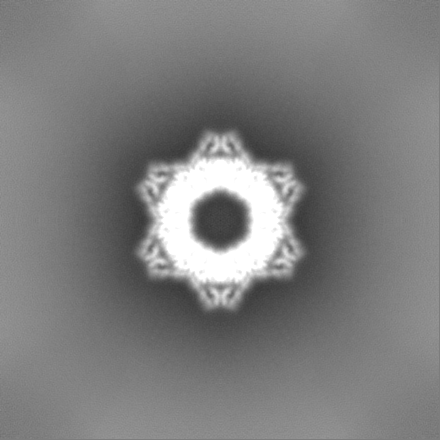
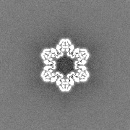
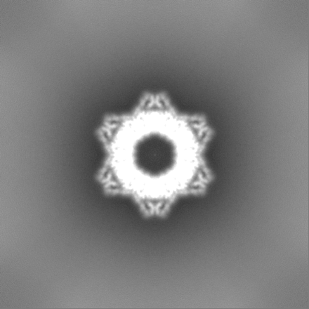
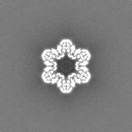
| Annotation | main map | ||||||||||||||||||||||||||||||||||||
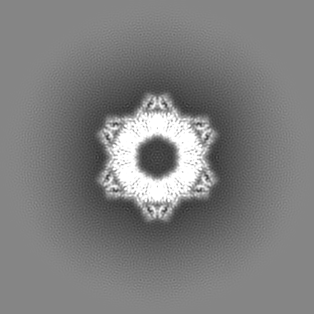
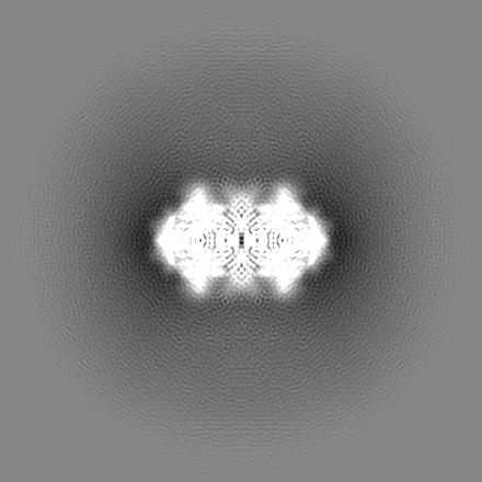
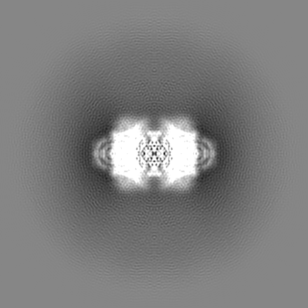
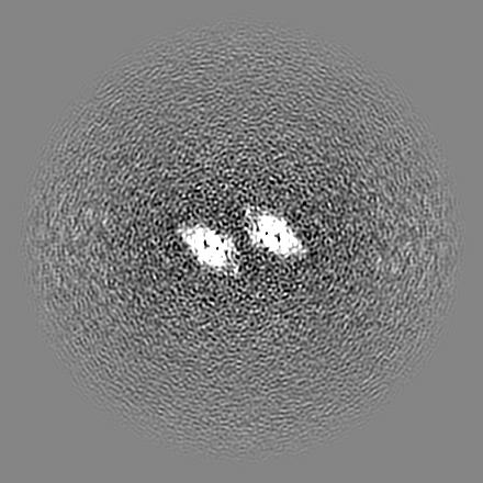
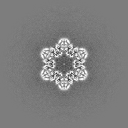
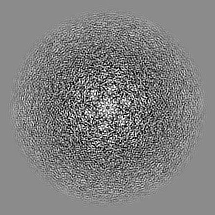
| Projections & slices | Image control
Images are generated by Spider. | ||||||||||||||||||||||||||||||||||||
| Voxel size | X=Y=Z: 0.989 Å | ||||||||||||||||||||||||||||||||||||
| Density |
| ||||||||||||||||||||||||||||||||||||
| Symmetry | Space group: 1 | ||||||||||||||||||||||||||||||||||||
| Details | EMDB XML:
|
 Z (Sec.)
 Y (Row.)
 X (Col.)


-Supplemental data
-Half map: half map
| File | emd_37384_half_map_1.map | ||||||||||||
|---|---|---|---|---|---|---|---|---|---|---|---|---|---|
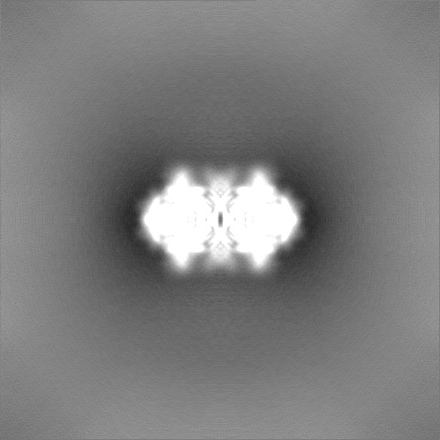
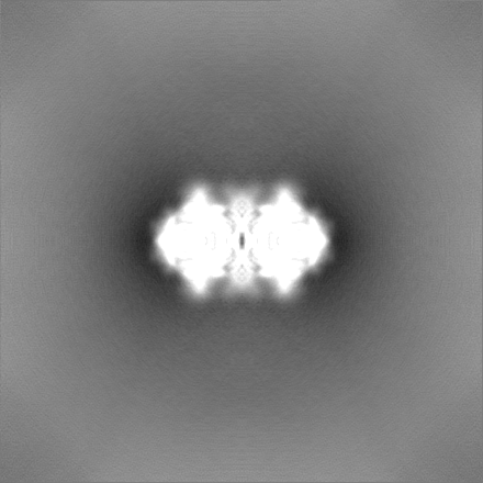
| Annotation | half map | ||||||||||||
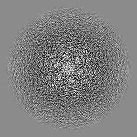
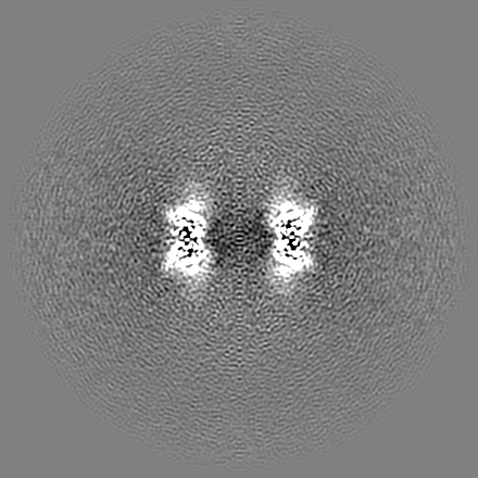
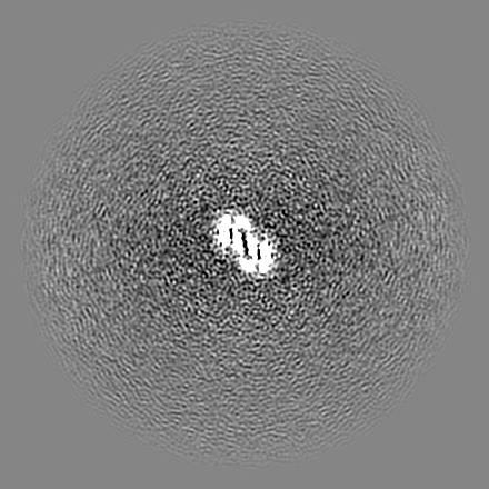
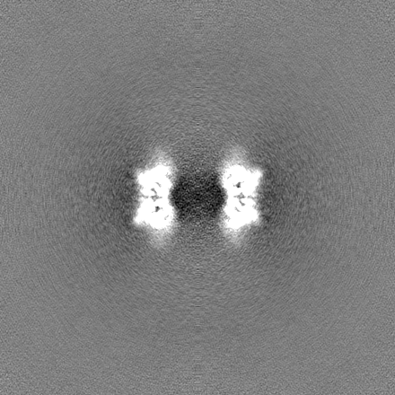
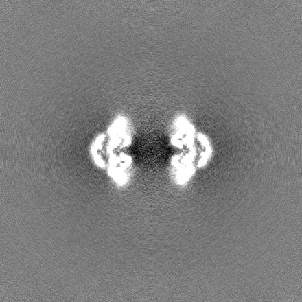
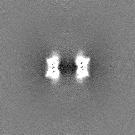
| Projections & Slices |
| ||||||||||||
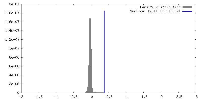
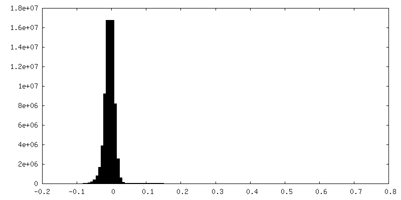
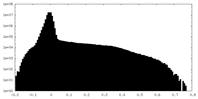
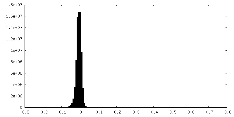
| Density Histograms |
-Half map: half map
| File | emd_37384_half_map_2.map | ||||||||||||
|---|---|---|---|---|---|---|---|---|---|---|---|---|---|
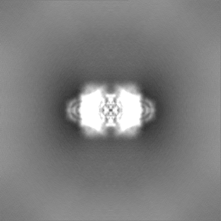
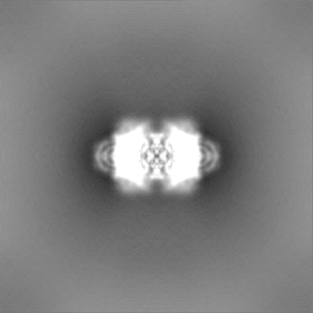
| Annotation | half map | ||||||||||||
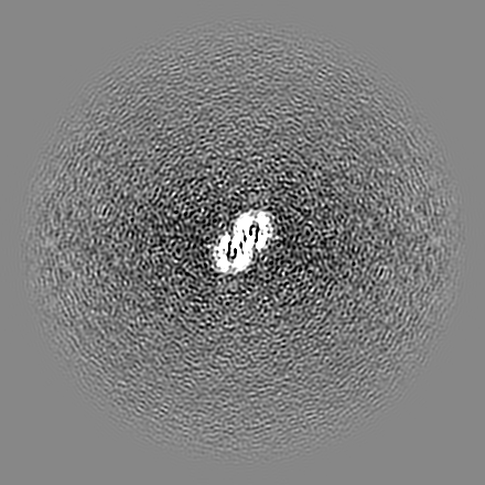
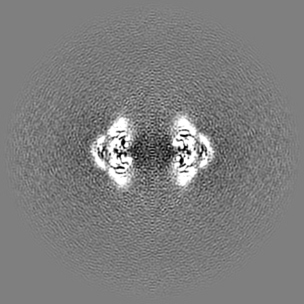
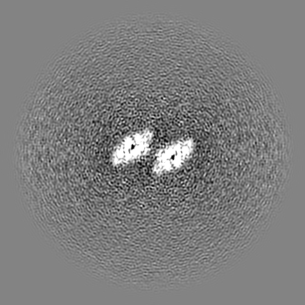
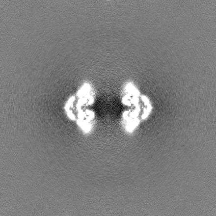
| Projections & Slices |
| ||||||||||||
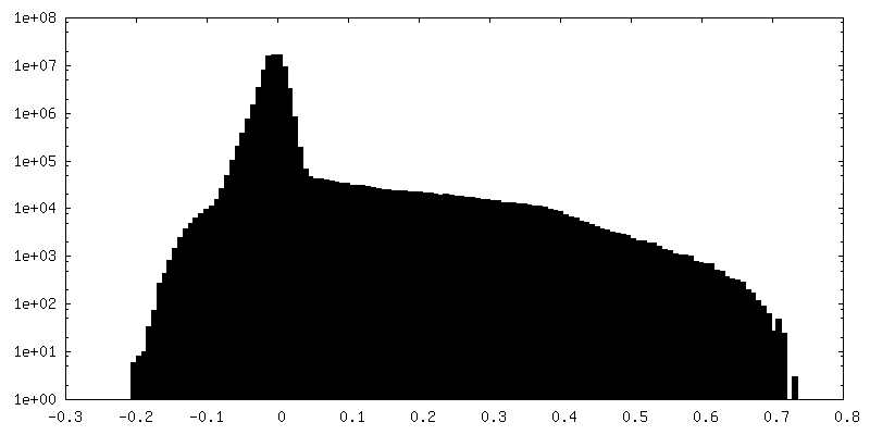
| Density Histograms |
- Sample components
-Entire : bacterial protein
| Entire | Name: bacterial protein |
|---|---|
| Components |
|
-Supramolecule #1: bacterial protein
| Supramolecule | Name: bacterial protein / type: complex / ID: 1 / Parent: 0 / Macromolecule list: all |
|---|---|
| Source (natural) | Organism: Paenibacillus sp. 453mf (bacteria) |
-Macromolecule #1: SIR2-like domain-containing protein
| Macromolecule | Name: SIR2-like domain-containing protein / type: protein_or_peptide / ID: 1 / Number of copies: 12 / Enantiomer: LEVO |
|---|---|
| Source (natural) | Organism: Paenibacillus sp. 453mf (bacteria) |
| Molecular weight | Theoretical: 43.60948 KDa |
| Recombinant expression | Organism: |
| Sequence | String: MDHSITASYY DTTQQLSLLK HVLSEDKRPI AFIIAAGCPV SIRHNDAPLI PDVAGLTRKI SDSFGGNPDS LLMKIIQNLK TTIPNPTIE DILSYIRLLQ QIPMSGKIHD VENSVINALE ESICELIEEE VNVDLPGNAT PYHKIAAWIN SINREHQVEI F TTNYDLLM ...String: MDHSITASYY DTTQQLSLLK HVLSEDKRPI AFIIAAGCPV SIRHNDAPLI PDVAGLTRKI SDSFGGNPDS LLMKIIQNLK TTIPNPTIE DILSYIRLLQ QIPMSGKIHD VENSVINALE ESICELIEEE VNVDLPGNAT PYHKIAAWIN SINREHQVEI F TTNYDLLM EQALEELNVP YFDGFVGSKR AFFDIRTIEE NKLPSRWSKL WKLHGSINWQ LDKQTQTIWR GTPSKGCSLI HP SHLKYDQ SRKMPYLVMM DQLKLFLNQP SAILITCGYS YKDQHINEVL SQGLQTNPNA LIYGLQYDVL ENYQEAKDMA LKR SNLILL AKDRAIIGKK EGEWKPDPQS SQDNDPLLFF KLGDFQHLAS FLEEISQYDW SKQND UniProtKB: SIR2-like domain-containing protein |
-Experimental details
-Structure determination
| Method | cryo EM |
|---|---|
 Processing Processing | single particle reconstruction |
| Aggregation state | particle |
-Sample preparation
| Buffer | pH: 7 |
|---|---|
| Vitrification | Cryogen name: ETHANE |
- Electron microscopy
| Microscope | FEI TITAN KRIOS |
|---|---|
| Image recording | Film or detector model: GATAN K3 (6k x 4k) / Average electron dose: 40.0 e/Å2 |
| Electron beam | Acceleration voltage: 300 kV / Electron source:  FIELD EMISSION GUN FIELD EMISSION GUN |
| Electron optics | Illumination mode: SPOT SCAN / Imaging mode: BRIGHT FIELD / Nominal defocus max: 2.5 µm / Nominal defocus min: 0.5 µm |
| Experimental equipment |  Model: Titan Krios / Image courtesy: FEI Company |