Movie
Movie Controller

[English] 日本語
 Yorodumi
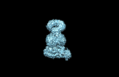
- EMDB-37324: Cryo-EM structure of Escherichia coli Str K12 FtsEX complex with ... -
+ Open data

- Basic information
| Entry |  | |||||||||
|---|---|---|---|---|---|---|---|---|---|---|
| Title | Cryo-EM structure of Escherichia coli Str K12 FtsEX complex with ATP-gamma-S in peptidisc | |||||||||
 Map data Map data | ||||||||||
 Sample Sample |
| |||||||||
 Keywords Keywords | complex / TRANSPORT PROTEIN | |||||||||
| Function / homology |  Function and homology information Function and homology informationdivision septum / divisome complex / Gram-negative-bacterium-type cell wall / peptidoglycan turnover / plasma membrane protein complex / division septum assembly / FtsZ-dependent cytokinesis / extrinsic component of membrane / cell division site / ATPase complex ...division septum / divisome complex / Gram-negative-bacterium-type cell wall / peptidoglycan turnover / plasma membrane protein complex / division septum assembly / FtsZ-dependent cytokinesis / extrinsic component of membrane / cell division site / ATPase complex / positive regulation of cell division / transmembrane transporter activity / transmembrane transport / cell division / response to antibiotic / ATP hydrolysis activity / ATP binding / membrane / plasma membrane / cytoplasm Similarity search - Function | |||||||||
| Biological species |  | |||||||||
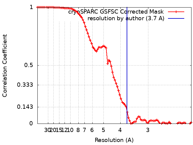
| Method | single particle reconstruction / cryo EM / Resolution: 3.7 Å | |||||||||
 Authors Authors | Li J / Xu X / He Y / Luo M | |||||||||
| Funding support |  Singapore, 1 items Singapore, 1 items
| |||||||||
 Citation Citation | Journal: To Be Published Title: Cryo-EM structure of Escherichia coli Str K12 FtsEX complex with ATP-gamma-S in peptidisc Authors: Li J / Xu X / He Y / Luo M | |||||||||
| History |
|
- Structure visualization
| Supplemental images |
|---|
- Downloads & links
-EMDB archive
| Map data | emd_37324.map.gz | 59.5 MB | EMDB map data format | |
|---|---|---|---|---|
| Header (meta data) | emd-37324-v30.xmlemd-37324.xml | 17.6 KB 17.6 KB | Display Display | EMDB header |
| FSC (resolution estimation) | emd_37324_fsc.xml | 8.5 KB | Display | FSC data file |
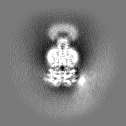
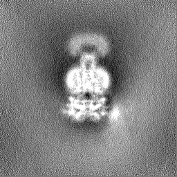
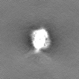
| Images |  emd_37324.png emd_37324.png | 26.4 KB | ||
| Filedesc metadata | emd-37324.cif.gz | 5.9 KB | ||
| Others | emd_37324_half_map_1.map.gzemd_37324_half_map_2.map.gz | 59.4 MB 59.4 MB | ||
| Archive directory |  http://ftp.pdbj.org/pub/emdb/structures/EMD-37324ftp://ftp.pdbj.org/pub/emdb/structures/EMD-37324 http://ftp.pdbj.org/pub/emdb/structures/EMD-37324ftp://ftp.pdbj.org/pub/emdb/structures/EMD-37324 | HTTPS FTP |
-Related structure data
| Related structure data |  8w6iMC M: atomic model generated by this map C: citing same article ( |
|---|---|
| Similar structure data |
-Links
| EMDB pages | EMDB (EBI/PDBe) / EMDataResource |
|---|---|
| Related items in Molecule of the Month |


-Map
| File | Download / File: emd_37324.map.gz / Format: CCP4 / Size: 64 MB / Type: IMAGE STORED AS FLOATING POINT NUMBER (4 BYTES) | ||||||||||||||||||||||||||||||||||||
|---|---|---|---|---|---|---|---|---|---|---|---|---|---|---|---|---|---|---|---|---|---|---|---|---|---|---|---|---|---|---|---|---|---|---|---|---|---|
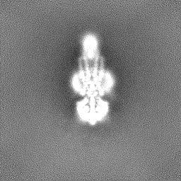
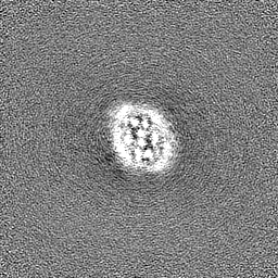
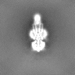
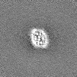
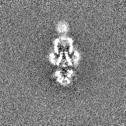
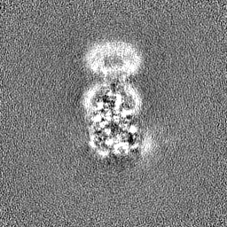
| Projections & slices | Image control
Images are generated by Spider. | ||||||||||||||||||||||||||||||||||||
| Voxel size | X=Y=Z: 1.06 Å | ||||||||||||||||||||||||||||||||||||
| Density |
| ||||||||||||||||||||||||||||||||||||
| Symmetry | Space group: 1 | ||||||||||||||||||||||||||||||||||||
| Details | EMDB XML:
|
 Z (Sec.)
 Y (Row.)
 X (Col.)


-Supplemental data
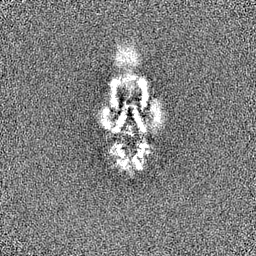
-Half map: #1
| File | emd_37324_half_map_1.map | ||||||||||||
|---|---|---|---|---|---|---|---|---|---|---|---|---|---|
| Projections & Slices |
| ||||||||||||
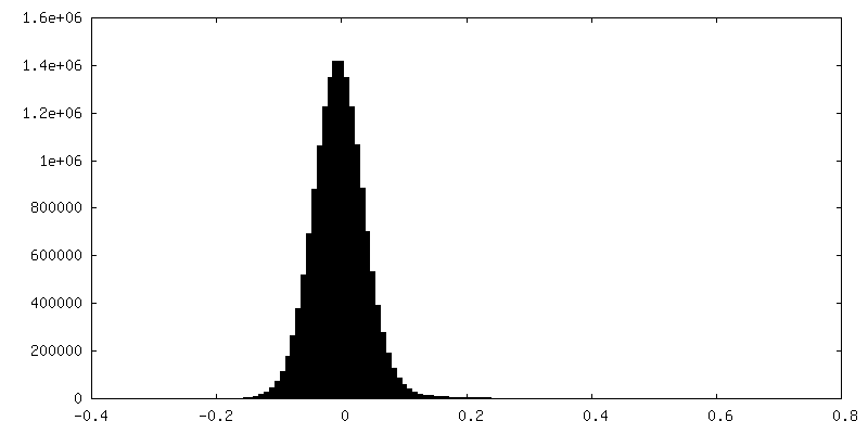
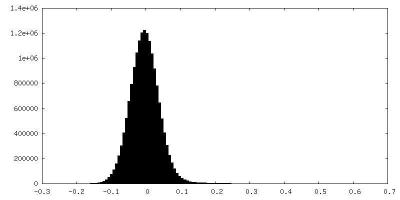
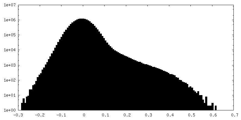
| Density Histograms |

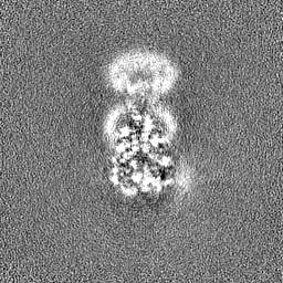
-Half map: #2
| File | emd_37324_half_map_2.map | ||||||||||||
|---|---|---|---|---|---|---|---|---|---|---|---|---|---|
| Projections & Slices |
| ||||||||||||
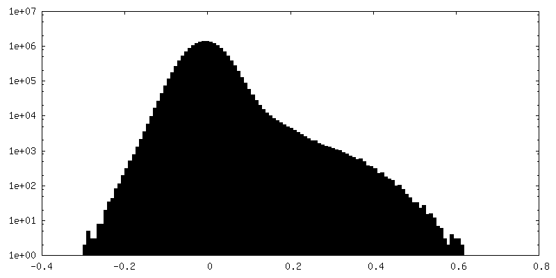
| Density Histograms |

- Sample components
-Entire : complex of FtsEX
| Entire | Name: complex of FtsEX |
|---|---|
| Components |
|
-Supramolecule #1: complex of FtsEX
| Supramolecule | Name: complex of FtsEX / type: complex / ID: 1 / Parent: 0 / Macromolecule list: #1-#2 |
|---|---|
| Source (natural) | Organism: |
-Macromolecule #1: Cell division ATP-binding protein FtsE
| Macromolecule | Name: Cell division ATP-binding protein FtsE / type: protein_or_peptide / ID: 1 / Number of copies: 2 / Enantiomer: LEVO |
|---|---|
| Source (natural) | Organism: |
| Molecular weight | Theoretical: 24.476279 KDa |
| Recombinant expression | Organism: |
| Sequence | String: MIRFEHVSKA YLGGRQALQG VTFHMQPGEM AFLTGHSGAG KSTLLKLICG IERPSAGKIW FSGHDITRLK NREVPFLRRQ IGMIFQDHH LLMDRTVYDN VAIPLIIAGA SGDDIRRRVS AALDKVGLLD KAKNFPIQLS GGEQQRVGIA RAVVNKPAVL L ADEPTGNL ...String: MIRFEHVSKA YLGGRQALQG VTFHMQPGEM AFLTGHSGAG KSTLLKLICG IERPSAGKIW FSGHDITRLK NREVPFLRRQ IGMIFQDHH LLMDRTVYDN VAIPLIIAGA SGDDIRRRVS AALDKVGLLD KAKNFPIQLS GGEQQRVGIA RAVVNKPAVL L ADEPTGNL DDALSEGILR LFEEFNRVGV TVLMATHDIN LISRRSYRML TLSDGHLHGG VGHE UniProtKB: Cell division ATP-binding protein FtsE |
-Macromolecule #2: Cell division protein FtsX
| Macromolecule | Name: Cell division protein FtsX / type: protein_or_peptide / ID: 2 / Number of copies: 2 / Enantiomer: LEVO |
|---|---|
| Source (natural) | Organism: |
| Molecular weight | Theoretical: 38.5835 KDa |
| Recombinant expression | Organism: |
| Sequence | String: MNKRDAINHI RQFGGRLDRF RKSVGGSGDG GRNAPKRAKS SPKPVNRKTN VFNEQVRYAF HGALQDLKSK PFATFLTVMV IAISLTLPS VCYMVYKNVN QAATQYYPSP QITVYLQKTL DDDAAAGVVA QLQAEQGVEK VNYLSREDAL GEFRNWSGFG G ALDMLEEN ...String: MNKRDAINHI RQFGGRLDRF RKSVGGSGDG GRNAPKRAKS SPKPVNRKTN VFNEQVRYAF HGALQDLKSK PFATFLTVMV IAISLTLPS VCYMVYKNVN QAATQYYPSP QITVYLQKTL DDDAAAGVVA QLQAEQGVEK VNYLSREDAL GEFRNWSGFG G ALDMLEEN PLPAVAVVIP KLDFQGTESL NTLRDRITQI NGIDEVRMDD SWFARLAALT GLVGRVSAMI GVLMVAAVFL VI GNSVRLS IFARRDSINV QKLIGATDGF ILRPFLYGGA LLGFSGALLS LILSEILVLR LSSAVAEVAQ VFGTKFDING LSF DECLLL LLVCSMIGWV AAWLATVQHL RHFTPE UniProtKB: Cell division protein FtsX |
-Macromolecule #3: PHOSPHOTHIOPHOSPHORIC ACID-ADENYLATE ESTER
| Macromolecule | Name: PHOSPHOTHIOPHOSPHORIC ACID-ADENYLATE ESTER / type: ligand / ID: 3 / Number of copies: 2 / Formula: AGS |
|---|---|
| Molecular weight | Theoretical: 523.247 Da |
| Chemical component information |  ChemComp-AGS: |
-Experimental details
-Structure determination
| Method | cryo EM |
|---|---|
 Processing Processing | single particle reconstruction |
| Aggregation state | particle |
-Sample preparation
| Buffer | pH: 8 |
|---|---|
| Vitrification | Cryogen name: ETHANE |
- Electron microscopy
| Microscope | FEI TITAN KRIOS |
|---|---|
| Image recording | Film or detector model: GATAN K3 (6k x 4k) / Number grids imaged: 1 / Average exposure time: 6.02 sec. / Average electron dose: 38.837 e/Å2 |
| Electron beam | Acceleration voltage: 300 kV / Electron source:  FIELD EMISSION GUN FIELD EMISSION GUN |
| Electron optics | C2 aperture diameter: 70.0 µm / Calibrated defocus max: 2.5 µm / Calibrated defocus min: 1.0 µm / Illumination mode: SPOT SCAN / Imaging mode: BRIGHT FIELD / Cs: 2.7 mm / Nominal defocus max: 2.5 µm / Nominal defocus min: 1.0 µm |
| Experimental equipment |  Model: Titan Krios / Image courtesy: FEI Company |