 Movie
Movie Controller


+ Open data

- Basic information
| Entry |  | |||||||||
|---|---|---|---|---|---|---|---|---|---|---|
| Title | Gi bound CCR8 in ligand free state | |||||||||
 Map data Map data | ||||||||||
 Sample Sample |
| |||||||||
 Keywords Keywords | GPCR / Gi / Complex / SIGNALING PROTEIN | |||||||||
| Function / homology |  Function and homology information Function and homology informationchemokine receptor activity / C-C chemokine receptor activity / C-C chemokine binding / Chemokine receptors bind chemokines / adenylate cyclase inhibitor activity / positive regulation of protein localization to cell cortex / coreceptor activity / Adenylate cyclase inhibitory pathway / T cell migration / D2 dopamine receptor binding ...chemokine receptor activity / C-C chemokine receptor activity / C-C chemokine binding / Chemokine receptors bind chemokines / adenylate cyclase inhibitor activity / positive regulation of protein localization to cell cortex / coreceptor activity / Adenylate cyclase inhibitory pathway / T cell migration / D2 dopamine receptor binding / response to prostaglandin E / adenylate cyclase regulator activity / G protein-coupled serotonin receptor binding / adenylate cyclase-inhibiting serotonin receptor signaling pathway / cellular response to forskolin / regulation of mitotic spindle organization / cell chemotaxis / Regulation of insulin secretion / positive regulation of cholesterol biosynthetic process / calcium-mediated signaling / negative regulation of insulin secretion / G protein-coupled receptor binding / response to peptide hormone / adenylate cyclase-inhibiting G protein-coupled receptor signaling pathway / adenylate cyclase-modulating G protein-coupled receptor signaling pathway / G-protein beta/gamma-subunit complex binding / centriolar satellite / Olfactory Signaling Pathway / Activation of the phototransduction cascade / G beta:gamma signalling through PLC beta / Presynaptic function of Kainate receptors / Thromboxane signalling through TP receptor / G protein-coupled acetylcholine receptor signaling pathway / Activation of G protein gated Potassium channels / Inhibition of voltage gated Ca2+ channels via Gbeta/gamma subunits / G-protein activation / chemotaxis / Prostacyclin signalling through prostacyclin receptor / G beta:gamma signalling through CDC42 / Glucagon signaling in metabolic regulation / G beta:gamma signalling through BTK / Synthesis, secretion, and inactivation of Glucagon-like Peptide-1 (GLP-1) / ADP signalling through P2Y purinoceptor 12 / photoreceptor disc membrane / Sensory perception of sweet, bitter, and umami (glutamate) taste / Glucagon-type ligand receptors / Adrenaline,noradrenaline inhibits insulin secretion / Vasopressin regulates renal water homeostasis via Aquaporins / GDP binding / Glucagon-like Peptide-1 (GLP1) regulates insulin secretion / G alpha (z) signalling events / cellular response to catecholamine stimulus / ADP signalling through P2Y purinoceptor 1 / ADORA2B mediated anti-inflammatory cytokines production / G beta:gamma signalling through PI3Kgamma / adenylate cyclase-activating dopamine receptor signaling pathway / Cooperation of PDCL (PhLP1) and TRiC/CCT in G-protein beta folding / GPER1 signaling / Inactivation, recovery and regulation of the phototransduction cascade / cellular response to prostaglandin E stimulus / G-protein beta-subunit binding / heterotrimeric G-protein complex / G alpha (12/13) signalling events / sensory perception of taste / extracellular vesicle / signaling receptor complex adaptor activity / Thrombin signalling through proteinase activated receptors (PARs) / retina development in camera-type eye / G protein activity / positive regulation of cytosolic calcium ion concentration / GTPase binding / Ca2+ pathway / fibroblast proliferation / midbody / High laminar flow shear stress activates signaling by PIEZO1 and PECAM1:CDH5:KDR in endothelial cells / cell cortex / G alpha (i) signalling events / G alpha (s) signalling events / phospholipase C-activating G protein-coupled receptor signaling pathway / G alpha (q) signalling events / Hydrolases; Acting on acid anhydrides; Acting on GTP to facilitate cellular and subcellular movement / Ras protein signal transduction / Extra-nuclear estrogen signaling / cell population proliferation / cell adhesion / immune response / ciliary basal body / G protein-coupled receptor signaling pathway / lysosomal membrane / cell division / external side of plasma membrane / GTPase activity / synapse / centrosome / GTP binding / protein-containing complex binding / nucleolus / magnesium ion binding / Golgi apparatus / signal transduction Similarity search - Function | |||||||||
| Biological species |  Homo sapiens (human) / synthetic construct (others) Homo sapiens (human) / synthetic construct (others) | |||||||||
| Method | single particle reconstruction / cryo EM / Resolution: 3.3 Å | |||||||||
 Authors Authors | Jiang S / Lin X / Wu LJ / Xu F | |||||||||
| Funding support | 1 items
| |||||||||
 Citation Citation | Journal: Sci Adv / Year: 2024 Title: Unveiling the structural mechanisms of nonpeptide ligand recognition and activation in human chemokine receptor CCR8. Authors: Shan Jiang / Xi Lin / Lijie Wu / Ling Wang / Yiran Wu / Ziyi Xu / Fei Xu /  Abstract: The human CC chemokine receptor 8 (CCR8) is an emerging therapeutic target for cancer immunotherapy and autoimmune diseases. Understanding the molecular recognition of CCR8, particularly with ...The human CC chemokine receptor 8 (CCR8) is an emerging therapeutic target for cancer immunotherapy and autoimmune diseases. Understanding the molecular recognition of CCR8, particularly with nonpeptide ligands, is valuable for drug development. Here, we report three cryo-electron microscopy structures of human CCR8 complexed with G trimers in the ligand-free state or activated by nonpeptide agonists LMD-009 and ZK 756326. A conserved YYE motif in the orthosteric binding pocket is shown to play a crucial role in the chemokine and nonpeptide ligand recognition. Structural and functional analyses indicate that the lack of conservation in Y114 and Y172 among the CC chemokine receptors could potentially contribute to the selectivity of the nonpeptide ligand binding to CCR8. These findings present the characterization of the molecular interaction between a nonpeptide agonist and a chemokine receptor, aiding the development of therapeutics targeting related diseases through a structure-based approach. | |||||||||
| History |
|
- Structure visualization
| Supplemental images |
|---|

- Downloads & links
-EMDB archive
| Map data | emd_37209.map.gz | 56.9 MB | EMDB map data format | |
|---|---|---|---|---|
| Header (meta data) | emd-37209-v30.xmlemd-37209.xml | 24.1 KB 24.1 KB | Display Display | EMDB header |
| FSC (resolution estimation) | emd_37209_fsc.xml | 8.4 KB | Display | FSC data file |
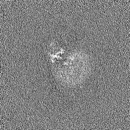
| Images | 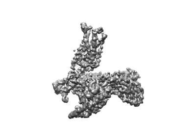 emd_37209.png emd_37209.png | 38 KB | ||
| Filedesc metadata | emd-37209.cif.gz | 7.3 KB | ||
| Others | emd_37209_additional_1.map.gzemd_37209_half_map_1.map.gzemd_37209_half_map_2.map.gz | 32.2 MB 59.5 MB 59.5 MB | ||
| Archive directory |  http://ftp.pdbj.org/pub/emdb/structures/EMD-37209ftp://ftp.pdbj.org/pub/emdb/structures/EMD-37209 http://ftp.pdbj.org/pub/emdb/structures/EMD-37209ftp://ftp.pdbj.org/pub/emdb/structures/EMD-37209 | HTTPS FTP |
-Validation report
| Summary document | emd_37209_validation.pdf.gz | 1 MB | Display | EMDB validaton report |
|---|---|---|---|---|
| Full document | emd_37209_full_validation.pdf.gz | 1 MB | Display | |
| Data in XML | emd_37209_validation.xml.gz | 16.2 KB | Display | |
| Data in CIF | emd_37209_validation.cif.gz | 20.6 KB | Display | |
| Arichive directory | https://ftp.pdbj.org/pub/emdb/validation_reports/EMD-37209ftp://ftp.pdbj.org/pub/emdb/validation_reports/EMD-37209 | HTTPS FTP |
-Related structure data
| Related structure data |  8kfzMC  8kfxC  8kfyC M: atomic model generated by this map C: citing same article ( |
|---|---|
| Similar structure data |

-Links
| EMDB pages | EMDB (EBI/PDBe) / EMDataResource |
|---|---|
| Related items in Molecule of the Month |


-Map
| File | Download / File: emd_37209.map.gz / Format: CCP4 / Size: 64 MB / Type: IMAGE STORED AS FLOATING POINT NUMBER (4 BYTES) | ||||||||||||||||||||||||||||||||||||
|---|---|---|---|---|---|---|---|---|---|---|---|---|---|---|---|---|---|---|---|---|---|---|---|---|---|---|---|---|---|---|---|---|---|---|---|---|---|
| Projections & slices | Image control
Images are generated by Spider. | ||||||||||||||||||||||||||||||||||||
| Voxel size | X=Y=Z: 1.04 Å | ||||||||||||||||||||||||||||||||||||
| Density |
| ||||||||||||||||||||||||||||||||||||
| Symmetry | Space group: 1 | ||||||||||||||||||||||||||||||||||||
| Details | EMDB XML:
|
 Z (Sec.)
 Y (Row.)
 X (Col.)


-Supplemental data
-Additional map: #1
| File | emd_37209_additional_1.map | ||||||||||||
|---|---|---|---|---|---|---|---|---|---|---|---|---|---|
| Projections & Slices |
| ||||||||||||
| Density Histograms |


-Half map: #2
| File | emd_37209_half_map_1.map | ||||||||||||
|---|---|---|---|---|---|---|---|---|---|---|---|---|---|
| Projections & Slices |
| ||||||||||||
| Density Histograms |


-Half map: #1
| File | emd_37209_half_map_2.map | ||||||||||||
|---|---|---|---|---|---|---|---|---|---|---|---|---|---|
| Projections & Slices |
| ||||||||||||
| Density Histograms |


- Sample components
-Entire : Hetero Complex
| Entire | Name: Hetero Complex |
|---|---|
| Components |
|
-Supramolecule #1: Hetero Complex
| Supramolecule | Name: Hetero Complex / type: complex / ID: 1 / Parent: 0 / Macromolecule list: all |
|---|---|
| Source (natural) | Organism: Homo sapiens (human) |
-Macromolecule #1: C-C chemokine receptor type 8,LgBiT fusion protein,Recombinant Hu...
| Macromolecule | Name: C-C chemokine receptor type 8,LgBiT fusion protein,Recombinant Human Rhinovirus type: protein_or_peptide / ID: 1 Details: Author stated: 1-16:hemagglutinin tag; 17-24:Flag tag;383-397: GS linker; 398-555: LgBiT fusion protein; 558-565: Recombinant Human Rhinovirus (HRV3C) Protease;566-575: 10xHis tag. Number of copies: 1 / Enantiomer: LEVO |
|---|---|
| Source (natural) | Organism: synthetic construct (others) |
| Molecular weight | Theoretical: 65.061355 KDa |
| Recombinant expression | Organism:   Spodoptera frugiperda (fall armyworm) Spodoptera frugiperda (fall armyworm) |
| Sequence | String: MKTIIALSYI FCLVFADYKD DDDAGRAMDY TLDLSVTTVT DYYYPDIFSS PCDAELIQTN GKLLLAVFYC LLFVFSLLGN SLVILVLVV CKKLRSITDV YLLNLALSDL LFVFSFPFQT YYLLDQWVFG TVMCKVVSGF YYIGFYSSMF FITLMSVDRY L AVVHAVYA ...String: MKTIIALSYI FCLVFADYKD DDDAGRAMDY TLDLSVTTVT DYYYPDIFSS PCDAELIQTN GKLLLAVFYC LLFVFSLLGN SLVILVLVV CKKLRSITDV YLLNLALSDL LFVFSFPFQT YYLLDQWVFG TVMCKVVSGF YYIGFYSSMF FITLMSVDRY L AVVHAVYA LKVRTIRMGT TLCLAVWLTA IMATIPLLVF YQVASEDGVL QCYSFYNQQT LKWKIFTNFK MNILGLLIPF TI FMFCYIK ILHQLKRCQN HNKTKAIRLV LIVVIASLLF WVPFNVVLFL TSLHSMHILD GCSISQQLTY ATHVTEIISF THC CVNPVI YAFVGEKFKK HLSEIFQKSC SQIFNYLGRQ MPRESCEKSS SCQQHSSRSS SVDYILGSSG GGGSGGGGSS GVFT LEDFV GDWEQTAAYN LDQVLEQGGV SSLLQNLAVS VTPIQRIVRS GENALKIDIH VIIPYEGLSA DQMAQIEEVF KVVYP VDDH HFKVILPYGT LVIDGVTPNM LNYFGRPYEG IAVFDGKKIT VTGTLWNGNK IIDERLITPD GSMLFRVTIN SEFLEV LFQ GPHHHHHHHH HH UniProtKB: C-C chemokine receptor type 8 |
-Macromolecule #2: Guanine nucleotide-binding protein G(i) subunit alpha-1
| Macromolecule | Name: Guanine nucleotide-binding protein G(i) subunit alpha-1 type: protein_or_peptide / ID: 2 / Number of copies: 1 / Enantiomer: LEVO |
|---|---|
| Source (natural) | Organism: Homo sapiens (human) |
| Molecular weight | Theoretical: 40.472082 KDa |
| Recombinant expression | Organism: Spodoptera frugiperda (fall armyworm) |
| Sequence | String: MGCTLSAEDK AAVERSKMID RNLREDGEKA AREVKLLLLG AGESGKNTIV KQMKIIHEAG YSEEECKQYK AVVYSNTIQS IIAIIRAMG RLKIDFGDSA RADDARQLFV LAGAAEEGFM TAELAGVIKR LWKDSGVQAC FNRSREYQLN DSAAYYLNDL D RIAQPNYI ...String: MGCTLSAEDK AAVERSKMID RNLREDGEKA AREVKLLLLG AGESGKNTIV KQMKIIHEAG YSEEECKQYK AVVYSNTIQS IIAIIRAMG RLKIDFGDSA RADDARQLFV LAGAAEEGFM TAELAGVIKR LWKDSGVQAC FNRSREYQLN DSAAYYLNDL D RIAQPNYI PTQQDVLRTR VKTTGIVETH FTFKDLHFKM FDVGAQRSER KKWIHCFEGV TAIIFCVALS DYDLVLAEDE EM NRMHESM KLFDSICNNK WFTDTSIILF LNKKDLFEEK IKKSPLTICY PEYAGSNTYE EAAAYIQCQF EDLNKRKDTK EIY THFTCS TDTKNVQFVF DAVTDVIIKN NLKDCGLF UniProtKB: Guanine nucleotide-binding protein G(i) subunit alpha-1 |
-Macromolecule #3: Guanine nucleotide-binding protein G(I)/G(S)/G(T) subunit beta-1
| Macromolecule | Name: Guanine nucleotide-binding protein G(I)/G(S)/G(T) subunit beta-1 type: protein_or_peptide / ID: 3 / Number of copies: 1 / Enantiomer: LEVO |
|---|---|
| Source (natural) | Organism: Homo sapiens (human) |
| Molecular weight | Theoretical: 39.728426 KDa |
| Recombinant expression | Organism: Spodoptera frugiperda (fall armyworm) |
| Sequence | String: MSELDQLRQE AEQLKNQIRD ARKACADATL SQITNNIDPV GRIQMRTRRT LRGHLAKIYA MHWGTDSRLL VSASQDGKLI IWDSYTTNK VHAIPLRSSW VMTCAYAPSG NYVACGGLDN ICSIYNLKTR EGNVRVSREL AGHTGYLSCC RFLDDNQIVT S SGDTTCAL ...String: MSELDQLRQE AEQLKNQIRD ARKACADATL SQITNNIDPV GRIQMRTRRT LRGHLAKIYA MHWGTDSRLL VSASQDGKLI IWDSYTTNK VHAIPLRSSW VMTCAYAPSG NYVACGGLDN ICSIYNLKTR EGNVRVSREL AGHTGYLSCC RFLDDNQIVT S SGDTTCAL WDIETGQQTT TFTGHTGDVM SLSLAPDTRL FVSGACDASA KLWDVREGMC RQTFTGHESD INAICFFPNG NA FATGSDD ATCRLFDLRA DQELMTYSHD NIICGITSVS FSKSGRLLLA GYDDFNCNVW DALKADRAGV LAGHDNRVSC LGV TDDGMA VATGSWDSFL KIWNGSSGGG GSGGGGSSGV SGWRLFKKIS UniProtKB: Guanine nucleotide-binding protein G(I)/G(S)/G(T) subunit beta-1 |
-Macromolecule #4: Guanine nucleotide-binding protein G(I)/G(S)/G(O) subunit gamma-2
| Macromolecule | Name: Guanine nucleotide-binding protein G(I)/G(S)/G(O) subunit gamma-2 type: protein_or_peptide / ID: 4 / Number of copies: 1 / Enantiomer: LEVO |
|---|---|
| Source (natural) | Organism: Homo sapiens (human) |
| Molecular weight | Theoretical: 7.861143 KDa |
| Recombinant expression | Organism: Spodoptera frugiperda (fall armyworm) |
| Sequence | String: MASNNTASIA QARKLVEQLK MEANIDRIKV SKAAADLMAY CEAHAKEDPL LTPVPASENP FREKKFFCAI L UniProtKB: Guanine nucleotide-binding protein G(I)/G(S)/G(O) subunit gamma-2 |
-Macromolecule #5: scFv16
| Macromolecule | Name: scFv16 / type: protein_or_peptide / ID: 5 / Number of copies: 1 / Enantiomer: LEVO |
|---|---|
| Source (natural) | Organism: Homo sapiens (human) |
| Molecular weight | Theoretical: 31.870629 KDa |
| Recombinant expression | Organism: Spodoptera frugiperda (fall armyworm) |
| Sequence | String: MLLVNQSHQG FNKEHTSKMV SAIVLYVLLA AAAHSAFADV QLVESGGGLV QPGGSRKLSC SASGFAFSSF GMHWVRQAPE KGLEWVAYI SSGSGTIYYA DTVKGRFTIS RDDPKNTLFL QMTSLRSEDT AMYYCVRSIY YYGSSPFDFW GQGTTLTVSS G GGGSGGGG ...String: MLLVNQSHQG FNKEHTSKMV SAIVLYVLLA AAAHSAFADV QLVESGGGLV QPGGSRKLSC SASGFAFSSF GMHWVRQAPE KGLEWVAYI SSGSGTIYYA DTVKGRFTIS RDDPKNTLFL QMTSLRSEDT AMYYCVRSIY YYGSSPFDFW GQGTTLTVSS G GGGSGGGG SGGGGSDIVM TQATSSVPVT PGESVSISCR SSKSLLHSNG NTYLYWFLQR PGQSPQLLIY RMSNLASGVP DR FSGSGSG TAFTLTISRL EAEDVGVYYC MQHLEYPLTF GAGTKLELKA AAHHHHHHHH |
-Experimental details
-Structure determination
| Method | cryo EM |
|---|---|
 Processing Processing | single particle reconstruction |
| Aggregation state | particle |
-Sample preparation
| Buffer | pH: 7.14 |
|---|---|
| Vitrification | Cryogen name: ETHANE |
- Electron microscopy
| Microscope | FEI TITAN KRIOS |
|---|---|
| Image recording | Film or detector model: GATAN K3 BIOQUANTUM (6k x 4k) / Average electron dose: 60.0 e/Å2 |
| Electron beam | Acceleration voltage: 300 kV / Electron source:  FIELD EMISSION GUN FIELD EMISSION GUN |
| Electron optics | Illumination mode: FLOOD BEAM / Imaging mode: BRIGHT FIELD / Nominal defocus max: 2.2 µm / Nominal defocus min: 0.7000000000000001 µm |
| Experimental equipment |  Model: Titan Krios / Image courtesy: FEI Company |

