Movie
Movie Controller


+ Open data

- Basic information
| Entry | 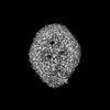 | |||||||||
|---|---|---|---|---|---|---|---|---|---|---|
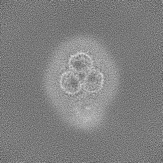
| Title | DDM-bound complex of OmpC3-MlaA-MlaC | |||||||||
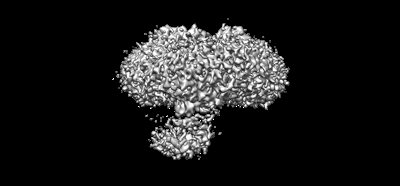
 Map data Map data | Full map of DDM-bound complex of OmpC3-MlaA-MlaC | |||||||||
 Sample Sample |
| |||||||||
 Keywords Keywords | bacteria / outer membrane / phospholipid / lipid asymmetry / membrane protein / protein complex structure / channel / LIPID TRANSPORT | |||||||||
| Biological species |  | |||||||||
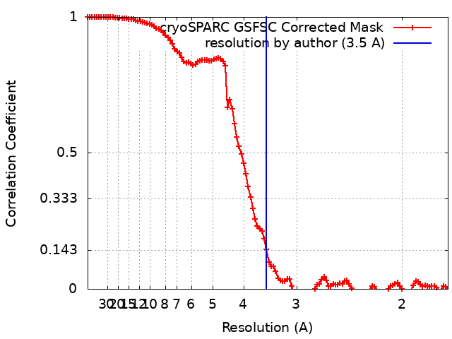
| Method | single particle reconstruction / cryo EM / Resolution: 3.5 Å | |||||||||
 Authors Authors | Yeow J / Luo M / Chng SS | |||||||||
| Funding support |  Singapore, 1 items Singapore, 1 items
| |||||||||
 Citation Citation | Journal: To Be Published Title: Molecular mechanism of phospholipid transport at the bacterial outer membrane interface Authors: Yeow J / Luo M / Chng SS | |||||||||
| History |
|
- Structure visualization
| Supplemental images |
|---|
- Downloads & links
-EMDB archive
| Map data | emd_36823.map.gz | 62.5 MB |  EMDB map data format EMDB map data format | |
|---|---|---|---|---|
| Header (meta data) | emd-36823-v30.xmlemd-36823.xml | 16.7 KB 16.7 KB | Display Display | EMDB header |
| FSC (resolution estimation) | emd_36823_fsc.xml | 10.6 KB | Display | FSC data file |
| Images |  emd_36823.png emd_36823.png | 36.9 KB | ||
| Filedesc metadata | emd-36823.cif.gz | 4.7 KB | ||
| Others | emd_36823_half_map_1.map.gzemd_36823_half_map_2.map.gz | 115.9 MB 116 MB | ||
| Archive directory |  http://ftp.pdbj.org/pub/emdb/structures/EMD-36823ftp://ftp.pdbj.org/pub/emdb/structures/EMD-36823 http://ftp.pdbj.org/pub/emdb/structures/EMD-36823ftp://ftp.pdbj.org/pub/emdb/structures/EMD-36823 | HTTPS FTP |
-Links
| EMDB pages | EMDB (EBI/PDBe) / EMDataResource |
|---|
-Map
| File | Download / File: emd_36823.map.gz / Format: CCP4 / Size: 125 MB / Type: IMAGE STORED AS FLOATING POINT NUMBER (4 BYTES) | ||||||||||||||||||||||||||||||||||||
|---|---|---|---|---|---|---|---|---|---|---|---|---|---|---|---|---|---|---|---|---|---|---|---|---|---|---|---|---|---|---|---|---|---|---|---|---|---|
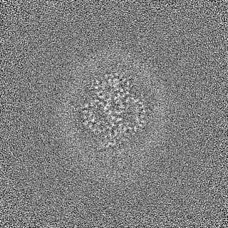
| Annotation | Full map of DDM-bound complex of OmpC3-MlaA-MlaC | ||||||||||||||||||||||||||||||||||||
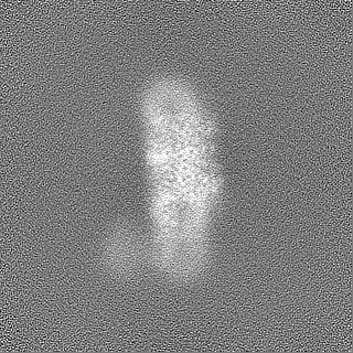
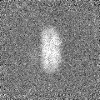
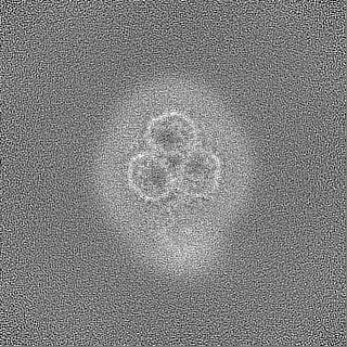
| Projections & slices | Image control
Images are generated by Spider. | ||||||||||||||||||||||||||||||||||||
| Voxel size | X=Y=Z: 0.834 Å | ||||||||||||||||||||||||||||||||||||
| Density |
| ||||||||||||||||||||||||||||||||||||
| Symmetry | Space group: 1 | ||||||||||||||||||||||||||||||||||||
| Details | EMDB XML:
|
 Z (Sec.)
 Y (Row.)
 X (Col.)


-Supplemental data
-Half map: Half map B of DDM-bound complex of OmpC3-MlaA-MlaC
| File | emd_36823_half_map_1.map | ||||||||||||
|---|---|---|---|---|---|---|---|---|---|---|---|---|---|
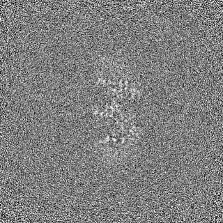
| Annotation | Half map B of DDM-bound complex of OmpC3-MlaA-MlaC | ||||||||||||
| Projections & Slices |
| ||||||||||||
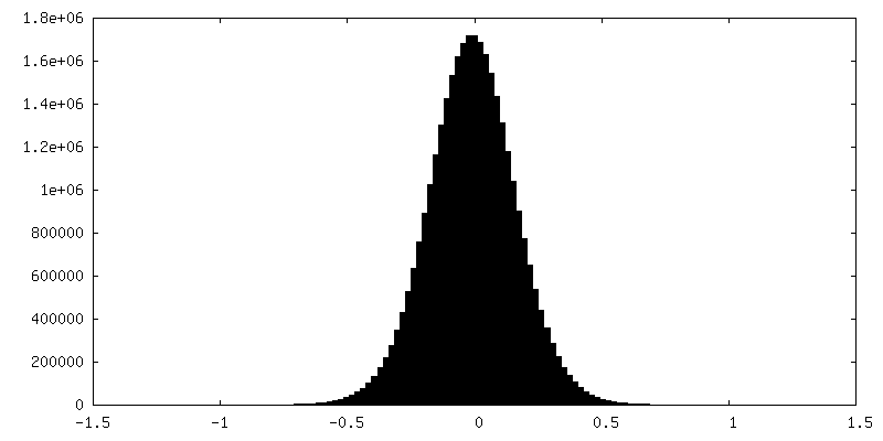
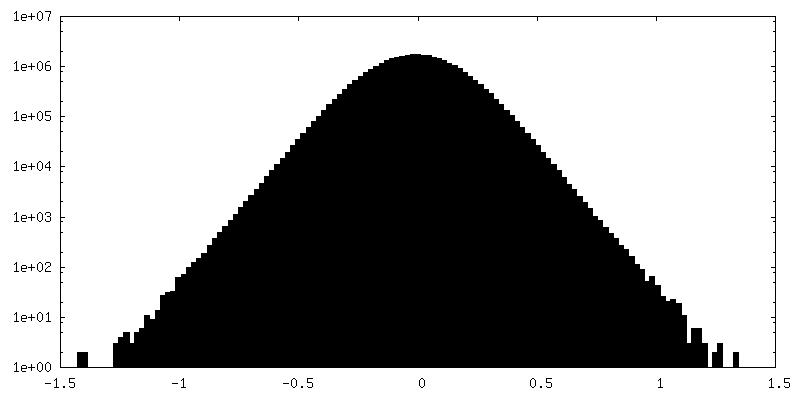
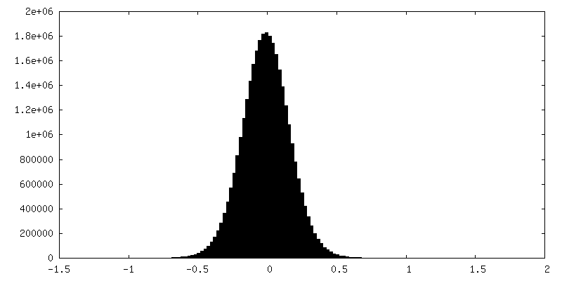
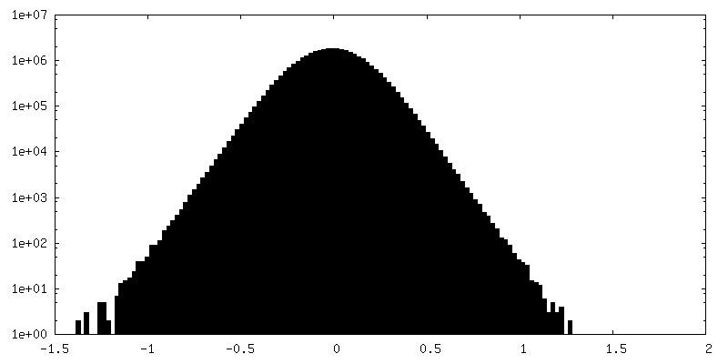
| Density Histograms |

-Half map: Half map A of DDM-bound complex of OmpC3-MlaA-MlaC
| File | emd_36823_half_map_2.map | ||||||||||||
|---|---|---|---|---|---|---|---|---|---|---|---|---|---|
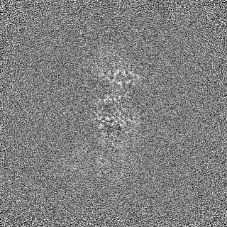
| Annotation | Half map A of DDM-bound complex of OmpC3-MlaA-MlaC | ||||||||||||
| Projections & Slices |
| ||||||||||||
| Density Histograms |

- Sample components
-Entire : DDM-bound outer membrane complex of OmpC-MlaA with periplasmic MlaC
| Entire | Name: DDM-bound outer membrane complex of OmpC-MlaA with periplasmic MlaC |
|---|---|
| Components |
|
-Supramolecule #1: DDM-bound outer membrane complex of OmpC-MlaA with periplasmic MlaC
| Supramolecule | Name: DDM-bound outer membrane complex of OmpC-MlaA with periplasmic MlaC type: complex / ID: 1 / Parent: 0 / Macromolecule list: #1-#2 Details: DDM-bound outer membrane complex of OmpC with disulfide-trapped MlaA and MlaC |
|---|---|
| Source (natural) | Organism: |
| Molecular weight | Theoretical: 28 KDa |
-Supramolecule #2: Outer membrane porin C
| Supramolecule | Name: Outer membrane porin C / type: complex / ID: 2 / Parent: 1 / Macromolecule list: #1 / Details: OmpC |
|---|---|
| Source (natural) | Organism: |
-Supramolecule #3: Intermembrane phospholipid transport system lipoprotein MlaA
| Supramolecule | Name: Intermembrane phospholipid transport system lipoprotein MlaA type: complex / ID: 3 / Parent: 1 / Macromolecule list: #2 / Details: MlaA |
|---|
-Experimental details
-Structure determination
| Method | cryo EM |
|---|---|
 Processing Processing | single particle reconstruction |
| Aggregation state | particle |
-Sample preparation
| Concentration | 12 mg/mL | |||||||||
|---|---|---|---|---|---|---|---|---|---|---|
| Buffer | pH: 8 Component:
Details: Tris-buffered saline (TBS) buffer (20 mM Tris HCl pH 8.0, 150 mM NaCl) | |||||||||
| Vitrification | Cryogen name: ETHANE / Chamber humidity: 100 % / Chamber temperature: 277 K / Instrument: FEI VITROBOT MARK IV |
- Electron microscopy
| Microscope | FEI TITAN KRIOS |
|---|---|
| Specialist optics | Energy filter - Name: GIF Tridiem 4K / Energy filter - Slit width: 20 eV Details: Gatan GIF post-column energy filter operated in zero-loss mode |
| Image recording | Film or detector model: GATAN K3 (6k x 4k) / Digitization - Dimensions - Width: 5760 pixel / Digitization - Dimensions - Height: 4092 pixel / Number grids imaged: 1 / Number real images: 5816 / Average exposure time: 5.99 sec. / Average electron dose: 90.0 e/Å2 Details: Images were collected in movie-mode at 50 frames per image |
| Electron beam | Acceleration voltage: 300 kV / Electron source:  FIELD EMISSION GUN FIELD EMISSION GUN |
| Electron optics | Illumination mode: SPOT SCAN / Imaging mode: BRIGHT FIELD / Cs: 2.7 mm / Nominal defocus max: 2.0 µm / Nominal defocus min: 0.8 µm / Nominal magnification: 105000 |
| Sample stage | Specimen holder model: FEI TITAN KRIOS AUTOGRID HOLDER / Cooling holder cryogen: NITROGEN |
| Experimental equipment |  Model: Titan Krios / Image courtesy: FEI Company |