Japan Agency for Medical Research and Development (AMED)
JP21am0101093
Japan
Japan Agency for Medical Research and Development (AMED)
JP22ama121037
Japan
Japan Science and Technology
JPMJCR20H8
Japan
Japan Society for the Promotion of Science (JSPS)
JPJSCCA20190008
Japan
Japan Society for the Promotion of Science (JSPS)
20H05773
Japan
Japan Society for the Promotion of Science (JSPS)
JP20H05873
Japan
Citation
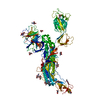
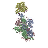
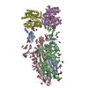
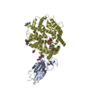
Journal: Nat Commun / Year: 2024 Title: Virological characteristics of the SARS-CoV-2 Omicron XBB.1.5 variant. Authors: Tomokazu Tamura / Takashi Irie / Sayaka Deguchi / Hisano Yajima / Masumi Tsuda / Hesham Nasser / Keita Mizuma / Arnon Plianchaisuk / Saori Suzuki / Keiya Uriu / Mst Monira Begum / Ryo ...Authors: Tomokazu Tamura / Takashi Irie / Sayaka Deguchi / Hisano Yajima / Masumi Tsuda / Hesham Nasser / Keita Mizuma / Arnon Plianchaisuk / Saori Suzuki / Keiya Uriu / Mst Monira Begum / Ryo Shimizu / Michael Jonathan / Rigel Suzuki / Takashi Kondo / Hayato Ito / Akifumi Kamiyama / Kumiko Yoshimatsu / Maya Shofa / Rina Hashimoto / Yuki Anraku / Kanako Terakado Kimura / Shunsuke Kita / Jiei Sasaki / Kaori Sasaki-Tabata / Katsumi Maenaka / Naganori Nao / Lei Wang / Yoshitaka Oda / / Terumasa Ikeda / Akatsuki Saito / Keita Matsuno / Jumpei Ito / Shinya Tanaka / Kei Sato / Takao Hashiguchi / Kazuo Takayama / Takasuke Fukuhara / Abstract: Circulation of SARS-CoV-2 Omicron XBB has resulted in the emergence of XBB.1.5, a new Variant of Interest. Our phylogenetic analysis suggests that XBB.1.5 evolved from XBB.1 by acquiring the S486P ...Circulation of SARS-CoV-2 Omicron XBB has resulted in the emergence of XBB.1.5, a new Variant of Interest. Our phylogenetic analysis suggests that XBB.1.5 evolved from XBB.1 by acquiring the S486P spike (S) mutation, subsequent to the acquisition of a nonsense mutation in ORF8. Neutralization assays showed similar abilities of immune escape between XBB.1.5 and XBB.1. We determine the structural basis for the interaction between human ACE2 and the S protein of XBB.1.5, showing similar overall structures between the S proteins of XBB.1 and XBB.1.5. We provide the intrinsic pathogenicity of XBB.1 and XBB.1.5 in hamsters. Importantly, we find that the ORF8 nonsense mutation of XBB.1.5 resulted in impairment of MHC suppression. In vivo experiments using recombinant viruses reveal that the XBB.1.5 mutations are involved with reduced virulence of XBB.1.5. Together, our study identifies the two viral functions defined the difference between XBB.1 and XBB.1.5.
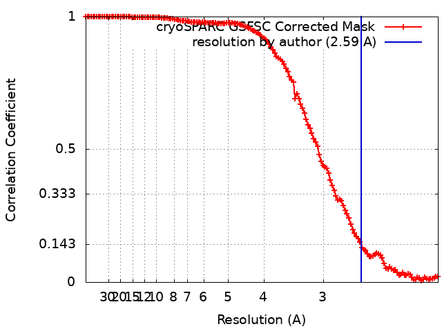
Cryogen name: ETHANE / Chamber humidity: 100 % / Chamber temperature: 291 K / Instrument: FEI VITROBOT MARK IV / Details: blotting time 5 s and blotting force 5..
-
Electron microscopy
Microscope
TFS KRIOS
Specialist optics
Energy filter - Name: GIF Bioquantum / Energy filter - Slit width: 20 eV
Image recording
Film or detector model: GATAN K3 (6k x 4k) / Digitization - Dimensions - Width: 5760 pixel / Digitization - Dimensions - Height: 4092 pixel / Number real images: 3522 / Average exposure time: 1.5 sec. / Average electron dose: 50.4 e/Å2
Electron beam
Acceleration voltage: 300 kV / Electron source: FIELD EMISSION GUN
In the structure databanks used in Yorodumi, some data are registered as the other names, "COVID-19 virus" and "2019-nCoV". Here are the details of the virus and the list of structure data.
Jan 31, 2019. EMDB accession codes are about to change! (news from PDBe EMDB page)
EMDB accession codes are about to change! (news from PDBe EMDB page)
The allocation of 4 digits for EMDB accession codes will soon come to an end. Whilst these codes will remain in use, new EMDB accession codes will include an additional digit and will expand incrementally as the available range of codes is exhausted. The current 4-digit format prefixed with “EMD-” (i.e. EMD-XXXX) will advance to a 5-digit format (i.e. EMD-XXXXX), and so on. It is currently estimated that the 4-digit codes will be depleted around Spring 2019, at which point the 5-digit format will come into force.
The EM Navigator/Yorodumi systems omit the EMD- prefix.
Related info.:Q: What is EMD? / ID/Accession-code notation in Yorodumi/EM Navigator
Yorodumi is a browser for structure data from EMDB, PDB, SASBDB, etc.
This page is also the successor to EM Navigator detail page, and also detail information page/front-end page for Omokage search.
The word "yorodu" (or yorozu) is an old Japanese word meaning "ten thousand". "mi" (miru) is to see.
Related info.:EMDB / PDB / SASBDB / Comparison of 3 databanks / Yorodumi Search / Aug 31, 2016. New EM Navigator & Yorodumi / Yorodumi Papers / Jmol/JSmol / Function and homology information / Changes in new EM Navigator and Yorodumi
 Movie
Movie Controller

 Yorodumi
 Open data
 Basic information
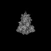
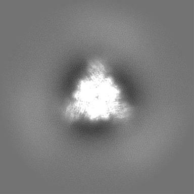
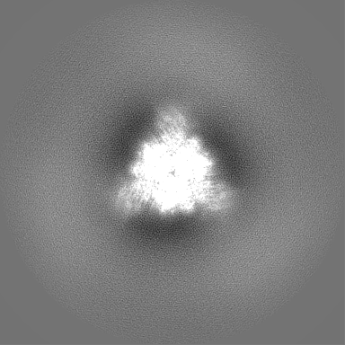
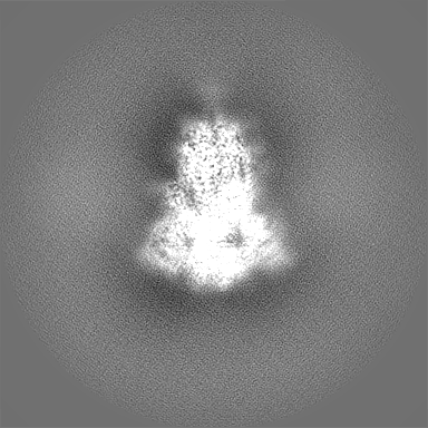
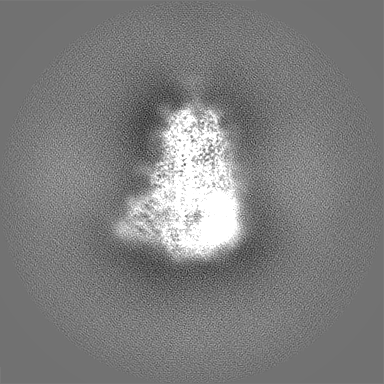
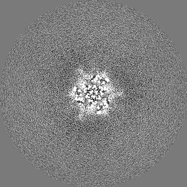
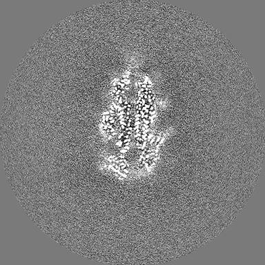
 Map data
 Sample
 Keywords
 Function and homology information
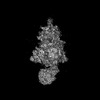
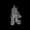
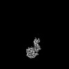
 Severe acute respiratory syndrome coronavirus 2
 Authors
 Japan, 6 items
  Citation
 Structure visualization
 Downloads & links
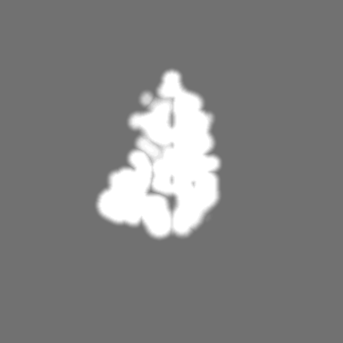
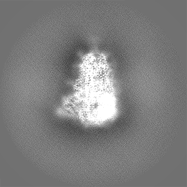
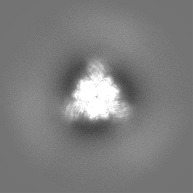
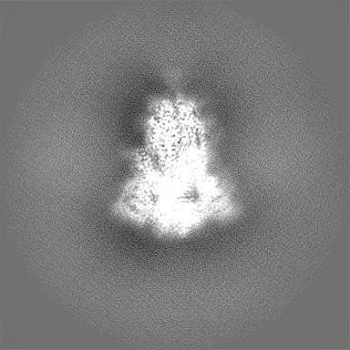
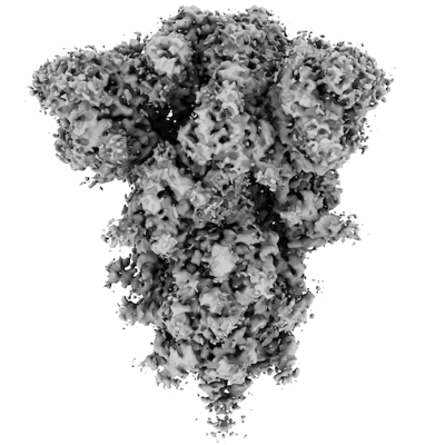 emd_36724.png
 http://ftp.pdbj.org/pub/emdb/structures/EMD-36724


 Z (Sec.)
 Y (Row.)
 X (Col.)


 Sample components
 Homo sapiens (human)
 Processing
 Electron microscopy
 FIELD EMISSION GUN