[English] 日本語
 Yorodumi
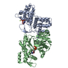
- EMDB-36455: Cryo-EM structure of a Legionella effector complexed with actin a... -
+ Open data
- Basic information
| Entry |  | |||||||||||||||
|---|---|---|---|---|---|---|---|---|---|---|---|---|---|---|---|---|
| Title | Cryo-EM structure of a Legionella effector complexed with actin and ATP | |||||||||||||||
 Map data Map data | ||||||||||||||||
 Sample Sample |
| |||||||||||||||
 Keywords Keywords | AMPylation / TOXIN | |||||||||||||||
| Function / homology |  Function and homology information Function and homology informationcytoskeletal motor activator activity / myosin heavy chain binding / tropomyosin binding / actin filament bundle / troponin I binding / filamentous actin / mesenchyme migration / skeletal muscle myofibril / actin filament bundle assembly / striated muscle thin filament ...cytoskeletal motor activator activity / myosin heavy chain binding / tropomyosin binding / actin filament bundle / troponin I binding / filamentous actin / mesenchyme migration / skeletal muscle myofibril / actin filament bundle assembly / striated muscle thin filament / skeletal muscle thin filament assembly / actin monomer binding / skeletal muscle fiber development / stress fiber / titin binding / actin filament polymerization / actin filament / filopodium / Hydrolases; Acting on acid anhydrides; Acting on acid anhydrides to facilitate cellular and subcellular movement / calcium-dependent protein binding / lamellipodium / cell body / hydrolase activity / protein domain specific binding / calcium ion binding / positive regulation of gene expression / magnesium ion binding / ATP binding / identical protein binding / cytoplasm Similarity search - Function | |||||||||||||||
| Biological species |  Legionella sainthelensi (bacteria) / Legionella sainthelensi (bacteria) /  | |||||||||||||||
| Method | single particle reconstruction / cryo EM / Resolution: 3.04 Å | |||||||||||||||
 Authors Authors | Zhou XT / Wang XF / Tan JX / Zhu YQ | |||||||||||||||
| Funding support |  China, 4 items China, 4 items
| |||||||||||||||
 Citation Citation |  Journal: Nature / Year: 2024 Journal: Nature / Year: 2024Title: Legionella effector LnaB is a phosphoryl-AMPylase that impairs phosphosignalling. Authors: Ting Wang / Xiaonan Song / Jiaxing Tan / Wei Xian / Xingtong Zhou / Mingru Yu / Xiaofei Wang / Yan Xu / Ting Wu / Keke Yuan / Yu Ran / Bing Yang / Gaofeng Fan / Xiaoyun Liu / Yan Zhou / Yongqun Zhu /  Abstract: AMPylation is a post-translational modification in which AMP is added to the amino acid side chains of proteins. Here we show that, with ATP as the ligand and actin as the host activator, the ...AMPylation is a post-translational modification in which AMP is added to the amino acid side chains of proteins. Here we show that, with ATP as the ligand and actin as the host activator, the effector protein LnaB of Legionella pneumophila exhibits AMPylase activity towards the phosphoryl group of phosphoribose on PR-Ub that is generated by the SidE family of effectors, and deubiquitinases DupA and DupB in an E1- and E2-independent ubiquitination process. The product of LnaB is further hydrolysed by an ADP-ribosylhydrolase, MavL, to Ub, thereby preventing the accumulation of PR-Ub and ADPR-Ub and protecting canonical ubiquitination in host cells. LnaB represents a large family of AMPylases that adopt a common structural fold, distinct from those of the previously known AMPylases, and LnaB homologues are found in more than 20 species of bacterial pathogens. Moreover, LnaB also exhibits robust phosphoryl AMPylase activity towards phosphorylated residues and produces unique ADPylation modifications in proteins. During infection, LnaB AMPylates the conserved phosphorylated tyrosine residues in the activation loop of the Src family of kinases, which dampens downstream phosphorylation signalling in the host. Structural studies reveal the actin-dependent activation and catalytic mechanisms of the LnaB family of AMPylases. This study identifies, to our knowledge, an unprecedented molecular regulation mechanism in bacterial pathogenesis and protein phosphorylation. | |||||||||||||||
| History |
|
- Structure visualization
| Supplemental images |
|---|
- Downloads & links
-EMDB archive
| Map data |  emd_36455.map.gz emd_36455.map.gz | 59.7 MB |  EMDB map data format EMDB map data format | |
|---|---|---|---|---|
| Header (meta data) |  emd-36455-v30.xml emd-36455-v30.xml emd-36455.xml emd-36455.xml | 17.7 KB 17.7 KB | Display Display |  EMDB header EMDB header |
| Images |  emd_36455.png emd_36455.png | 48 KB | ||
| Filedesc metadata |  emd-36455.cif.gz emd-36455.cif.gz | 6.5 KB | ||
| Others |  emd_36455_half_map_1.map.gz emd_36455_half_map_1.map.gz emd_36455_half_map_2.map.gz emd_36455_half_map_2.map.gz | 59.4 MB 59.4 MB | ||
| Archive directory |  http://ftp.pdbj.org/pub/emdb/structures/EMD-36455 http://ftp.pdbj.org/pub/emdb/structures/EMD-36455 ftp://ftp.pdbj.org/pub/emdb/structures/EMD-36455 ftp://ftp.pdbj.org/pub/emdb/structures/EMD-36455 | HTTPS FTP |
-Validation report
| Summary document |  emd_36455_validation.pdf.gz emd_36455_validation.pdf.gz | 766.7 KB | Display |  EMDB validaton report EMDB validaton report |
|---|---|---|---|---|
| Full document |  emd_36455_full_validation.pdf.gz emd_36455_full_validation.pdf.gz | 766.3 KB | Display | |
| Data in XML |  emd_36455_validation.xml.gz emd_36455_validation.xml.gz | 12.3 KB | Display | |
| Data in CIF |  emd_36455_validation.cif.gz emd_36455_validation.cif.gz | 14.4 KB | Display | |
| Arichive directory |  https://ftp.pdbj.org/pub/emdb/validation_reports/EMD-36455 https://ftp.pdbj.org/pub/emdb/validation_reports/EMD-36455 ftp://ftp.pdbj.org/pub/emdb/validation_reports/EMD-36455 ftp://ftp.pdbj.org/pub/emdb/validation_reports/EMD-36455 | HTTPS FTP |
-Related structure data
| Related structure data |  8jo4MC  8jo3C  8xepC M: atomic model generated by this map C: citing same article ( |
|---|---|
| Similar structure data | Similarity search - Function & homology  F&H Search F&H Search |
- Links
Links
| EMDB pages |  EMDB (EBI/PDBe) / EMDB (EBI/PDBe) /  EMDataResource EMDataResource |
|---|---|
| Related items in Molecule of the Month |
- Map
Map
| File |  Download / File: emd_36455.map.gz / Format: CCP4 / Size: 64 MB / Type: IMAGE STORED AS FLOATING POINT NUMBER (4 BYTES) Download / File: emd_36455.map.gz / Format: CCP4 / Size: 64 MB / Type: IMAGE STORED AS FLOATING POINT NUMBER (4 BYTES) | ||||||||||||||||||||||||||||||||||||
|---|---|---|---|---|---|---|---|---|---|---|---|---|---|---|---|---|---|---|---|---|---|---|---|---|---|---|---|---|---|---|---|---|---|---|---|---|---|
| Projections & slices | Image control
Images are generated by Spider. | ||||||||||||||||||||||||||||||||||||
| Voxel size | X=Y=Z: 0.93 Å | ||||||||||||||||||||||||||||||||||||
| Density |
| ||||||||||||||||||||||||||||||||||||
| Symmetry | Space group: 1 | ||||||||||||||||||||||||||||||||||||
| Details | EMDB XML:
|
-Supplemental data
-Half map: #2
| File | emd_36455_half_map_1.map | ||||||||||||
|---|---|---|---|---|---|---|---|---|---|---|---|---|---|
| Projections & Slices |
| ||||||||||||
| Density Histograms |
-Half map: #1
| File | emd_36455_half_map_2.map | ||||||||||||
|---|---|---|---|---|---|---|---|---|---|---|---|---|---|
| Projections & Slices |
| ||||||||||||
| Density Histograms |
- Sample components
-Entire : Cryo-EM structure of a Legionella effector complexed with actin a...
| Entire | Name: Cryo-EM structure of a Legionella effector complexed with actin and ATP |
|---|---|
| Components |
|
-Supramolecule #1: Cryo-EM structure of a Legionella effector complexed with actin a...
| Supramolecule | Name: Cryo-EM structure of a Legionella effector complexed with actin and ATP type: complex / ID: 1 / Parent: 0 / Macromolecule list: #1-#2 |
|---|---|
| Source (natural) | Organism:  Legionella sainthelensi (bacteria) Legionella sainthelensi (bacteria) |
-Macromolecule #1: Substrate of the Dot/Icm secretion system
| Macromolecule | Name: Substrate of the Dot/Icm secretion system / type: protein_or_peptide / ID: 1 / Number of copies: 1 / Enantiomer: LEVO |
|---|---|
| Source (natural) | Organism:  Legionella sainthelensi (bacteria) Legionella sainthelensi (bacteria) |
| Molecular weight | Theoretical: 50.352203 KDa |
| Recombinant expression | Organism:  |
| Sequence | String: MSYTKIESSL LLALDYPKLD ESDFILLLTK FLEKKLGNND NYPTFSKQIQ KYYLEQEYKK AIENILRLCQ ENETLLGTNL VQRLITKSS QVTSNPKDNE SRRFYEVLYA EHLESILRKD FDCSIFDELN EAYNEVRPEY TVNDLTKINT FEEARKLILA F VMLNDNVE ...String: MSYTKIESSL LLALDYPKLD ESDFILLLTK FLEKKLGNND NYPTFSKQIQ KYYLEQEYKK AIENILRLCQ ENETLLGTNL VQRLITKSS QVTSNPKDNE SRRFYEVLYA EHLESILRKD FDCSIFDELN EAYNEVRPEY TVNDLTKINT FEEARKLILA F VMLNDNVE LGLKAQSAIY QKKDRSREEL GQVLTANPGI MKPNSPNFAD NTVPIKKIDK IAIDEKKAGG YSKTNPQVPF VA SLSGTTY SLVVVLQKYM DKHKTDPNLE KKINNIVMLW TSAYIKDGYA SYKEVIDIFK DAHIQSIFAR ANIKLDYAII DDT DHEFHR AQEYTQGIAT KAMMHQELVQ KVQEKSMREE KQKQMLLFCG VLVNQLNQDP KSKEKYPEQL ESLNALYKNW NAGT IKNQE FKKECTQVCT EIQIKEQNTP LEHHHHHH UniProtKB: Substrate of the Dot/Icm secretion system |
-Macromolecule #2: Actin, alpha skeletal muscle
| Macromolecule | Name: Actin, alpha skeletal muscle / type: protein_or_peptide / ID: 2 / Number of copies: 1 / Enantiomer: LEVO EC number: Hydrolases; Acting on acid anhydrides; Acting on acid anhydrides to facilitate cellular and subcellular movement |
|---|---|
| Source (natural) | Organism:  |
| Molecular weight | Theoretical: 42.096953 KDa |
| Sequence | String: MCDEDETTAL VCDNGSGLVK AGFAGDDAPR AVFPSIVGRP RHQGVMVGMG QKDSYVGDEA QSKRGILTLK YPIEHGIITN WDDMEKIWH HTFYNELRVA PEEHPTLLTE APLNPKANRE KMTQIMFETF NVPAMYVAIQ AVLSLYASGR TTGIVLDSGD G VTHNVPIY ...String: MCDEDETTAL VCDNGSGLVK AGFAGDDAPR AVFPSIVGRP RHQGVMVGMG QKDSYVGDEA QSKRGILTLK YPIEHGIITN WDDMEKIWH HTFYNELRVA PEEHPTLLTE APLNPKANRE KMTQIMFETF NVPAMYVAIQ AVLSLYASGR TTGIVLDSGD G VTHNVPIY EGYALPHAIM RLDLAGRDLT DYLMKILTER GYSFVTTAER EIVRDIKEKL CYVALDFENE MATAASSSSL EK SYELPDG QVITIGNERF RCPETLFQPS FIGMESAGIH ETTYNSIMKC DIDIRKDLYA NNVMSGGTTM YPGIADRMQK EIT ALAPST MKIKIIAPPE RKYSVWIGGS ILASLSTFQQ MWITKQEYDE AGPSIVHRKC F UniProtKB: Actin, alpha skeletal muscle |
-Macromolecule #3: ADENOSINE-5'-TRIPHOSPHATE
| Macromolecule | Name: ADENOSINE-5'-TRIPHOSPHATE / type: ligand / ID: 3 / Number of copies: 2 / Formula: ATP |
|---|---|
| Molecular weight | Theoretical: 507.181 Da |
| Chemical component information |  ChemComp-ATP: |
-Macromolecule #4: MAGNESIUM ION
| Macromolecule | Name: MAGNESIUM ION / type: ligand / ID: 4 / Number of copies: 1 / Formula: MG |
|---|---|
| Molecular weight | Theoretical: 24.305 Da |
-Macromolecule #5: CALCIUM ION
| Macromolecule | Name: CALCIUM ION / type: ligand / ID: 5 / Number of copies: 1 / Formula: CA |
|---|---|
| Molecular weight | Theoretical: 40.078 Da |
-Experimental details
-Structure determination
| Method | cryo EM |
|---|---|
 Processing Processing | single particle reconstruction |
| Aggregation state | particle |
- Sample preparation
| Buffer | pH: 8 |
|---|---|
| Grid | Model: Quantifoil R0.6/1 / Material: COPPER / Mesh: 300 / Pretreatment - Type: GLOW DISCHARGE / Pretreatment - Time: 180 sec. |
| Vitrification | Cryogen name: ETHANE |
- Electron microscopy
| Microscope | FEI TITAN KRIOS |
|---|---|
| Image recording | Film or detector model: FEI FALCON IV (4k x 4k) / Average electron dose: 50.0 e/Å2 |
| Electron beam | Acceleration voltage: 300 kV / Electron source:  FIELD EMISSION GUN FIELD EMISSION GUN |
| Electron optics | Illumination mode: SPOT SCAN / Imaging mode: BRIGHT FIELD / Nominal defocus max: 1.6 µm / Nominal defocus min: 0.8 µm |
| Experimental equipment |  Model: Titan Krios / Image courtesy: FEI Company |
- Image processing
| Startup model | Type of model: NONE |
|---|---|
| Final reconstruction | Applied symmetry - Point group: C1 (asymmetric) / Resolution.type: BY AUTHOR / Resolution: 3.04 Å / Resolution method: FSC 0.143 CUT-OFF / Number images used: 171800 |
| Initial angle assignment | Type: MAXIMUM LIKELIHOOD |
| Final angle assignment | Type: MAXIMUM LIKELIHOOD |
-Atomic model buiding 1
| Initial model | Chain - Source name: Other / Chain - Initial model type: in silico model / Details: ModelAngelo built model |
|---|---|
| Output model |  PDB-8jo4: |
 Movie
Movie Controller


 Z (Sec.)
 Y (Row.)
 X (Col.)


