 Movie
Movie Controller


+ Open data

- Basic information
| Entry |  | |||||||||
|---|---|---|---|---|---|---|---|---|---|---|
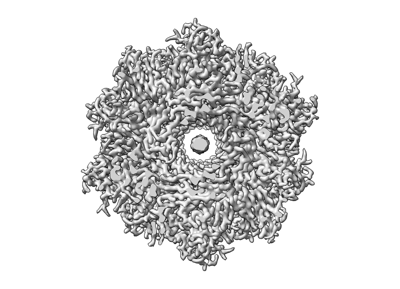
| Title | Fibril form of serine peptidase Vpr | |||||||||
 Map data Map data | ||||||||||
 Sample Sample |
| |||||||||
 Keywords Keywords | Serine peptidase / fibril / PROTEIN FIBRIL | |||||||||
| Function / homology |  Function and homology information Function and homology information | |||||||||
| Biological species |  | |||||||||
| Method | helical reconstruction / cryo EM / Resolution: 2.9 Å | |||||||||
 Authors Authors | Cao Q / Cheng Y | |||||||||
| Funding support |  China, 1 items China, 1 items
| |||||||||
 Citation Citation | Journal: Nat Commun / Year: 2023 Title: Serine peptidase Vpr forms enzymatically active fibrils outside Bacillus bacteria revealed by cryo-EM. Authors: Yijia Cheng / Jianting Han / Meinai Song / Shuqin Zhang / Qin Cao / Abstract: Bacteria develop a variety of extracellular fibrous structures crucial for their survival, such as flagella and pili. In this study, we use cryo-EM to identify protein fibrils surrounding lab- ...Bacteria develop a variety of extracellular fibrous structures crucial for their survival, such as flagella and pili. In this study, we use cryo-EM to identify protein fibrils surrounding lab-cultured Bacillus amyloiquefaciens and discover an unreported fibril species in addition to the flagellar fibrils. These previously unknown fibrils are composed of Vpr, an extracellular serine peptidase. We find that Vpr assembles into fibrils in an enzymatically active form, potentially representing a strategy of enriching Vpr activities around bacterial cells. Vpr fibrils are also observed under other culture conditions and around other Bacillus bacteria, such as Bacillus subtilis, which may suggest a general mechanism across all Bacillus bacterial groups. Taken together, our study reveals fibrils outside the bacterial cell and sheds light on the physiological role of these extracellular fibrils. | |||||||||
| History |
|
- Structure visualization
| Supplemental images |
|---|
- Downloads & links
-EMDB archive
| Map data | emd_36428.map.gz | 109.1 MB | EMDB map data format | |
|---|---|---|---|---|
| Header (meta data) | emd-36428-v30.xmlemd-36428.xml | 13.4 KB 13.4 KB | Display Display | EMDB header |
| FSC (resolution estimation) | emd_36428_fsc.xml | 11.3 KB | Display | FSC data file |
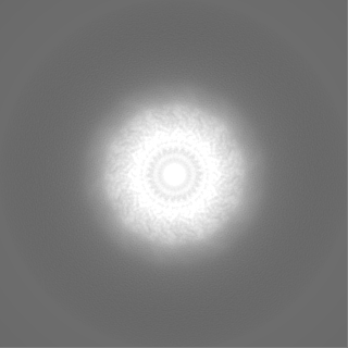
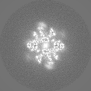
| Images |  emd_36428.png emd_36428.png | 88.2 KB | ||
| Filedesc metadata | emd-36428.cif.gz | 5.4 KB | ||
| Others | emd_36428_half_map_1.map.gzemd_36428_half_map_2.map.gz | 96.2 MB 96.2 MB | ||
| Archive directory |  http://ftp.pdbj.org/pub/emdb/structures/EMD-36428ftp://ftp.pdbj.org/pub/emdb/structures/EMD-36428 http://ftp.pdbj.org/pub/emdb/structures/EMD-36428ftp://ftp.pdbj.org/pub/emdb/structures/EMD-36428 | HTTPS FTP |
-Validation report
| Summary document | emd_36428_validation.pdf.gz | 1017.8 KB | Display | EMDB validaton report |
|---|---|---|---|---|
| Full document | emd_36428_full_validation.pdf.gz | 1017.3 KB | Display | |
| Data in XML | emd_36428_validation.xml.gz | 18.7 KB | Display | |
| Data in CIF | emd_36428_validation.cif.gz | 24.1 KB | Display | |
| Arichive directory | https://ftp.pdbj.org/pub/emdb/validation_reports/EMD-36428ftp://ftp.pdbj.org/pub/emdb/validation_reports/EMD-36428 | HTTPS FTP |
-Related structure data
| Related structure data |  8jmwMC  8jmvC M: atomic model generated by this map C: citing same article ( |
|---|---|
| Similar structure data |

-Links
| EMDB pages | EMDB (EBI/PDBe) / EMDataResource |
|---|---|
| Related items in Molecule of the Month |


-Map
| File | Download / File: emd_36428.map.gz / Format: CCP4 / Size: 125 MB / Type: IMAGE STORED AS FLOATING POINT NUMBER (4 BYTES) | ||||||||||||||||||||
|---|---|---|---|---|---|---|---|---|---|---|---|---|---|---|---|---|---|---|---|---|---|
| Voxel size | X=Y=Z: 1.05 Å | ||||||||||||||||||||
| Density |
| ||||||||||||||||||||
| Symmetry | Space group: 1 | ||||||||||||||||||||
| Details | EMDB XML:
|
-Supplemental data
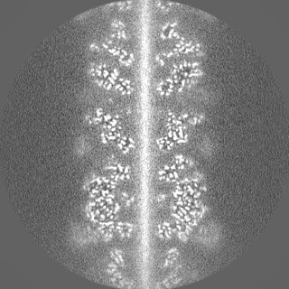
-Half map: #2
| File | emd_36428_half_map_1.map | ||||||||||||
|---|---|---|---|---|---|---|---|---|---|---|---|---|---|
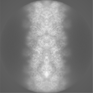
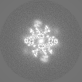
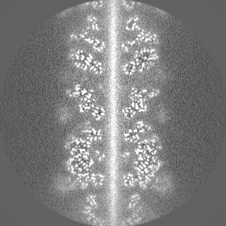
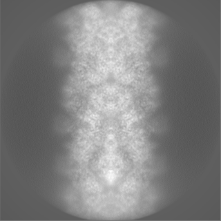
| Projections & Slices |
| ||||||||||||
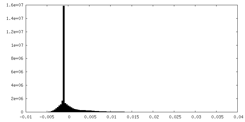
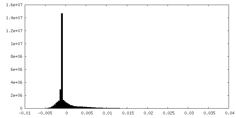
| Density Histograms |
 Z
Z Y
Y X
X

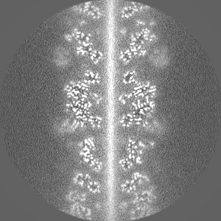
-Half map: #1
| File | emd_36428_half_map_2.map | ||||||||||||
|---|---|---|---|---|---|---|---|---|---|---|---|---|---|
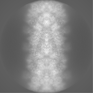
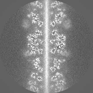
| Projections & Slices |
| ||||||||||||
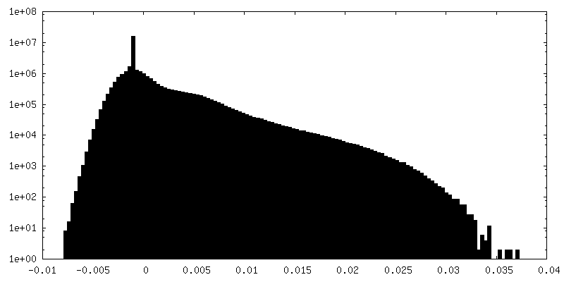
| Density Histograms |


- Sample components
-Entire : Bacillus amyloliquefaciens
| Entire | Name: |
|---|---|
| Components |
|
-Supramolecule #1: Bacillus amyloliquefaciens
| Supramolecule | Name: Bacillus amyloliquefaciens / type: cell / ID: 1 / Parent: 0 / Macromolecule list: all |
|---|---|
| Source (natural) | Organism: |
-Macromolecule #1: S8 family serine peptidase
| Macromolecule | Name: S8 family serine peptidase / type: protein_or_peptide / ID: 1 / Number of copies: 18 / Enantiomer: LEVO |
|---|---|
| Source (natural) | Organism: |
| Molecular weight | Theoretical: 85.965602 KDa |
| Sequence | String: LKKGIIRYLL PAFVLSFTLS TSSQAAPASK PQTPDLEKAE VFGDIDMTTG KQTTVIVELK EKSLAEAKEL GKAQTKSKLK SERSKVKKK ALKTIKHGKI NREYEQVFSG FSMKLPANEI PKLLSDQDVK AVYPNVTYHT DQLKDKDITL SKDAVSPQMD D SAPYIGAN ...String: LKKGIIRYLL PAFVLSFTLS TSSQAAPASK PQTPDLEKAE VFGDIDMTTG KQTTVIVELK EKSLAEAKEL GKAQTKSKLK SERSKVKKK ALKTIKHGKI NREYEQVFSG FSMKLPANEI PKLLSDQDVK AVYPNVTYHT DQLKDKDITL SKDAVSPQMD D SAPYIGAN DAWKLGYTGK GVKVAIIDTG VEYKHPDLKK NFGQYKGYDF VDNDYDPEET PSGDPRGAST DHGTHVAGTV AA NGTIKGV APDATLLAYR VLGPGGSGTT ENVIAGIERA VQDGADVMNL SLGNSVNNPD WATSTALDWA MSEGVTAVTS NGN SGPNNW TVGSPGTSRE AISVGATQLP LNEYAVSFGS YSSAKVMGYN KEDDIKALNK KETELIEAGI GEQKDFEGKD LKGK VAVVK RGSIAFVDKA DNAKKAGAIG MVVYNNAPGE IEANVPGMSV PTVKLSSEDG EKLVSQLKAG GTKATFHLSV AKSLT EQMA DFSSRGPVMD TWMIKPDVSA PGVNIVSTIP THDPADPYGY GSKQGTSMAS PHVAGAAAVI KQAKPKWSPE QIKAAL MNT AETLTDADGD VYPHNAQGAG SIRIMKAIKA DSLVAPGSYS YGTFMKDKGN ETKKETFTIE NQSSIRKSYQ LEYSFNG TG ITVSGTDRVV IPAHQTGKVN AKVKVNAKKV KAGTYEGTVT VREGGKTVAK VPTLLIVKEP DYPRVTSIDV QDGTTQGT Y QIETYLPAGA EELAFLVYDS NLDFVGQAGI YKKQDKGYQY FDWNGKVNGD TALPAGEYYM LAYAANKGKS SQVLTEKPF IIE UniProtKB: S8 family serine peptidase |
-Experimental details
-Structure determination
| Method | cryo EM |
|---|---|
 Processing Processing | helical reconstruction |
| Aggregation state | cell |
-Sample preparation
| Buffer | pH: 7.4 |
|---|---|
| Vitrification | Cryogen name: ETHANE |
- Electron microscopy
| Microscope | FEI TITAN KRIOS |
|---|---|
| Image recording | Film or detector model: GATAN K2 QUANTUM (4k x 4k) / Detector mode: SUPER-RESOLUTION / Average electron dose: 40.0 e/Å2 |
| Electron beam | Acceleration voltage: 300 kV / Electron source:  FIELD EMISSION GUN FIELD EMISSION GUN |
| Electron optics | Illumination mode: FLOOD BEAM / Imaging mode: BRIGHT FIELD / Nominal defocus max: 4.6000000000000005 µm / Nominal defocus min: 0.6 µm |
| Experimental equipment |  Model: Titan Krios / Image courtesy: FEI Company |
-Image processing
| Final reconstruction | Applied symmetry - Helical parameters - Δz: 20.46 Å Applied symmetry - Helical parameters - Δ&Phi: -68.75 ° Applied symmetry - Helical parameters - Axial symmetry: C2 (2 fold cyclic) Resolution.type: BY AUTHOR / Resolution: 2.9 Å / Resolution method: FSC 0.143 CUT-OFF / Number images used: 76671 |
|---|---|
| Startup model | Type of model: NONE |
| Final angle assignment | Type: NOT APPLICABLE |
| FSC plot (resolution estimation) | 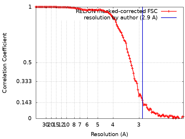 |
