Movie
Movie Controller


+ Open data

- Basic information
| Entry |  | |||||||||
|---|---|---|---|---|---|---|---|---|---|---|
| Title | membrane proteins | |||||||||
 Map data Map data | ||||||||||
 Sample Sample |
| |||||||||
 Keywords Keywords | enzyme / acetylation / MEMBRANE PROTEIN | |||||||||
| Function / homology | :  Function and homology information Function and homology information | |||||||||
| Biological species |  Homo sapiens (human) Homo sapiens (human) | |||||||||
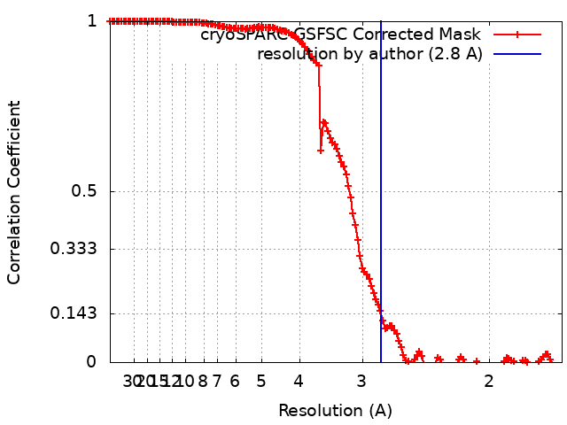
| Method | single particle reconstruction / cryo EM / Resolution: 2.8 Å | |||||||||
 Authors Authors | Yu J / Ge JP / Xu RS | |||||||||
| Funding support |  China, 1 items China, 1 items
| |||||||||
 Citation Citation | Journal: To Be Published Title: membrane proteins Authors: Yu J / Ge JP / Xu RS | |||||||||
| History |
|
- Structure visualization
| Supplemental images |
|---|
- Downloads & links
-EMDB archive
| Map data | emd_36386.map.gz | 230 MB | EMDB map data format | |
|---|---|---|---|---|
| Header (meta data) | emd-36386-v30.xmlemd-36386.xml | 15.2 KB 15.2 KB | Display Display | EMDB header |
| FSC (resolution estimation) | emd_36386_fsc.xml | 13.2 KB | Display | FSC data file |
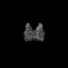
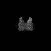
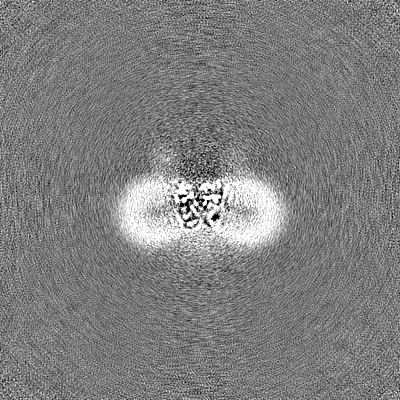
| Images |  emd_36386.png emd_36386.png | 92.7 KB | ||
| Filedesc metadata | emd-36386.cif.gz | 5.7 KB | ||
| Others | emd_36386_half_map_1.map.gzemd_36386_half_map_2.map.gz | 226.1 MB 226.1 MB | ||
| Archive directory |  http://ftp.pdbj.org/pub/emdb/structures/EMD-36386ftp://ftp.pdbj.org/pub/emdb/structures/EMD-36386 http://ftp.pdbj.org/pub/emdb/structures/EMD-36386ftp://ftp.pdbj.org/pub/emdb/structures/EMD-36386 | HTTPS FTP |
-Validation report
| Summary document | emd_36386_validation.pdf.gz | 852.7 KB | Display | EMDB validaton report |
|---|---|---|---|---|
| Full document | emd_36386_full_validation.pdf.gz | 852.3 KB | Display | |
| Data in XML | emd_36386_validation.xml.gz | 21.7 KB | Display | |
| Data in CIF | emd_36386_validation.cif.gz | 27.6 KB | Display | |
| Arichive directory | https://ftp.pdbj.org/pub/emdb/validation_reports/EMD-36386ftp://ftp.pdbj.org/pub/emdb/validation_reports/EMD-36386 | HTTPS FTP |
-Related structure data
| Related structure data |  8jl1MC  8jkvC  8jl3C  8jl4C M: atomic model generated by this map C: citing same article ( |
|---|---|
| Similar structure data |
-Links
| EMDB pages | EMDB (EBI/PDBe) / EMDataResource |
|---|
-Map
| File | Download / File: emd_36386.map.gz / Format: CCP4 / Size: 244.1 MB / Type: IMAGE STORED AS FLOATING POINT NUMBER (4 BYTES) | ||||||||||||||||||||||||||||||||||||
|---|---|---|---|---|---|---|---|---|---|---|---|---|---|---|---|---|---|---|---|---|---|---|---|---|---|---|---|---|---|---|---|---|---|---|---|---|---|
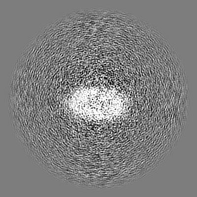
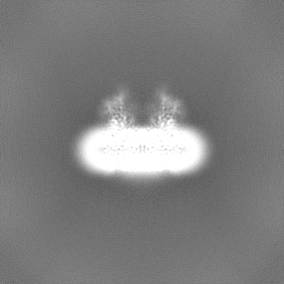
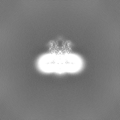
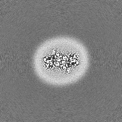
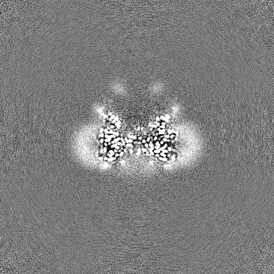
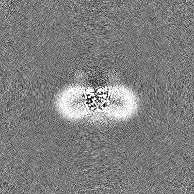
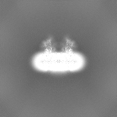
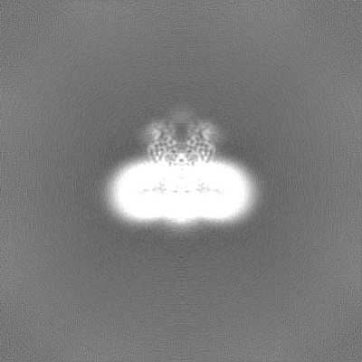
| Projections & slices | Image control
Images are generated by Spider. | ||||||||||||||||||||||||||||||||||||
| Voxel size | X=Y=Z: 0.83 Å | ||||||||||||||||||||||||||||||||||||
| Density |
| ||||||||||||||||||||||||||||||||||||
| Symmetry | Space group: 1 | ||||||||||||||||||||||||||||||||||||
| Details | EMDB XML:
|
 Z (Sec.)
 Y (Row.)
 X (Col.)


-Supplemental data
-Half map: #1
| File | emd_36386_half_map_1.map | ||||||||||||
|---|---|---|---|---|---|---|---|---|---|---|---|---|---|
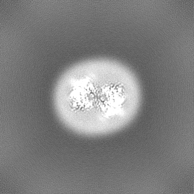
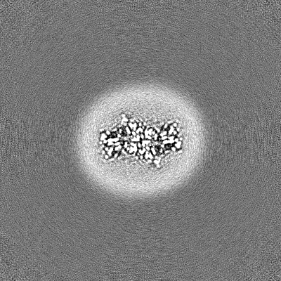
| Projections & Slices |
| ||||||||||||
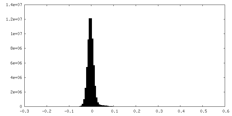
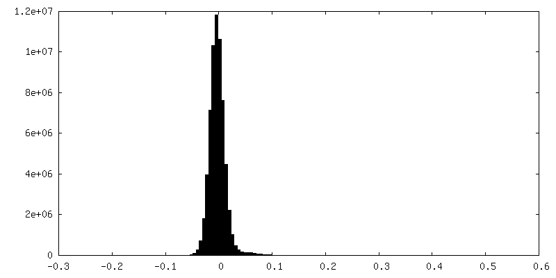
| Density Histograms |

-Half map: #2
| File | emd_36386_half_map_2.map | ||||||||||||
|---|---|---|---|---|---|---|---|---|---|---|---|---|---|
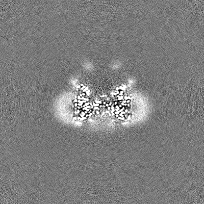
| Projections & Slices |
| ||||||||||||
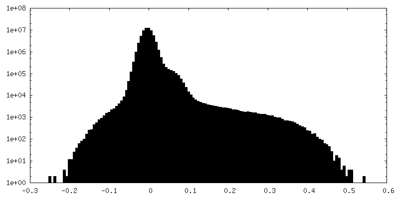
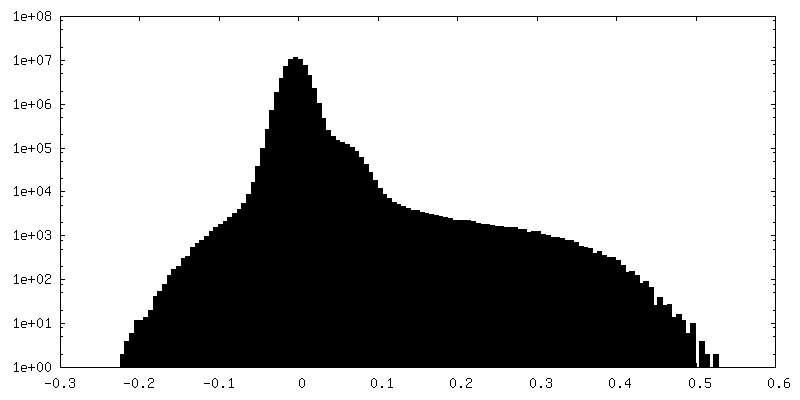
| Density Histograms |
- Sample components
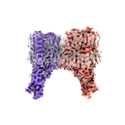
+Entire : HGSNAT
+Supramolecule #1: HGSNAT
+Macromolecule #1: Heparan-alpha-glucosaminide N-acetyltransferase
+Macromolecule #3: ACETYL COENZYME *A

+Macromolecule #4: 2-acetamido-2-deoxy-beta-D-glucopyranose

+Macromolecule #5: DODECANE

+Macromolecule #6: N-OCTANE

+Macromolecule #7: TETRADECANE

+Macromolecule #8: CHOLESTEROL

+Macromolecule #9: DECANE

+Macromolecule #10: HEXADECANE

-Experimental details
-Structure determination
| Method | cryo EM |
|---|---|
 Processing Processing | single particle reconstruction |
| Aggregation state | particle |
-Sample preparation
| Buffer | pH: 8 |
|---|---|
| Vitrification | Cryogen name: ETHANE |
- Electron microscopy
| Microscope | FEI TITAN KRIOS |
|---|---|
| Image recording | Film or detector model: GATAN K3 (6k x 4k) / Average electron dose: 50.0 e/Å2 |
| Electron beam | Acceleration voltage: 300 kV / Electron source:  FIELD EMISSION GUN FIELD EMISSION GUN |
| Electron optics | Illumination mode: FLOOD BEAM / Imaging mode: BRIGHT FIELD / Nominal defocus max: 2.0 µm / Nominal defocus min: 0.8 µm |
| Experimental equipment |  Model: Titan Krios / Image courtesy: FEI Company |