 Movie
Movie Controller

[English] 日本語
 Yorodumi
- EMDB-36257: Immune complex of W328-6H2 IgG binding the RBD of SARS-CoV-1 2p s... -
+ Open data

- Basic information
| Entry |  | |||||||||
|---|---|---|---|---|---|---|---|---|---|---|
| Title | Immune complex of W328-6H2 IgG binding the RBD of SARS-CoV-1 2p spike protein | |||||||||
 Map data Map data | Immune complex of W328-6H2 IgG binding the RBD of SARS-CoV-1 2p spike protein | |||||||||
 Sample Sample |
| |||||||||
 Keywords Keywords | Complex / SARS-CoV-1 / antibody / IgG / dimer / Homo sapiens / RBD / VIRAL PROTEIN | |||||||||
| Biological species |  Homo sapiens (human) / Homo sapiens (human) /  Severe acute respiratory syndrome coronavirus Severe acute respiratory syndrome coronavirus | |||||||||
| Method | single particle reconstruction / negative staining / Resolution: 25.0 Å | |||||||||
 Authors Authors | Nan XY / Li YJ | |||||||||
| Funding support | 1 items
| |||||||||
 Citation Citation | Journal: Nat Commun / Year: 2024 Title: Exploring distinct modes of inter-spike cross-linking for enhanced neutralization by SARS-CoV-2 antibodies. Authors: Xuanyu Nan / Yujie Li / Rui Zhang / Ruoke Wang / Niannian Lv / Jiayi Li / Yuanfang Chen / Bini Zhou / Yangjunqi Wang / Ziyi Wang / Jiayi Zhu / Jing Chen / Jinqian Li / Wenlong Chen / Qi ...Authors: Xuanyu Nan / Yujie Li / Rui Zhang / Ruoke Wang / Niannian Lv / Jiayi Li / Yuanfang Chen / Bini Zhou / Yangjunqi Wang / Ziyi Wang / Jiayi Zhu / Jing Chen / Jinqian Li / Wenlong Chen / Qi Zhang / Xuanling Shi / Changwen Zhao / Chunying Chen / Zhihua Liu / Yuliang Zhao / Dongsheng Liu / Xinquan Wang / Li-Tang Yan / Taisheng Li / Linqi Zhang / Yuhe R Yang /  Abstract: The emergence of severe acute respiratory syndrome coronavirus 2 (SARS-CoV-2) and its Omicron subvariants drastically amplifies transmissibility, infectivity, and immune escape, mainly due to their ...The emergence of severe acute respiratory syndrome coronavirus 2 (SARS-CoV-2) and its Omicron subvariants drastically amplifies transmissibility, infectivity, and immune escape, mainly due to their resistance to most neutralizing antibodies. Thus, exploring the mechanisms underlying antibody evasion is crucial. Although the full-length native form of antibody, immunoglobulin G (IgG), offers valuable insights into the neutralization, structural investigations primarily focus on the fragment of antigen-binding (Fab). Here, we employ single-particle cryo-electron microscopy (cryo-EM) to characterize a W328-6H2 antibody, in its native IgG form complexed with severe acute respiratory syndrome (SARS), severe acute respiratory syndrome coronavirus 2 wild-type (WT) and Omicron variant BA.1 spike protein (S). Three high-resolution structures reveal that the full-length IgG forms a centered head-to-head dimer of trimer when binds fully stoichiometrically with both SARS and WT S, while adopting a distinct offset configuration with Omicron BA.1 S. Combined with functional assays, our results suggest that, beyond the binding affinity between the RBD epitope and Fab, the higher-order architectures of S trimer and full-length IgG play an additional role in neutralization, enriching our understanding of enhanced neutralization by SARS-CoV-2 antibodies. | |||||||||
| History |
|
- Structure visualization
| Supplemental images |
|---|

- Downloads & links
-EMDB archive
| Map data | emd_36257.map.gz | 194.1 MB |  EMDB map data format EMDB map data format | |
|---|---|---|---|---|
| Header (meta data) | emd-36257-v30.xmlemd-36257.xml | 15.8 KB 15.8 KB | Display Display | EMDB header |
| Images |  emd_36257.png emd_36257.png | 12.9 KB | ||
| Filedesc metadata | emd-36257.cif.gz | 4.2 KB | ||
| Others | emd_36257_half_map_1.map.gzemd_36257_half_map_2.map.gz | 194.1 MB 194.1 MB | ||
| Archive directory |  http://ftp.pdbj.org/pub/emdb/structures/EMD-36257ftp://ftp.pdbj.org/pub/emdb/structures/EMD-36257 http://ftp.pdbj.org/pub/emdb/structures/EMD-36257ftp://ftp.pdbj.org/pub/emdb/structures/EMD-36257 | HTTPS FTP |
-Related structure data


-Links
| EMDB pages | EMDB (EBI/PDBe) / EMDataResource |
|---|
-Map
| File | Download / File: emd_36257.map.gz / Format: CCP4 / Size: 244.1 MB / Type: IMAGE STORED AS FLOATING POINT NUMBER (4 BYTES) | ||||||||||||||||||||||||||||||||||||
|---|---|---|---|---|---|---|---|---|---|---|---|---|---|---|---|---|---|---|---|---|---|---|---|---|---|---|---|---|---|---|---|---|---|---|---|---|---|
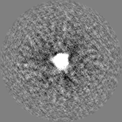
| Annotation | Immune complex of W328-6H2 IgG binding the RBD of SARS-CoV-1 2p spike protein | ||||||||||||||||||||||||||||||||||||
| Projections & slices | Image control
Images are generated by Spider. | ||||||||||||||||||||||||||||||||||||
| Voxel size | X=Y=Z: 2.21 Å | ||||||||||||||||||||||||||||||||||||
| Density |
| ||||||||||||||||||||||||||||||||||||
| Symmetry | Space group: 1 | ||||||||||||||||||||||||||||||||||||
| Details | EMDB XML:
|
 Z (Sec.)
 Y (Row.)
 X (Col.)


-Supplemental data
-Half map: Immune complex of W328-6H2 IgG binding the RBD...
| File | emd_36257_half_map_1.map | ||||||||||||
|---|---|---|---|---|---|---|---|---|---|---|---|---|---|
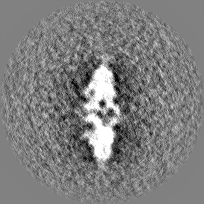
| Annotation | Immune complex of W328-6H2 IgG binding the RBD of SARS-CoV-1 2p spike protein | ||||||||||||
| Projections & Slices |
| ||||||||||||
| Density Histograms |


-Half map: Immune complex of W328-6H2 IgG binding the RBD...
| File | emd_36257_half_map_2.map | ||||||||||||
|---|---|---|---|---|---|---|---|---|---|---|---|---|---|
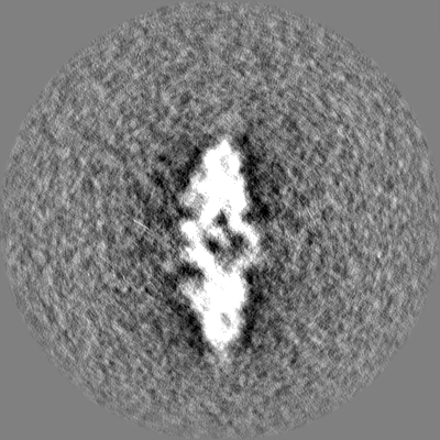
| Annotation | Immune complex of W328-6H2 IgG binding the RBD of SARS-CoV-1 2p spike protein | ||||||||||||
| Projections & Slices |
| ||||||||||||
| Density Histograms |


- Sample components
-Entire : SARS-CoV-1 2P in complex with W328-6H2 IgG
| Entire | Name: SARS-CoV-1 2P in complex with W328-6H2 IgG |
|---|---|
| Components |
|
-Supramolecule #1: SARS-CoV-1 2P in complex with W328-6H2 IgG
| Supramolecule | Name: SARS-CoV-1 2P in complex with W328-6H2 IgG / type: complex / ID: 1 / Parent: 0 / Macromolecule list: #1-#3 Details: Immune complex of W328-6H2 IgG binding the RBD of SARS-CoV-1 2p spike protein |
|---|---|
| Source (natural) | Organism: Homo sapiens (human) |
-Supramolecule #2: Severe acute respiratory syndrome coronavirus 1-2P
| Supramolecule | Name: Severe acute respiratory syndrome coronavirus 1-2P / type: complex / ID: 2 / Parent: 1 / Macromolecule list: #2 / Details: SARS-CoV-1 2P |
|---|---|
| Source (natural) | Organism: Severe acute respiratory syndrome coronavirus |
-Supramolecule #3: W328-6H2 IgG
| Supramolecule | Name: W328-6H2 IgG / type: complex / ID: 3 / Parent: 1 / Macromolecule list: #3 / Details: W328-6H2 IgG |
|---|---|
| Source (natural) | Organism: Homo sapiens (human) |
-Experimental details
-Structure determination
| Method | negative staining |
|---|---|
 Processing Processing | single particle reconstruction |
| Aggregation state | particle |
-Sample preparation
| Concentration | 0.015 mg/mL |
|---|---|
| Buffer | pH: 7.4 |
| Staining | Type: NEGATIVE / Material: Uranyl Acetate |
- Electron microscopy
| Microscope | JEOL 2100F |
|---|---|
| Image recording | Film or detector model: OTHER / Average electron dose: 25.0 e/Å2 |
| Electron beam | Acceleration voltage: 200 kV / Electron source:  FIELD EMISSION GUN FIELD EMISSION GUN |
| Electron optics | Illumination mode: FLOOD BEAM / Imaging mode: BRIGHT FIELD / Cs: 2.7 mm / Nominal defocus max: 1.5 µm / Nominal defocus min: 1.5 µm |
