 Movie
Movie Controller


+ Open data

- Basic information
| Entry |  | |||||||||
|---|---|---|---|---|---|---|---|---|---|---|
| Title | Cryo-EM structure of RDGC/Ca2+-CaM complex | |||||||||
 Map data Map data | ||||||||||
 Sample Sample |
| |||||||||
 Keywords Keywords | Phosphatase / metal binding. / HYDROLASE | |||||||||
| Function / homology |  Function and homology information Function and homology informationdetection of stimulus involved in sensory perception / negative regulation of opsin-mediated signaling pathway / calcium-dependent protein serine/threonine phosphatase activity / thermotaxis / protein-serine/threonine phosphatase / protein serine/threonine phosphatase activity / phototransduction / visual perception / calcium-mediated signaling / mitotic spindle ...detection of stimulus involved in sensory perception / negative regulation of opsin-mediated signaling pathway / calcium-dependent protein serine/threonine phosphatase activity / thermotaxis / protein-serine/threonine phosphatase / protein serine/threonine phosphatase activity / phototransduction / visual perception / calcium-mediated signaling / mitotic spindle / manganese ion binding / calmodulin binding / iron ion binding / calcium ion binding / nucleus / cytoplasm / cytosol Similarity search - Function | |||||||||
| Biological species |  | |||||||||
| Method | single particle reconstruction / cryo EM / Resolution: 2.79 Å | |||||||||
 Authors Authors | Liu ZM / Liu W / Wu C / Liu J | |||||||||
| Funding support |  China, 1 items China, 1 items
| |||||||||
 Citation Citation | Journal: To Be Published Title: Cryo-EM structure of RDGC/Ca2+-CaM complex Authors: Liu ZM / Liu W / Wu C / Liu J | |||||||||
| History |
|
- Structure visualization
| Supplemental images |
|---|

- Downloads & links
-EMDB archive
| Map data | emd_36219.map.gz | 203.9 MB | EMDB map data format | |
|---|---|---|---|---|
| Header (meta data) | emd-36219-v30.xmlemd-36219.xml | 18 KB 18 KB | Display Display | EMDB header |
| FSC (resolution estimation) | emd_36219_fsc.xml | 12.7 KB | Display | FSC data file |
| Images |  emd_36219.png emd_36219.png | 69 KB | ||
| Filedesc metadata | emd-36219.cif.gz | 6.1 KB | ||
| Others | emd_36219_half_map_1.map.gzemd_36219_half_map_2.map.gz | 200.4 MB 200.4 MB | ||
| Archive directory |  http://ftp.pdbj.org/pub/emdb/structures/EMD-36219ftp://ftp.pdbj.org/pub/emdb/structures/EMD-36219 http://ftp.pdbj.org/pub/emdb/structures/EMD-36219ftp://ftp.pdbj.org/pub/emdb/structures/EMD-36219 | HTTPS FTP |
-Related structure data
| Related structure data |  8jfyMC M: atomic model generated by this map C: citing same article ( |
|---|---|
| Similar structure data |
-Links
| EMDB pages | EMDB (EBI/PDBe) / EMDataResource |
|---|---|
| Related items in Molecule of the Month |


-Map
| File | Download / File: emd_36219.map.gz / Format: CCP4 / Size: 216 MB / Type: IMAGE STORED AS FLOATING POINT NUMBER (4 BYTES) | ||||||||||||||||||||||||||||||||||||
|---|---|---|---|---|---|---|---|---|---|---|---|---|---|---|---|---|---|---|---|---|---|---|---|---|---|---|---|---|---|---|---|---|---|---|---|---|---|
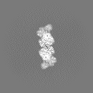
| Projections & slices | Image control
Images are generated by Spider. | ||||||||||||||||||||||||||||||||||||
| Voxel size | X=Y=Z: 0.855 Å | ||||||||||||||||||||||||||||||||||||
| Density |
| ||||||||||||||||||||||||||||||||||||
| Symmetry | Space group: 1 | ||||||||||||||||||||||||||||||||||||
| Details | EMDB XML:
|
 Z (Sec.)
 Y (Row.)
 X (Col.)


-Supplemental data
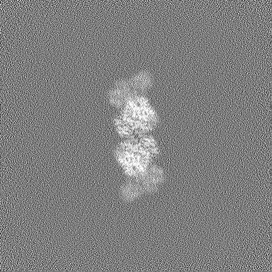
-Half map: #2
| File | emd_36219_half_map_1.map | ||||||||||||
|---|---|---|---|---|---|---|---|---|---|---|---|---|---|
| Projections & Slices |
| ||||||||||||
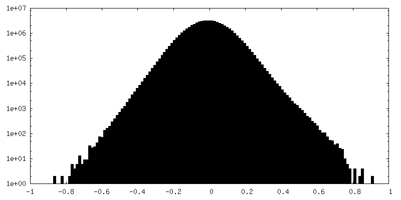
| Density Histograms |


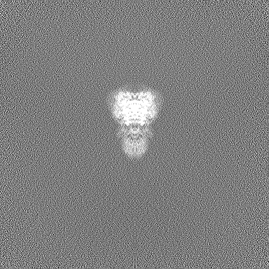
-Half map: #1
| File | emd_36219_half_map_2.map | ||||||||||||
|---|---|---|---|---|---|---|---|---|---|---|---|---|---|
| Projections & Slices |
| ||||||||||||
| Density Histograms |


- Sample components
-Entire : Cryo-EM structure of RDGC/Ca2+-CaM complex
| Entire | Name: Cryo-EM structure of RDGC/Ca2+-CaM complex |
|---|---|
| Components |
|
-Supramolecule #1: Cryo-EM structure of RDGC/Ca2+-CaM complex
| Supramolecule | Name: Cryo-EM structure of RDGC/Ca2+-CaM complex / type: complex / ID: 1 / Parent: 0 / Macromolecule list: #1-#2 |
|---|---|
| Source (natural) | Organism: |
-Macromolecule #1: AT15141p
| Macromolecule | Name: AT15141p / type: protein_or_peptide / ID: 1 / Number of copies: 2 / Enantiomer: LEVO |
|---|---|
| Source (natural) | Organism: |
| Molecular weight | Theoretical: 16.82552 KDa |
| Recombinant expression | Organism:  Spodoptera frugiperda (fall armyworm) Spodoptera frugiperda (fall armyworm) |
| Sequence | String: MADQLTEEQI AEFKEAFSLF DKDGDGTITT KELGTVMRSL GQNPTEAELQ DMINEVDADG NGTIDFPEFL TMMARKMKDT DSEEEIREA FRVFDKDGNG FISAAELRHV MTNLGEKLTD EEVDEMIREA DIDGDGQVNY EEFVTMMTSK UniProtKB: AT15141p |
-Macromolecule #2: Serine/threonine-protein phosphatase rdgC
| Macromolecule | Name: Serine/threonine-protein phosphatase rdgC / type: protein_or_peptide / ID: 2 / Number of copies: 2 / Enantiomer: LEVO / EC number: protein-serine/threonine phosphatase |
|---|---|
| Source (natural) | Organism: |
| Molecular weight | Theoretical: 68.828453 KDa |
| Recombinant expression | Organism: Spodoptera frugiperda (fall armyworm) |
| Sequence | String: MDENAIRAAI FIQKWYRRHQ ARREMQRRCN WQIFQNLEYA SEQDQAELYK FFNDLIKHMP QAAGRKNQYQ GSDDKDDLVE EFGDIVNAK IELPIRKNHI DLLIDVFRKK RGNRLHPKYV ALILREAAKS LKQLPNISPV STAVSQQVTV CGDLHGKLDD L LVVLHKNG ...String: MDENAIRAAI FIQKWYRRHQ ARREMQRRCN WQIFQNLEYA SEQDQAELYK FFNDLIKHMP QAAGRKNQYQ GSDDKDDLVE EFGDIVNAK IELPIRKNHI DLLIDVFRKK RGNRLHPKYV ALILREAAKS LKQLPNISPV STAVSQQVTV CGDLHGKLDD L LVVLHKNG LPSSSNPYVF NGDFVDRGKR GLEVLLLLLS LYLAFPNAVF LNRGNHEDSV MNARYGFIRE VESKYPRNHK RI LAFIDEV YRWLPLGSVL NSRVLIVHGG FSDSTSLDLI KSIDRGKYVS ILRPPLTDGE PLDKTEWQQI FDIMWSDPQA TMG CVPNTL RGAGVWFGPD VTDNFLQRHR LSYVIRSHEC KPNGHEFMHD NKIITIFSAS NYYAIGSNKG AYIRLNNQLM PHFV QYISA ASQTKRLSFK QRMGIVESSA LKELAVRMRD HRDELEDEFR KYDPKDSGYI SISHWCKVME NVTKLGLPWR LLRDK LAPG TDSQKVNYNR TLDLLDTDVI LEAEADGMSV MDALYANKAS LVAIFNIIDA DNSGEITLDE FETAIDLLVA HMPGAY SKA EMLEKCRMMD LNGDGKVDLN EFLEAFRLSD LHRKEQ UniProtKB: Serine/threonine-protein phosphatase rdgC |
-Macromolecule #3: CALCIUM ION
| Macromolecule | Name: CALCIUM ION / type: ligand / ID: 3 / Number of copies: 12 / Formula: CA |
|---|---|
| Molecular weight | Theoretical: 40.078 Da |
-Macromolecule #4: ZINC ION
| Macromolecule | Name: ZINC ION / type: ligand / ID: 4 / Number of copies: 2 / Formula: ZN |
|---|---|
| Molecular weight | Theoretical: 65.409 Da |
-Macromolecule #5: FE (III) ION
| Macromolecule | Name: FE (III) ION / type: ligand / ID: 5 / Number of copies: 2 / Formula: FE |
|---|---|
| Molecular weight | Theoretical: 55.845 Da |
-Macromolecule #6: PHOSPHATE ION
| Macromolecule | Name: PHOSPHATE ION / type: ligand / ID: 6 / Number of copies: 2 / Formula: PO4 |
|---|---|
| Molecular weight | Theoretical: 94.971 Da |
| Chemical component information |  ChemComp-PO4: |
-Experimental details
-Structure determination
| Method | cryo EM |
|---|---|
 Processing Processing | single particle reconstruction |
| Aggregation state | particle |
-Sample preparation
| Buffer | pH: 7 |
|---|---|
| Vitrification | Cryogen name: ETHANE |
- Electron microscopy
| Microscope | FEI TITAN KRIOS |
|---|---|
| Image recording | Film or detector model: GATAN K3 BIOQUANTUM (6k x 4k) / Average electron dose: 50.0 e/Å2 |
| Electron beam | Acceleration voltage: 300 kV / Electron source:  FIELD EMISSION GUN FIELD EMISSION GUN |
| Electron optics | Illumination mode: FLOOD BEAM / Imaging mode: BRIGHT FIELD / Nominal defocus max: 1.6 µm / Nominal defocus min: 1.0 µm |
| Experimental equipment |  Model: Titan Krios / Image courtesy: FEI Company |

