 Movie
Movie Controller

[English] 日本語
 Yorodumi
- EMDB-3600: Contracted sheath of a Pseudomonas aeruginosa type six secretion ... -
+ Open data

- Basic information
| Entry | Database: EMDB / ID: EMD-3600 | |||||||||
|---|---|---|---|---|---|---|---|---|---|---|
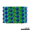
| Title | Contracted sheath of a Pseudomonas aeruginosa type six secretion system consisting of TssB1 and TssC1 | |||||||||
 Map data Map data | ||||||||||
 Sample Sample |
| |||||||||
 Keywords Keywords | type six secretion system / structural protein | |||||||||
| Function / homology |  Function and homology information Function and homology informationType VI secretion system TssC-like / TssC1, N-terminal / TssC1, C-terminal / EvpB/VC_A0108, tail sheath N-terminal domain / EvpB/VC_A0108, tail sheath gpW/gp25-like domain / Type VI secretion system sheath protein TssB1 / Type VI secretion system, VipA, VC_A0107 or Hcp2 Similarity search - Domain/homology | |||||||||
| Biological species |   Pseudomonas aeruginosa (bacteria) Pseudomonas aeruginosa (bacteria) | |||||||||
| Method | helical reconstruction / cryo EM / Resolution: 3.28 Å | |||||||||
 Authors Authors | Salih O / He S | |||||||||
 Citation Citation | Journal: Structure / Year: 2018 Title: Atomic Structure of Type VI Contractile Sheath from Pseudomonas aeruginosa. Authors: Osman Salih / Shaoda He / Sara Planamente / Lasse Stach / James T MacDonald / Eleni Manoli / Sjors H W Scheres / Alain Filloux / Paul S Freemont /  Abstract: Pseudomonas aeruginosa has three type VI secretion systems (T6SSs), H1-, H2-, and H3-T6SS, each belonging to a distinct group. The two T6SS components, TssB/VipA and TssC/VipB, assemble to form ...Pseudomonas aeruginosa has three type VI secretion systems (T6SSs), H1-, H2-, and H3-T6SS, each belonging to a distinct group. The two T6SS components, TssB/VipA and TssC/VipB, assemble to form tubules that conserve structural/functional homology with tail sheaths of contractile bacteriophages and pyocins. Here, we used cryoelectron microscopy to solve the structure of the H1-T6SS P. aeruginosa TssB1C1 sheath at 3.3 Å resolution. Our structure allowed us to resolve some features of the T6SS sheath that were not resolved in the Vibrio cholerae VipAB and Francisella tularensis IglAB structures. Comparison with sheath structures from other contractile machines, including T4 phage and R-type pyocins, provides a better understanding of how these systems have conserved similar functions/mechanisms despite evolution. We used the P. aeruginosa R2 pyocin as a structural template to build an atomic model of the TssB1C1 sheath in its extended conformation, allowing us to propose a coiled-spring-like mechanism for T6SS sheath contraction. | |||||||||
| History |
|
- Structure visualization
| Movie |
Movie viewer |
|---|---|
| Structure viewer | EM map: SurfViewMolmilJmol/JSmol |
| Supplemental images |

- Downloads & links
-EMDB archive
| Map data | emd_3600.map.gz | 15.7 MB | EMDB map data format | |
|---|---|---|---|---|
| Header (meta data) | emd-3600-v30.xmlemd-3600.xml | 11.5 KB 11.5 KB | Display Display | EMDB header |
| Images | 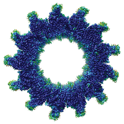 emd_3600.png emd_3600.png | 241.2 KB | ||
| Filedesc metadata | emd-3600.cif.gz | 5.2 KB | ||
| Archive directory |  http://ftp.pdbj.org/pub/emdb/structures/EMD-3600ftp://ftp.pdbj.org/pub/emdb/structures/EMD-3600 http://ftp.pdbj.org/pub/emdb/structures/EMD-3600ftp://ftp.pdbj.org/pub/emdb/structures/EMD-3600 | HTTPS FTP |
-Related structure data
| Related structure data | 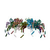 5n8nMC M: atomic model generated by this map C: citing same article ( |
|---|---|
| Similar structure data |
-Links
| EMDB pages | EMDB (EBI/PDBe) / EMDataResource |
|---|
-Map
| File | Download / File: emd_3600.map.gz / Format: CCP4 / Size: 59.6 MB / Type: IMAGE STORED AS FLOATING POINT NUMBER (4 BYTES) | ||||||||||||||||||||||||||||||||||||||||||||||||||||||||||||
|---|---|---|---|---|---|---|---|---|---|---|---|---|---|---|---|---|---|---|---|---|---|---|---|---|---|---|---|---|---|---|---|---|---|---|---|---|---|---|---|---|---|---|---|---|---|---|---|---|---|---|---|---|---|---|---|---|---|---|---|---|---|
| Projections & slices | Image control
Images are generated by Spider. | ||||||||||||||||||||||||||||||||||||||||||||||||||||||||||||
| Voxel size | X=Y=Z: 1.34 Å | ||||||||||||||||||||||||||||||||||||||||||||||||||||||||||||
| Density |
| ||||||||||||||||||||||||||||||||||||||||||||||||||||||||||||
| Symmetry | Space group: 1 | ||||||||||||||||||||||||||||||||||||||||||||||||||||||||||||
| Details | EMDB XML:
CCP4 map header:
| ||||||||||||||||||||||||||||||||||||||||||||||||||||||||||||
 Z (Sec.)
 Y (Row.)
 X (Col.)


-Supplemental data
- Sample components
-Entire : contracted sheath of a Pseudomonas aeruginosa T6SS consisting of ...
| Entire | Name: contracted sheath of a Pseudomonas aeruginosa T6SS consisting of TssB1 and TssC1 |
|---|---|
| Components |
|
-Supramolecule #1: contracted sheath of a Pseudomonas aeruginosa T6SS consisting of ...
| Supramolecule | Name: contracted sheath of a Pseudomonas aeruginosa T6SS consisting of TssB1 and TssC1 type: complex / ID: 1 / Parent: 0 / Macromolecule list: all |
|---|
-Supramolecule #2: AO964_32215, PAERUG_E15_London_28_01_14_04448, PAERUG_P32_London_...
| Supramolecule | Name: AO964_32215, PAERUG_E15_London_28_01_14_04448, PAERUG_P32_London_17_VIM_2_10_11_02574, PAMH19_0082 type: complex / ID: 2 / Parent: 1 / Macromolecule list: #1 |
|---|---|
| Source (natural) | Organism: Pseudomonas aeruginosa (bacteria) |
-Supramolecule #3: U769_00445
| Supramolecule | Name: U769_00445 / type: complex / ID: 3 / Parent: 1 / Macromolecule list: #2 |
|---|---|
| Source (natural) | Organism: Pseudomonas aeruginosa (bacteria) |
-Macromolecule #1: Type VI secretion protein, family
| Macromolecule | Name: Type VI secretion protein, family / type: protein_or_peptide / ID: 1 / Number of copies: 15 / Enantiomer: LEVO |
|---|---|
| Source (natural) | Organism: Pseudomonas aeruginosa (bacteria) |
| Molecular weight | Theoretical: 14.634741 KDa |
| Recombinant expression | Organism: |
| Sequence | String: TTSSQKFIAR NRAPRVQIEY DVELYGAEKK VQLPFVMGVM ADLAGKPAEP QAAVADRKFL EIDVDNFDAR LKAMKPRVAF NVPNVLTGE GNLSLDITFE SMDDFSPAAV ARKVDSLNKL LEARTQLANL LTY UniProtKB: Type VI secretion system contractile sheath small subunit |
-Macromolecule #2: EvpB family type VI secretion protein
| Macromolecule | Name: EvpB family type VI secretion protein / type: protein_or_peptide / ID: 2 / Number of copies: 15 / Enantiomer: LEVO |
|---|---|
| Source (natural) | Organism: Pseudomonas aeruginosa (bacteria) |
| Molecular weight | Theoretical: 51.721125 KDa |
| Recombinant expression | Organism: |
| Sequence | String: REAVETAVRT LAEHALEQTS LISNDAIKSI ESIIAALDAK LTAQVNLIMH HADFQQLESA WRGLHYLVNN TETDEQLKIR VLNISKPEL HKTLKKFKGT TWDQSPIFKK LYEEEYGQFG GEPYGCLVGD YYFDQSPPDV ELLGEMAKIS AAMHAPFISA A SPTVMGMG ...String: REAVETAVRT LAEHALEQTS LISNDAIKSI ESIIAALDAK LTAQVNLIMH HADFQQLESA WRGLHYLVNN TETDEQLKIR VLNISKPEL HKTLKKFKGT TWDQSPIFKK LYEEEYGQFG GEPYGCLVGD YYFDQSPPDV ELLGEMAKIS AAMHAPFISA A SPTVMGMG SWQELSNPRD LTKIFTTPEY AGWRSLRESE DSRYIGLTMP RFLARLPYGA KTDPVEEFAF EEETDGADSS KY AWANSAY AMAVNINRSF KLYGWCSRIR GVESGGEVQG LPAHTFPTDD GGVDMKCPTE IAISDRREAE LAKNGFMPLL HKK NTDFAA FIGAQSLQKP AEYDDPDATA NANLAARLPY LFATCRFAHY LKCIVRDKIG SFKEKDEMQR WLQDWILNYV DGDP AHSTE TTKAQHPLAA AEVVVEEVEG NPGYYNSKFF LRPHYQLEGL TVSLRLVSKL PSAKEA UniProtKB: UNIPROTKB: A0A0E1AL03 |
-Experimental details
-Structure determination
| Method | cryo EM |
|---|---|
 Processing Processing | helical reconstruction |
| Aggregation state | helical array |
-Sample preparation
| Concentration | 1 mg/mL |
|---|---|
| Buffer | pH: 9 |
| Vitrification | Cryogen name: ETHANE |
- Electron microscopy
| Microscope | FEI TITAN KRIOS |
|---|---|
| Image recording | Film or detector model: FEI FALCON II (4k x 4k) / Average electron dose: 30.0 e/Å2 |
| Electron beam | Acceleration voltage: 300 kV / Electron source:  FIELD EMISSION GUN FIELD EMISSION GUN |
| Electron optics | Illumination mode: FLOOD BEAM / Imaging mode: BRIGHT FIELD |
| Experimental equipment |  Model: Titan Krios / Image courtesy: FEI Company |
-Image processing
| Final reconstruction | Applied symmetry - Helical parameters - Δz: 20.2 Å Applied symmetry - Helical parameters - Δ&Phi: 27.7 ° Applied symmetry - Helical parameters - Axial symmetry: C1 (asymmetric) Resolution.type: BY AUTHOR / Resolution: 3.28 Å / Resolution method: FSC 0.143 CUT-OFF / Number images used: 71264 |
|---|---|
| Startup model | Type of model: NONE |
| Final angle assignment | Type: NOT APPLICABLE |
