National Natural Science Foundation of China (NSFC)
#31970974
China
Citation
Journal: Proc Natl Acad Sci U S A / Year: 2023 Title: Vimentin regulates nuclear segmentation in neutrophils. Authors: Jiaqi Liu / Zhixun Li / Meijing Li / Wenjing Du / Wolfgang Baumeister / Jing Yang / Qiang Guo / Abstract: Granulocytes are indispensable for various immune responses. Unlike other cell types in the body, the nuclei of granulocytes, particularly neutrophils, are heavily segmented into multiple lobes. ...Granulocytes are indispensable for various immune responses. Unlike other cell types in the body, the nuclei of granulocytes, particularly neutrophils, are heavily segmented into multiple lobes. Although this distinct morphological feature has long been observed, the underlying mechanism remains incompletely characterized. In this study, we utilize cryo-electron tomography to examine the nuclei of mouse neutrophils, revealing the cytoplasmic enrichment of intermediate filaments on the concave regions of the nuclear envelope. Aided by expression profiling and immuno-electron microscopy, we then elucidate that the intermediate-filament protein vimentin is responsible for such perinuclear structures. Of importance, exogenously expressed vimentin in nonimmune cells is sufficient to form cytoplasmic filaments wrapping on the concave nuclear surface. Moreover, genetic deletion of the protein causes a significant reduction of the number of nuclear lobes in neutrophils and eosinophils, mimicking the hematological condition of the Pelger-Huët anomaly. These results have uncovered a new component establishing the nuclear segmentation of granulocytes.
Focused ion beam - Instrument: OTHER / Focused ion beam - Ion: OTHER / Focused ion beam - Voltage: 30 / Focused ion beam - Current: 0.5 / Focused ion beam - Duration: 1800 / Focused ion beam - Temperature: 100 K / Focused ion beam - Initial thickness: 1000 / Focused ion beam - Final thickness: 180 Focused ion beam - Details: The value given for _em_focused_ion_beam.instrument is Aquilos 2. This is not in a list of allowed values {'DB235', 'OTHER'} so OTHER is written into the XML file.
-
Electron microscopy
Microscope
FEI TITAN KRIOS
Image recording
Film or detector model: GATAN K3 (6k x 4k) / Average exposure time: 1.43 sec. / Average electron dose: 1.9 e/Å2
Electron beam
Acceleration voltage: 300 kV / Electron source: FIELD EMISSION GUN
In the structure databanks used in Yorodumi, some data are registered as the other names, "COVID-19 virus" and "2019-nCoV". Here are the details of the virus and the list of structure data.
Jan 31, 2019. EMDB accession codes are about to change! (news from PDBe EMDB page)
EMDB accession codes are about to change! (news from PDBe EMDB page)
The allocation of 4 digits for EMDB accession codes will soon come to an end. Whilst these codes will remain in use, new EMDB accession codes will include an additional digit and will expand incrementally as the available range of codes is exhausted. The current 4-digit format prefixed with “EMD-” (i.e. EMD-XXXX) will advance to a 5-digit format (i.e. EMD-XXXXX), and so on. It is currently estimated that the 4-digit codes will be depleted around Spring 2019, at which point the 5-digit format will come into force.
The EM Navigator/Yorodumi systems omit the EMD- prefix.
Related info.:Q: What is EMD? / ID/Accession-code notation in Yorodumi/EM Navigator
Yorodumi is a browser for structure data from EMDB, PDB, SASBDB, etc.
This page is also the successor to EM Navigator detail page, and also detail information page/front-end page for Omokage search.
The word "yorodu" (or yorozu) is an old Japanese word meaning "ten thousand". "mi" (miru) is to see.
Related info.:EMDB / PDB / SASBDB / Comparison of 3 databanks / Yorodumi Search / Aug 31, 2016. New EM Navigator & Yorodumi / Yorodumi Papers / Jmol/JSmol / Function and homology information / Changes in new EM Navigator and Yorodumi
 Movie
Movie Controller


 Open data
 Basic information
 Map data
 Sample
 Keywords
 Authors
 China, 1 items
  Citation
 Structure visualization
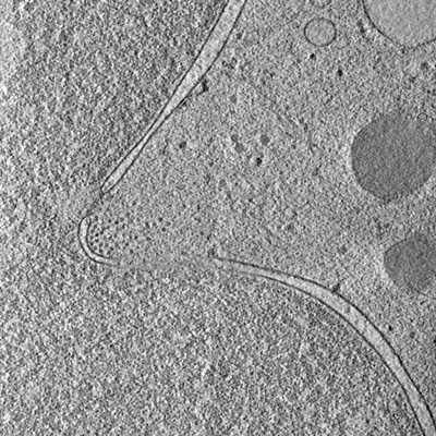
 Downloads & links
 EMDB map data format
 emd_35979.png
 http://ftp.pdbj.org/pub/emdb/structures/EMD-35979
 Z (Sec.)
 Y (Row.)
 X (Col.)


 Sample components
 Processing
 Electron microscopy
 FIELD EMISSION GUN
