 Movie
Movie Controller


+ Open data

- Basic information
| Entry | 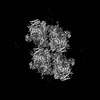 | ||||||||||||
|---|---|---|---|---|---|---|---|---|---|---|---|---|---|
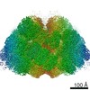
| Title | Structure of in situ PSII from P. purpureum lamellae by GisSPA | ||||||||||||
 Map data Map data | |||||||||||||
 Sample Sample |
| ||||||||||||
 Keywords Keywords | in situ / MEMBRANE PROTEIN | ||||||||||||
| Biological species |  Porphyridium purpureum (eukaryote) Porphyridium purpureum (eukaryote) | ||||||||||||
| Method | single particle reconstruction / cryo EM / Resolution: 3.9 Å | ||||||||||||
 Authors Authors | Zhang X / Cheng J | ||||||||||||
| Funding support |  China, 1 items China, 1 items
| ||||||||||||
 Citation Citation | Journal: Nat Commun / Year: 2023 Title: Determining protein structures in cellular lamella at pseudo-atomic resolution by GisSPA. Authors: Jing Cheng / Tong Liu / Xin You / Fa Zhang / Sen-Fang Sui / Xiaohua Wan / Xinzheng Zhang / Abstract: Cryo-electron tomography is a major tool used to study the structure of protein complexes in situ. However, the throughput of tilt-series image data collection is still quite low. Here, we show that ...Cryo-electron tomography is a major tool used to study the structure of protein complexes in situ. However, the throughput of tilt-series image data collection is still quite low. Here, we show that GisSPA, a GPU accelerated program, can translationally and rotationally localize the target protein complex in cellular lamellae, as prepared with a focused ion beam, using single cryo-electron microscopy images without tilt-series, and reconstruct the protein complex at near-atomic resolution. GisSPA allows high-throughput data collection without the acquisition of tilt-series images and reconstruction of the tomogram, which is essential for high-resolution reconstruction of asymmetric or low-symmetry protein complexes. We demonstrate the power of GisSPA with 3.4-Å and 3.9-Å resolutions of resolving phycobilisome and tetrameric photosystem II complex structures in cellular lamellae, respectively. In this work, we present GisSPA as a practical tool that facilitates high-resolution in situ protein structure determination. | ||||||||||||
| History |
|
- Structure visualization
| Supplemental images |
|---|

- Downloads & links
-EMDB archive
| Map data | emd_35976.map.gz | 59.1 MB |  EMDB map data format EMDB map data format | |
|---|---|---|---|---|
| Header (meta data) | emd-35976-v30.xmlemd-35976.xml | 13 KB 13 KB | Display Display | EMDB header |
| FSC (resolution estimation) | emd_35976_fsc.xml | 9.1 KB | Display | FSC data file |
| Images |  emd_35976.png emd_35976.png | 134.9 KB | ||
| Masks | emd_35976_msk_1.map | 64 MB | Mask map | |
| Others | emd_35976_half_map_1.map.gzemd_35976_half_map_2.map.gz | 59.3 MB 59.3 MB | ||
| Archive directory |  http://ftp.pdbj.org/pub/emdb/structures/EMD-35976ftp://ftp.pdbj.org/pub/emdb/structures/EMD-35976 http://ftp.pdbj.org/pub/emdb/structures/EMD-35976ftp://ftp.pdbj.org/pub/emdb/structures/EMD-35976 | HTTPS FTP |
-Validation report
| Summary document | emd_35976_validation.pdf.gz | 1.2 MB | Display | EMDB validaton report |
|---|---|---|---|---|
| Full document | emd_35976_full_validation.pdf.gz | 1.2 MB | Display | |
| Data in XML | emd_35976_validation.xml.gz | 16.3 KB | Display | |
| Data in CIF | emd_35976_validation.cif.gz | 21.5 KB | Display | |
| Arichive directory | https://ftp.pdbj.org/pub/emdb/validation_reports/EMD-35976ftp://ftp.pdbj.org/pub/emdb/validation_reports/EMD-35976 | HTTPS FTP |
-Links
| EMDB pages | EMDB (EBI/PDBe) / EMDataResource |
|---|
-Map
| File | Download / File: emd_35976.map.gz / Format: CCP4 / Size: 64 MB / Type: IMAGE STORED AS FLOATING POINT NUMBER (4 BYTES) | ||||||||||||||||||||||||||||||||||||
|---|---|---|---|---|---|---|---|---|---|---|---|---|---|---|---|---|---|---|---|---|---|---|---|---|---|---|---|---|---|---|---|---|---|---|---|---|---|
| Projections & slices | Image control
Images are generated by Spider. | ||||||||||||||||||||||||||||||||||||
| Voxel size | X=Y=Z: 1.632 Å | ||||||||||||||||||||||||||||||||||||
| Density |
| ||||||||||||||||||||||||||||||||||||
| Symmetry | Space group: 1 | ||||||||||||||||||||||||||||||||||||
| Details | EMDB XML:
|
 Z (Sec.)
 Y (Row.)
 X (Col.)


-Supplemental data
-Mask #1
| File | emd_35976_msk_1.map | ||||||||||||
|---|---|---|---|---|---|---|---|---|---|---|---|---|---|
| Projections & Slices |
| ||||||||||||
| Density Histograms |


-Half map: #2
| File | emd_35976_half_map_1.map | ||||||||||||
|---|---|---|---|---|---|---|---|---|---|---|---|---|---|
| Projections & Slices |
| ||||||||||||
| Density Histograms |


-Half map: #1
| File | emd_35976_half_map_2.map | ||||||||||||
|---|---|---|---|---|---|---|---|---|---|---|---|---|---|
| Projections & Slices |
| ||||||||||||
| Density Histograms |


- Sample components
-Entire : dimeric PSII complex
| Entire | Name: dimeric PSII complex |
|---|---|
| Components |
|
-Supramolecule #1: dimeric PSII complex
| Supramolecule | Name: dimeric PSII complex / type: complex / ID: 1 / Parent: 0 / Macromolecule list: #1 |
|---|---|
| Source (natural) | Organism: Porphyridium purpureum (eukaryote) |
-Experimental details
-Structure determination
| Method | cryo EM |
|---|---|
 Processing Processing | single particle reconstruction |
| Aggregation state | cell |
-Sample preparation
| Buffer | pH: 7 |
|---|---|
| Vitrification | Cryogen name: ETHANE |
- Electron microscopy
| Microscope | FEI TITAN KRIOS |
|---|---|
| Image recording | Film or detector model: GATAN K3 (6k x 4k) / Average electron dose: 35.0 e/Å2 |
| Electron beam | Acceleration voltage: 300 kV / Electron source:  FIELD EMISSION GUN FIELD EMISSION GUN |
| Electron optics | Illumination mode: FLOOD BEAM / Imaging mode: BRIGHT FIELD / Cs: 2.7 mm / Nominal defocus max: 6.0 µm / Nominal defocus min: 2.0 µm |
| Sample stage | Cooling holder cryogen: NITROGEN |
| Experimental equipment |  Model: Titan Krios / Image courtesy: FEI Company |
+Image processing

-Atomic model buiding 1
| Refinement | Protocol: OTHER |
|---|
