 ムービー
ムービー コントローラー


+ データを開く

- 基本情報
基本情報
| 登録情報 | 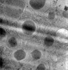 | |||||||||
|---|---|---|---|---|---|---|---|---|---|---|
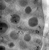
| タイトル | cryo-electron tomogram from mouse islets lift-out sample(TOMO_002) | |||||||||
 マップデータ マップデータ | ||||||||||
 試料 試料 |
| |||||||||
 キーワード キーワード | islet / vesicle / insulin / EXOCYTOSIS | |||||||||
| 生物種 |  | |||||||||
| 手法 | 電子線トモグラフィー法 / クライオ電子顕微鏡法 | |||||||||
 データ登録者 データ登録者 | Wu Y / Qin C / Du W / Chen L / Guo Q | |||||||||
| 資金援助 | 1件
| |||||||||
 引用 引用 | ジャーナル: J Struct Biol / 年: 2023 タイトル: A practical multicellular sample preparation pipeline broadens the application of in situ cryo-electron tomography. 著者: Yichun Wu / Changdong Qin / Wenjing Du / Zhenxi Guo / Liangyi Chen / Qiang Guo /  要旨: The structural studies of macromolecules in their physiological context, particularly in tissue, is constrained by the bottleneck of sample preparation. In this study, we present a practical pipeline ...The structural studies of macromolecules in their physiological context, particularly in tissue, is constrained by the bottleneck of sample preparation. In this study, we present a practical pipeline for preparing multicellular samples for cryo-electron tomography. The pipeline comprises sample isolation, vitrification, and lift-out-based lamella preparation using commercially available instruments. We demonstrate the efficacy of our pipeline by visualizing pancreatic β cells from mouse islets at the molecular level. This pipeline enables the determination of the properties of insulin crystals in situ for the first time, using unperturbed samples. | |||||||||
| 履歴 |
|
- 構造の表示
構造の表示
| 添付画像 |
|---|

- ダウンロードとリンク
-EMDBアーカイブ
| マップデータ | emd_35653.map.gz | 940.6 MB |  EMDBマップデータ形式 EMDBマップデータ形式 | |
|---|---|---|---|---|
| ヘッダ (付随情報) | emd-35653-v30.xmlemd-35653.xml | 8.5 KB 8.5 KB | 表示 表示 | EMDBヘッダ |
| 画像 | 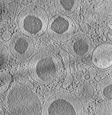 emd_35653.png emd_35653.png | 229.4 KB | ||
| アーカイブディレクトリ |  http://ftp.pdbj.org/pub/emdb/structures/EMD-35653ftp://ftp.pdbj.org/pub/emdb/structures/EMD-35653 http://ftp.pdbj.org/pub/emdb/structures/EMD-35653ftp://ftp.pdbj.org/pub/emdb/structures/EMD-35653 | HTTPS FTP |
-検証レポート
| 文書・要旨 | emd_35653_validation.pdf.gz | 464.6 KB | 表示 | EMDB検証レポート |
|---|---|---|---|---|
| 文書・詳細版 | emd_35653_full_validation.pdf.gz | 464.1 KB | 表示 | |
| XML形式データ | emd_35653_validation.xml.gz | 5.1 KB | 表示 | |
| CIF形式データ | emd_35653_validation.cif.gz | 5.7 KB | 表示 | |
| アーカイブディレクトリ | https://ftp.pdbj.org/pub/emdb/validation_reports/EMD-35653ftp://ftp.pdbj.org/pub/emdb/validation_reports/EMD-35653 | HTTPS FTP |
-関連構造データ
-リンク
| EMDBのページ | EMDB (EBI/PDBe) / EMDataResource |
|---|
-マップ
| ファイル | ダウンロード / ファイル: emd_35653.map.gz / 形式: CCP4 / 大きさ: 1019.5 MB / タイプ: IMAGE STORED AS FLOATING POINT NUMBER (4 BYTES) | ||||||||||||||||||||||||||||||||
|---|---|---|---|---|---|---|---|---|---|---|---|---|---|---|---|---|---|---|---|---|---|---|---|---|---|---|---|---|---|---|---|---|---|
| 投影像・断面図 | 画像のコントロール
画像は Spider により作成 これらの図は立方格子座標系で作成されたものです | ||||||||||||||||||||||||||||||||
| ボクセルのサイズ | X=Y=Z: 15.3 Å | ||||||||||||||||||||||||||||||||
| 密度 |
| ||||||||||||||||||||||||||||||||
| 対称性 | 空間群: 1 | ||||||||||||||||||||||||||||||||
| 詳細 | EMDB XML:
|
 Z (Sec.)
 Y (Row.)
 X (Col.)


-添付データ
- 試料の構成要素
-全体 : mouse islets lift-out sample
| 全体 | 名称: mouse islets lift-out sample |
|---|---|
| 要素 |
|
-超分子 #1: mouse islets lift-out sample
| 超分子 | 名称: mouse islets lift-out sample / タイプ: tissue / ID: 1 / 親要素: 0 |
|---|---|
| 由来(天然) | 生物種: |
-実験情報
-構造解析
| 手法 | クライオ電子顕微鏡法 |
|---|---|
 解析 解析 | 電子線トモグラフィー法 |
| 試料の集合状態 | tissue |
-試料調製
| 緩衝液 | pH: 7.4 |
|---|---|
| 凍結 | 凍結剤: NITROGEN / 詳細: high pressure freezing. |
| 加圧凍結法 | 装置: OTHER 詳細: The value given for _em_high_pressure_freezing.instrument is Leica EM HP100. This is not in a list of allowed values {'BAL-TEC HPM 010', 'LEICA EM HPM100', 'LEICA EM PACT', 'LEICA EM PACT2', ...詳細: The value given for _em_high_pressure_freezing.instrument is Leica EM HP100. This is not in a list of allowed values {'BAL-TEC HPM 010', 'LEICA EM HPM100', 'LEICA EM PACT', 'LEICA EM PACT2', 'OTHER', 'EMS-002 RAPID IMMERSION FREEZER'} so OTHER is written into the XML file. |
| 切片作成 | 集束イオンビーム - 装置: OTHER / 集束イオンビーム - イオン: OTHER / 集束イオンビーム - 電圧: 30 / 集束イオンビーム - 電流: 0.03 / 集束イオンビーム - 時間: 1800 / 集束イオンビーム - 温度: 81 K / 集束イオンビーム - Initial thickness: 10000 / 集束イオンビーム - 最終 厚さ: 320 集束イオンビーム - 詳細: The value given for _em_focused_ion_beam.instrument is Aquilos. This is not in a list of allowed values {'DB235', 'OTHER'} so OTHER is written into the XML file. |
- 電子顕微鏡法
| 顕微鏡 | FEI TALOS ARCTICA |
|---|---|
| 撮影 | フィルム・検出器のモデル: GATAN K2 SUMMIT (4k x 4k) 平均電子線量: 2.89 e/Å2 |
| 電子線 | 加速電圧: 200 kV / 電子線源:  FIELD EMISSION GUN FIELD EMISSION GUN |
| 電子光学系 | 照射モード: FLOOD BEAM / 撮影モード: BRIGHT FIELD / 最大 デフォーカス(公称値): 7.0 µm / 最小 デフォーカス(公称値): 5.0 µm |
| 実験機器 |  モデル: Talos Arctica / 画像提供: FEI Company |
-画像解析
| 最終 再構成 | 使用した粒子像数: 38 |
|---|
