 Movie
Movie Controller

[English] 日本語
 Yorodumi
- EMDB-35382: Cryo-EM structure of GPR156A/B of G-protein free GPR156 (local refine) -
+ Open data

- Basic information
| Entry |  | |||||||||
|---|---|---|---|---|---|---|---|---|---|---|
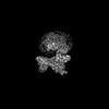
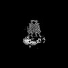
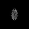
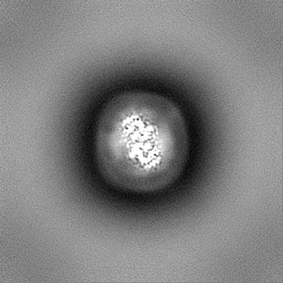
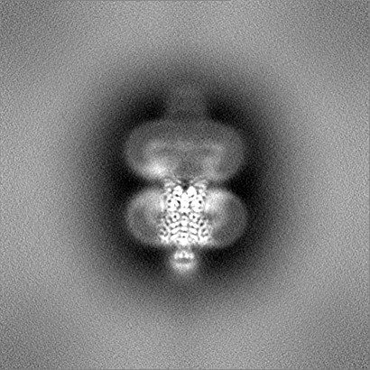
| Title | Cryo-EM structure of GPR156A/B of G-protein free GPR156 (local refine) | |||||||||
 Map data Map data | ||||||||||
 Sample Sample |
| |||||||||
 Keywords Keywords | Membrane protein / G-protein coupled receptor / Signal transduction / Phospholipid | |||||||||
| Function / homology |  Function and homology information Function and homology informationstereocilium bundle organization / G protein-coupled receptor heterodimeric complex / G protein-coupled GABA receptor activity / gamma-aminobutyric acid signaling pathway / plasma membrane Similarity search - Function | |||||||||
| Biological species |  Homo sapiens (human) Homo sapiens (human) | |||||||||
| Method | single particle reconstruction / cryo EM / Resolution: 2.62 Å | |||||||||
 Authors Authors | Shin J / Park J / Cho Y | |||||||||
| Funding support |  Korea, Republic Of, 1 items Korea, Republic Of, 1 items
| |||||||||
 Citation Citation | Journal: Nat Struct Mol Biol / Year: 2024 Title: Constitutive activation mechanism of a class C GPCR. Authors: Jinwoo Shin / Junhyeon Park / Jieun Jeong / Jordy Homing Lam / Xingyu Qiu / Di Wu / Kuglae Kim / Joo-Youn Lee / Carol V Robinson / Jaekyung Hyun / Vsevolod Katritch / Kwang Pyo Kim / Yunje Cho /   Abstract: Class C G-protein-coupled receptors (GPCRs) are activated through binding of agonists to the large extracellular domain (ECD) followed by rearrangement of the transmembrane domains (TMDs). GPR156, a ...Class C G-protein-coupled receptors (GPCRs) are activated through binding of agonists to the large extracellular domain (ECD) followed by rearrangement of the transmembrane domains (TMDs). GPR156, a class C orphan GPCR, is unique because it lacks an ECD and exhibits constitutive activity. Impaired GPR156-G signaling contributes to loss of hearing. Here we present the cryo-electron microscopy structures of human GPR156 in the G-free and G-coupled states. We found that an endogenous phospholipid molecule is located within each TMD of the GPR156 dimer. Asymmetric binding of Gα to the phospholipid-bound GPR156 dimer restructures the first and second intracellular loops and the carboxy-terminal part of the elongated transmembrane 7 (TM7) without altering dimer conformation. Our findings reveal that GPR156 is a transducer for phospholipid signaling. Constant binding of abundant phospholipid molecules and the G-protein-induced reshaping of the cytoplasmic face provide a basis for the constitutive activation of GPR156. | |||||||||
| History |
|
- Structure visualization
| Supplemental images |
|---|

- Downloads & links
-EMDB archive
| Map data | emd_35382.map.gz | 232.5 MB | EMDB map data format | |
|---|---|---|---|---|
| Header (meta data) | emd-35382-v30.xmlemd-35382.xml | 20.2 KB 20.2 KB | Display Display | EMDB header |
| Images | 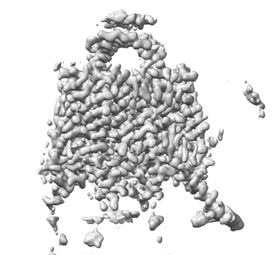 emd_35382.png emd_35382.png | 123.7 KB | ||
| Filedesc metadata | emd-35382.cif.gz | 6.7 KB | ||
| Others | emd_35382_half_map_1.map.gzemd_35382_half_map_2.map.gz | 243.6 MB 243.6 MB | ||
| Archive directory |  http://ftp.pdbj.org/pub/emdb/structures/EMD-35382ftp://ftp.pdbj.org/pub/emdb/structures/EMD-35382 http://ftp.pdbj.org/pub/emdb/structures/EMD-35382ftp://ftp.pdbj.org/pub/emdb/structures/EMD-35382 | HTTPS FTP |
-Related structure data
| Related structure data | 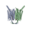 8ieiMC 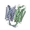 8iebC 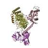 8iecC  8iedC 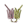 8iepC 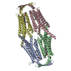 8ieqC M: atomic model generated by this map C: citing same article ( |
|---|---|
| Similar structure data |
-Links
| EMDB pages | EMDB (EBI/PDBe) / EMDataResource |
|---|---|
| Related items in Molecule of the Month |

-Map
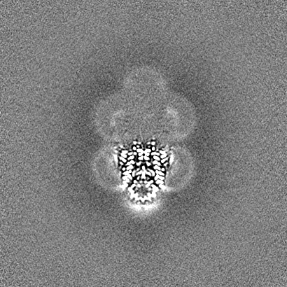
| File | Download / File: emd_35382.map.gz / Format: CCP4 / Size: 262.9 MB / Type: IMAGE STORED AS FLOATING POINT NUMBER (4 BYTES) | ||||||||||||||||||||||||||||||||||||
|---|---|---|---|---|---|---|---|---|---|---|---|---|---|---|---|---|---|---|---|---|---|---|---|---|---|---|---|---|---|---|---|---|---|---|---|---|---|
| Projections & slices | Image control
Images are generated by Spider. | ||||||||||||||||||||||||||||||||||||
| Voxel size | X=Y=Z: 0.81 Å | ||||||||||||||||||||||||||||||||||||
| Density |
| ||||||||||||||||||||||||||||||||||||
| Symmetry | Space group: 1 | ||||||||||||||||||||||||||||||||||||
| Details | EMDB XML:
|
 Z (Sec.)
 Y (Row.)
 X (Col.)


-Supplemental data
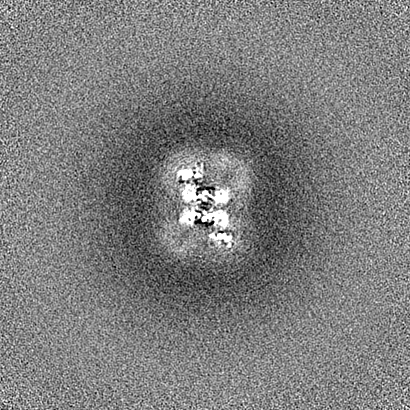
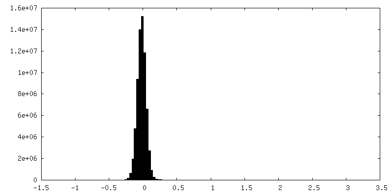
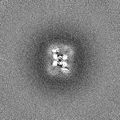
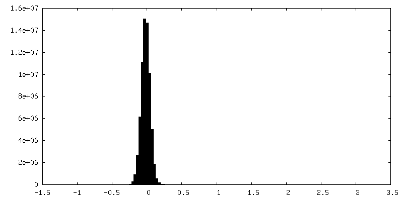
-Half map: #2
| File | emd_35382_half_map_1.map | ||||||||||||
|---|---|---|---|---|---|---|---|---|---|---|---|---|---|
| Projections & Slices |
| ||||||||||||
| Density Histograms |

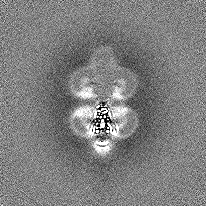
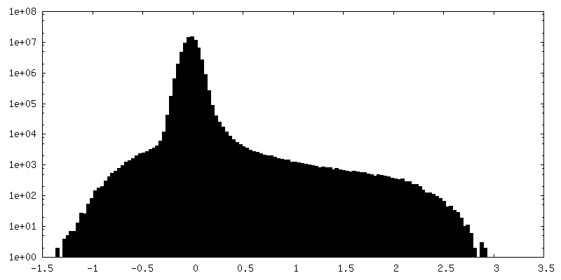
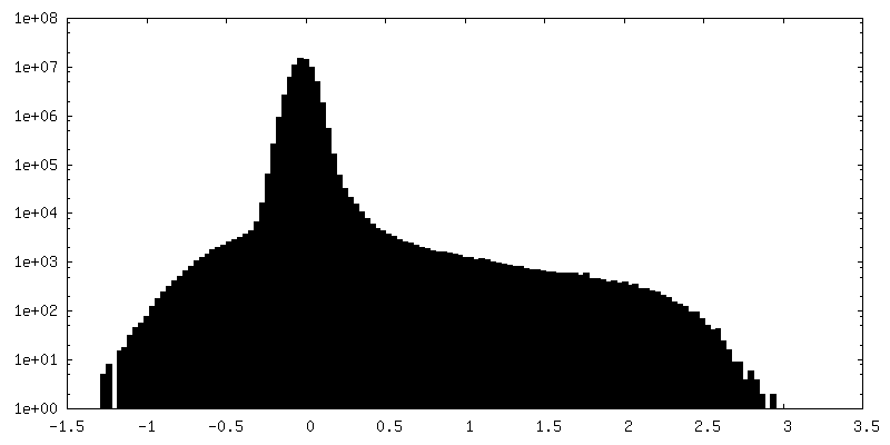
-Half map: #1
| File | emd_35382_half_map_2.map | ||||||||||||
|---|---|---|---|---|---|---|---|---|---|---|---|---|---|
| Projections & Slices |
| ||||||||||||
| Density Histograms |


- Sample components
-Entire : GPR156A/B
| Entire | Name: GPR156A/B |
|---|---|
| Components |
|
-Supramolecule #1: GPR156A/B
| Supramolecule | Name: GPR156A/B / type: complex / ID: 1 / Parent: 0 / Macromolecule list: #1 |
|---|---|
| Source (natural) | Organism: Homo sapiens (human) |
| Molecular weight | Theoretical: 131.2 kDa/nm |
-Macromolecule #1: Probable G-protein coupled receptor 156
| Macromolecule | Name: Probable G-protein coupled receptor 156 / type: protein_or_peptide / ID: 1 / Number of copies: 2 / Enantiomer: LEVO |
|---|---|
| Source (natural) | Organism: Homo sapiens (human) |
| Molecular weight | Theoretical: 65.628484 KDa |
| Recombinant expression | Organism: Homo sapiens (human) |
| Sequence | String: MEPEINCSEL CDSFPGQELD RRPLHDLCKT TITSSHHSSK TISSLSPVLL GIVWTFLSCG LLLILFFLAF TIHCRKNRIV KMSSPNLNI VTLLGSCLTY SSAYLFGIQD VLVGSSMETL IQTRLSMLCI GTSLVFGPIL GKSWRLYKVF TQRVPDKRVI I KDLQLLGL ...String: MEPEINCSEL CDSFPGQELD RRPLHDLCKT TITSSHHSSK TISSLSPVLL GIVWTFLSCG LLLILFFLAF TIHCRKNRIV KMSSPNLNI VTLLGSCLTY SSAYLFGIQD VLVGSSMETL IQTRLSMLCI GTSLVFGPIL GKSWRLYKVF TQRVPDKRVI I KDLQLLGL VAALLMADVI LLMTWVLTDP IQCLQILSVS MTVTGKDVSC TSTSTHFCAS RYSDVWIALI WGCKGLLLLY GA YLAGLTG HVSSPPVNQS LTIMVGVNLL VLAAGLLFVV TRYLHSWPNL VFGLTSGGIF VCTTTINCFI FIPQLKQWKA FEE ENQTIR RMAKYFSTPN KSFHTQYGEE ENCHPRGEKS SMERLLTEKN AVIESLQEQV NNAKEKIVRL MSAECTYDLP EGAA PPASS PNKDVQAVAS VHTLAAAQGP SGHLSDFQND PGMAARDSQC TSGPSSYAQS LEGPGKDSSF SPGKEEKISD SKDFS DHLD SGCSQKPWTE QSLGPERGDQ VPMNPSQSLL PERGGSDPQR QRHLENSEEP PERRSRVSSV IREKLQEVLQ DLGSGS GSG RGRGGSENLY FQGGSGSGGD YKDDDDKDYK DDDDK UniProtKB: Probable G-protein coupled receptor 156 |
-Macromolecule #2: [(2R)-3-[(E)-hexadec-9-enoyl]oxy-2-octadecanoyloxy-propyl] 2-(tri...
| Macromolecule | Name: [(2R)-3-[(E)-hexadec-9-enoyl]oxy-2-octadecanoyloxy-propyl] 2-(trimethylazaniumyl)ethyl phosphate type: ligand / ID: 2 / Number of copies: 2 / Formula: A1LYA |
|---|---|
| Molecular weight | Theoretical: 760.076 Da |
-Experimental details
-Structure determination
| Method | cryo EM |
|---|---|
 Processing Processing | single particle reconstruction |
| Aggregation state | particle |
-Sample preparation
| Buffer | pH: 7.5 |
|---|---|
| Vitrification | Cryogen name: ETHANE |
- Electron microscopy
| Microscope | FEI TITAN KRIOS |
|---|---|
| Image recording | Film or detector model: FEI FALCON IV (4k x 4k) / Average electron dose: 64.0 e/Å2 |
| Electron beam | Acceleration voltage: 300 kV / Electron source: OTHER |
| Electron optics | Illumination mode: OTHER / Imaging mode: OTHER / Nominal defocus max: 2.0 µm / Nominal defocus min: 1.0 µm |
| Experimental equipment |  Model: Titan Krios / Image courtesy: FEI Company |
