[日本語] English
- EMDB-35212: Cryo-EM structure of KCTD7 in complex with Cullin3 -

+
データを開く


IDまたはキーワード:

読み込み中...

-
基本情報

登録情報
データベース: EMDB / ID: EMD-35212
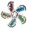
タイトルCryo-EM structure of KCTD7 in complex with Cullin3
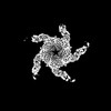
マップデータcryoEM map of KCTD7/CUL3
試料
  • 複合体: complex of KCTD7 and Cullin-3
    • 複合体: KCTD7
      • タンパク質・ペプチド: BTB/POZ domain-containing protein KCTD7
    • 複合体: Cullin-3
      • タンパク質・ペプチド: Cullin-3
キーワードCUL3 / ubiquitination / E3 ligase / SIGNALING PROTEIN
機能・相同性
機能・相同性情報


positive regulation of mitotic cell cycle phase transition / trophectodermal cellular morphogenesis / liver morphogenesis / POZ domain binding / nuclear protein quality control by the ubiquitin-proteasome system / regulation protein catabolic process at postsynapse / positive regulation of transporter activity / polar microtubule / anaphase-promoting complex-dependent catabolic process / COPII vesicle coating ...positive regulation of mitotic cell cycle phase transition / trophectodermal cellular morphogenesis / liver morphogenesis / POZ domain binding / nuclear protein quality control by the ubiquitin-proteasome system / regulation protein catabolic process at postsynapse / positive regulation of transporter activity / polar microtubule / anaphase-promoting complex-dependent catabolic process / COPII vesicle coating / intracellular glutamate homeostasis / stem cell division / Neddylation / RHOBTB3 ATPase cycle / Antigen processing: Ubiquitination & Proteasome degradation / embryonic cleavage / intracellular potassium ion homeostasis / cell projection organization / positive regulation of mitotic metaphase/anaphase transition / membrane hyperpolarization / fibroblast apoptotic process / Notch binding / RHOBTB1 GTPase cycle / negative regulation of Rho protein signal transduction / mitotic metaphase chromosome alignment / negative regulation of type I interferon production / ubiquitin ligase complex scaffold activity / Cul3-RING ubiquitin ligase complex / stress fiber assembly / positive regulation of cytokinesis / protein monoubiquitination / sperm flagellum / endoplasmic reticulum to Golgi vesicle-mediated transport / RHOBTB2 GTPase cycle / protein autoubiquitination / protein K48-linked ubiquitination / gastrulation / positive regulation of TORC1 signaling / intrinsic apoptotic signaling pathway / regulation of cellular response to insulin stimulus / cyclin binding / positive regulation of protein ubiquitination / integrin-mediated signaling pathway / Degradation of DVL / cellular response to amino acid stimulus / protein destabilization / Hedgehog 'on' state / protein homooligomerization / mitotic spindle / Wnt signaling pathway / spindle pole / protein polyubiquitination / Regulation of RAS by GAPs / G1/S transition of mitotic cell cycle / ubiquitin protein ligase activity / KEAP1-NFE2L2 pathway / Antigen processing: Ubiquitination & Proteasome degradation / cell migration / Neddylation / gene expression / ubiquitin-dependent protein catabolic process / proteasome-mediated ubiquitin-dependent protein catabolic process / postsynapse / Potential therapeutics for SARS / protein ubiquitination / inflammatory response / centrosome / glutamatergic synapse / ubiquitin protein ligase binding / positive regulation of cell population proliferation / Golgi apparatus / negative regulation of transcription by RNA polymerase II / extracellular exosome / nucleoplasm / identical protein binding / membrane / nucleus / plasma membrane / cytosol / cytoplasm
類似検索 - 分子機能
Potassium channel tetramerisation-type BTB domain / BTB/POZ domain / Cullin protein neddylation domain / Cullin, conserved site / Cullin family signature. / Cullin repeat-like-containing domain superfamily / Cullin protein, neddylation domain / Cullin / Cullin protein neddylation domain / Cullin ...Potassium channel tetramerisation-type BTB domain / BTB/POZ domain / Cullin protein neddylation domain / Cullin, conserved site / Cullin family signature. / Cullin repeat-like-containing domain superfamily / Cullin protein, neddylation domain / Cullin / Cullin protein neddylation domain / Cullin / Cullin, N-terminal / Cullin homology domain / Cullin homology domain superfamily / Cullin family / Cullin family profile. / Broad-Complex, Tramtrack and Bric a brac / BTB/POZ domain / SKP1/BTB/POZ domain superfamily / Winged helix DNA-binding domain superfamily / Winged helix-like DNA-binding domain superfamily
類似検索 - ドメイン・相同性
Cullin-3 / BTB/POZ domain-containing protein KCTD7
類似検索 - 構成要素
生物種Mus musculus (ハツカネズミ) / Homo sapiens (ヒト)
手法単粒子再構成法 / クライオ電子顕微鏡法 / 解像度: 2.8 Å
データ登録者Jiang W / Wang W / Zheng S
資金援助 中国, 1件
OrganizationGrant number
Ministry of Science and Technology (MoST, China) 中国
引用ジャーナル: Sci Adv / : 2023
タイトル: Structural basis for the ubiquitination of G protein βγ subunits by KCTD5/Cullin3 E3 ligase.
著者: Wentong Jiang / Wei Wang / Yinfei Kong / Sanduo Zheng /
要旨: G protein-coupled receptor (GPCR) signaling is precisely controlled to avoid overstimulation that results in detrimental consequences. Gβγ signaling is negatively regulated by a Cullin3 (Cul3)- ...G protein-coupled receptor (GPCR) signaling is precisely controlled to avoid overstimulation that results in detrimental consequences. Gβγ signaling is negatively regulated by a Cullin3 (Cul3)-dependent E3 ligase, KCTD5, which triggers ubiquitination and degradation of free Gβγ. Here, we report the cryo-electron microscopy structures of the KCTD5-Gβγ fusion complex and the KCTD7-Cul3 complex. KCTD5 in pentameric form engages symmetrically with five copies of Gβγ through its C-terminal domain. The unique pentameric assembly of the KCTD5/Cul3 E3 ligase places the ubiquitin-conjugating enzyme (E2) and the modification sites of Gβγ in close proximity and allows simultaneous transfer of ubiquitin from E2 to five Gβγ subunits. Moreover, we show that ubiquitination of Gβγ by KCTD5 is important for fine-tuning cyclic adenosine 3´,5´-monophosphate signaling of GPCRs. Our studies provide unprecedented insights into mechanisms of substrate recognition by unusual pentameric E3 ligases and highlight the KCTD family as emerging regulators of GPCR signaling.
履歴
登録2023年1月31日-
ヘッダ(付随情報) 公開2023年7月26日-
マップ公開2023年7月26日-
更新2024年7月3日-
現状2024年7月3日処理サイト: PDBj / 状態: 公開

-
構造の表示

添付画像

ダウンロードとリンク

-
マップ

ファイルダウンロード / ファイル: emd_35212.map.gz / 形式: CCP4 / 大きさ: 103 MB / タイプ: IMAGE STORED AS FLOATING POINT NUMBER (4 BYTES)
注釈cryoEM map of KCTD7/CUL3
投影像・断面図

画像のコントロール

大きさ
明度
コントラスト
その他
Z (Sec.)Y (Row.)X (Col.)
1.09 Å/pix.
x 300 pix.
= 326.1 Å
1.09 Å/pix.
x 300 pix.
= 326.1 Å
1.09 Å/pix.
x 300 pix.
= 326.1 Å

表面

投影像

断面 (1/3)

断面 (1/2)

断面 (2/3)

画像は Spider により作成

ボクセルのサイズX=Y=Z: 1.087 Å
密度
表面レベル登録者による: 0.275
最小 - 最大-0.4003876 - 1.247889
平均 (標準偏差)-0.0009664841 (±0.036459763)
対称性空間群: 1
詳細

EMDB XML:

マップ形状
Axis orderXYZ
Origin000
サイズ300300300
Spacing300300300
セルA=B=C: 326.1 Å
α=β=γ: 90.0 °

-
添付データ

-
追加マップ: cryoEM map of KCTD7/CUL3 after map sharpening

ファイルemd_35212_additional_1.map
注釈cryoEM map of KCTD7/CUL3 after map sharpening
投影像・断面図
ZYX

投影像

断面 (1/2)
密度ヒストグラム

-
ハーフマップ: half map of KCTD7/CUL3

ファイルemd_35212_half_map_1.map
注釈half map of KCTD7/CUL3
投影像・断面図
ZYX

投影像

断面 (1/2)
密度ヒストグラム

-
ハーフマップ: half map of KCTD7/CUL3

ファイルemd_35212_half_map_2.map
注釈half map of KCTD7/CUL3
投影像・断面図
ZYX

投影像

断面 (1/2)
密度ヒストグラム

-
試料の構成要素

-
全体 : complex of KCTD7 and Cullin-3

全体名称: complex of KCTD7 and Cullin-3
要素
  • 複合体: complex of KCTD7 and Cullin-3
    • 複合体: KCTD7
      • タンパク質・ペプチド: BTB/POZ domain-containing protein KCTD7
    • 複合体: Cullin-3
      • タンパク質・ペプチド: Cullin-3

-
超分子 #1: complex of KCTD7 and Cullin-3

超分子名称: complex of KCTD7 and Cullin-3 / タイプ: complex / ID: 1 / 親要素: 0 / 含まれる分子: all
由来(天然)生物種: Mus musculus (ハツカネズミ)

-
超分子 #2: KCTD7

超分子名称: KCTD7 / タイプ: complex / ID: 2 / 親要素: 1 / 含まれる分子: #1
由来(天然)生物種: Homo sapiens (ヒト)

-
超分子 #3: Cullin-3

超分子名称: Cullin-3 / タイプ: complex / ID: 3 / 親要素: 1 / 含まれる分子: #2

-
分子 #1: BTB/POZ domain-containing protein KCTD7

分子名称: BTB/POZ domain-containing protein KCTD7 / タイプ: protein_or_peptide / ID: 1 / コピー数: 5 / 光学異性体: LEVO
由来(天然)生物種: Mus musculus (ハツカネズミ)
分子量理論値: 34.146633 KDa
組換発現生物種: Spodoptera frugiperda (ツマジロクサヨトウ)
配列文字列: DYKDDDDKMV VVTGREPDSR HSDGAMSSSE AEDDFLEPAT PTATQAGHGL PLLPQEFPEV VPLNIGGAHF TTRLSTLRRY EDTMLAAMF SGRHYIPTDS EGRYFIDRDG THFGDVLNFL RSGDLPPREH VRAVYKEAQY YAIGPLLEQL ENMQPLKGEK V RQAFLGLM ...文字列:
DYKDDDDKMV VVTGREPDSR HSDGAMSSSE AEDDFLEPAT PTATQAGHGL PLLPQEFPEV VPLNIGGAHF TTRLSTLRRY EDTMLAAMF SGRHYIPTDS EGRYFIDRDG THFGDVLNFL RSGDLPPREH VRAVYKEAQY YAIGPLLEQL ENMQPLKGEK V RQAFLGLM PYYKDHLERI VEIARLRAVQ RKARFAKLKV CVFKEEMPIT PYECPLLNSL RFERSESDGQ LFEHHCEVDV SF GPWEAVA DVYDLLHCLV TDLSAQGLTV DHQCIGVCDK HLVNHYYCKR PIYEFKITWW

UniProtKB: BTB/POZ domain-containing protein KCTD7

-
分子 #2: Cullin-3

分子名称: Cullin-3 / タイプ: protein_or_peptide / ID: 2 / コピー数: 5 / 光学異性体: LEVO
由来(天然)生物種: Homo sapiens (ヒト)
分子量理論値: 44.146426 KDa
組換発現生物種: Escherichia coli (大腸菌)
配列文字列: MHHHHHHGSP MTMDEKYVNS IWDLLKNAIQ EIQRKNNSGL SFEELYRNAY TMVLHKHGEK LYTGLREVVT EHLINKVRED VLNSLNNNF LQTLNQAWND HQTAMVMIRD ILMYMDRVYV QQNNVENVYN LGLIIFRDQV VRYGCIRDHL RQTLLDMIAR E RKGEVVDR ...文字列:
MHHHHHHGSP MTMDEKYVNS IWDLLKNAIQ EIQRKNNSGL SFEELYRNAY TMVLHKHGEK LYTGLREVVT EHLINKVRED VLNSLNNNF LQTLNQAWND HQTAMVMIRD ILMYMDRVYV QQNNVENVYN LGLIIFRDQV VRYGCIRDHL RQTLLDMIAR E RKGEVVDR GAIRNACQML MILGLEGRSV YEEDFEAPFL EMSAEFFQME SQKFLAENSA SVYIKKVEAR INEEIERVMH CL DKSTEEP IVKVVERELI SKHMKTIVEM ENSGLVHMLK NGKTEDLGCM YKLFSRVPNG LKTMCECMSS YLREQGKALV SEE GEGKNP VDYRQGLDDL KSRFDRFLLE SFNNDRLFKQ TIAGDFEYFL NLNSRSPEYL

UniProtKB: Cullin-3

-
実験情報

-
構造解析

手法クライオ電子顕微鏡法
解析単粒子再構成法
試料の集合状態particle

-
試料調製

濃度0.32 mg/mL
緩衝液pH: 7.4 / 構成要素:
濃度名称
25.0 mMHEPEs
150.0 mMNaCl
グリッドモデル: Quantifoil R1.2/1.3 / 材質: GOLD / メッシュ: 400 / 支持フィルム - 材質: GRAPHENE OXIDE / 支持フィルム - トポロジー: HOLEY
凍結凍結剤: ETHANE / チャンバー内湿度: 100 % / チャンバー内温度: 281.15 K / 装置: FEI VITROBOT MARK IV

-
電子顕微鏡法

顕微鏡FEI TITAN KRIOS
撮影フィルム・検出器のモデル: GATAN K3 BIOQUANTUM (6k x 4k)
平均露光時間: 2.56 sec. / 平均電子線量: 50.0 e/Å2
電子線加速電圧: 300 kV / 電子線源: FIELD EMISSION GUN
電子光学系照射モード: FLOOD BEAM / 撮影モード: BRIGHT FIELD / 最大 デフォーカス(公称値): 2.4 µm / 最小 デフォーカス(公称値): 0.5 µm
実験機器
モデル: Titan Krios / 画像提供: FEI Company

+
画像解析

粒子像選択選択した数: 398489
初期モデルモデルのタイプ: INSILICO MODEL
最終 再構成使用したクラス数: 1 / アルゴリズム: FOURIER SPACE / 解像度のタイプ: BY AUTHOR / 解像度: 2.8 Å / 解像度の算出法: FSC 0.143 CUT-OFF / ソフトウェア - 名称: cryoSPARC / 使用した粒子像数: 115147
初期 角度割当タイプ: RANDOM ASSIGNMENT / ソフトウェア - 名称: cryoSPARC
最終 角度割当タイプ: MAXIMUM LIKELIHOOD / ソフトウェア - 名称: cryoSPARC
最終 3次元分類クラス数: 3 / ソフトウェア - 名称: cryoSPARC

+
万見について

-
お知らせ

-
2022年2月9日: EMDBエントリの付随情報ファイルのフォーマットが新しくなりました

EMDBエントリの付随情報ファイルのフォーマットが新しくなりました

  • EMDBのヘッダファイルのバージョン3が、公式のフォーマットとなりました。
  • これまでは公式だったバージョン1.9は、アーカイブから削除されます。

関連情報:EMDBヘッダ

外部リンク:wwPDBはEMDBデータモデルのバージョン3へ移行します

-
2020年8月12日: 新型コロナ情報

新型コロナ情報

URL: https://pdbj.org/emnavi/covid19.php

新ページ: EM Navigatorに新型コロナウイルスの特設ページを開設しました。

関連情報:Covid-19情報 / 2020年3月5日: 新型コロナウイルスの構造データ

+
2020年3月5日: 新型コロナウイルスの構造データ

新型コロナウイルスの構造データ

関連情報:万見生物種 / 2020年8月12日: 新型コロナ情報

外部リンク:COVID-19特集ページ - PDBj / 今月の分子2020年2月:コロナウイルスプロテーアーゼ

+
2019年1月31日: EMDBのIDの桁数の変更

EMDBのIDの桁数の変更

  • EMDBエントリに付与されているアクセスコード(EMDB-ID)は4桁の数字(例、EMD-1234)でしたが、間もなく枯渇します。これまでの4桁のID番号は4桁のまま変更されませんが、4桁の数字を使い切った後に発行されるIDは5桁以上の数字(例、EMD-12345)になります。5桁のIDは2019年の春頃から発行される見通しです。
  • EM Navigator/万見では、接頭語「EMD-」は省略されています。

関連情報:Q: 「EMD」とは何ですか? / 万見/EM NavigatorにおけるID/アクセスコードの表記

外部リンク:EMDB Accession Codes are Changing Soon! / PDBjへお問い合わせ

+
2017年7月12日: PDB大規模アップデート

PDB大規模アップデート

  • 新バージョンのPDBx/mmCIF辞書形式に基づくデータがリリースされました。
  • 今回の更新はバージョン番号が4から5になる大規模なもので、全エントリデータの書き換えが行われる「Remediation」というアップデートに該当します。
  • このバージョンアップで、電子顕微鏡の実験手法に関する多くの項目の書式が改定されました(例:em_softwareなど)。
  • EM NavigatorとYorodumiでも、この改定に基づいた表示内容になります。

外部リンク:wwPDB Remediation / OneDepデータ基準に準拠した、より強化された内容のモデル構造ファイルが、PDBアーカイブで公開されました。

-
万見 (Yorodumi)

幾万の構造データを、幾万の視点から

  • 万見(Yorodumi)は、EMDB/PDB/SASBDBなどの構造データを閲覧するためのページです。
  • EM Navigatorの詳細ページの後継、Omokage検索のフロントエンドも兼ねています。

関連情報:EMDB / PDB / SASBDB / 3つのデータバンクの比較 / 万見検索 / 2016年8月31日: 新しいEM Navigatorと万見 / 万見文献 / Jmol/JSmol / 機能・相同性情報 / 新しいEM Navigatorと万見の変更点

他の情報も見る