National Natural Science Foundation of China (NSFC)
31900046
China
Citation
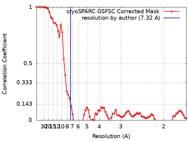
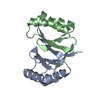
Journal: Proc Natl Acad Sci U S A / Year: 2024 Title: Molecular basis for curvature formation in SepF polymerization. Authors: Wenjing Liu / Chang Zhang / Huawei Zhang / Shaojie Ma / Jing Deng / Daping Wang / Ziwei Chang / Jun Yang / Abstract: The self-assembly of proteins into curved structures plays an important role in many cellular processes. One good example of this phenomenon is observed in the septum-forming protein (SepF), which ...The self-assembly of proteins into curved structures plays an important role in many cellular processes. One good example of this phenomenon is observed in the septum-forming protein (SepF), which forms polymerized structures with uniform curvatures. SepF is essential for regulating the thickness of the septum during bacteria cell division. In , SepF polymerization involves two distinct interfaces, the β-β and α-α interfaces, which define the assembly unit and contact interfaces, respectively. However, the mechanism of curvature formation in this step is not yet fully understood. In this study, we employed solid-state NMR (SSNMR) to compare the structures of cyclic wild-type SepF assemblies with linear assemblies resulting from a mutation of G137 on the β-β interface. Our results demonstrate that while the sequence differences arise from the internal assembly unit, the dramatic changes in the shape of the assemblies depend on the α-α interface between the units. We further provide atomic-level insights into how the angular variation of the α2 helix on the α-α interface affects the curvature of the assemblies, using a combination of SSNMR, cryo-electron microscopy, and simulation methods. Our findings shed light on the shape control of protein assemblies and emphasize the importance of interhelical contacts in retaining curvature.
In the structure databanks used in Yorodumi, some data are registered as the other names, "COVID-19 virus" and "2019-nCoV". Here are the details of the virus and the list of structure data.
Jan 31, 2019. EMDB accession codes are about to change! (news from PDBe EMDB page)
EMDB accession codes are about to change! (news from PDBe EMDB page)
The allocation of 4 digits for EMDB accession codes will soon come to an end. Whilst these codes will remain in use, new EMDB accession codes will include an additional digit and will expand incrementally as the available range of codes is exhausted. The current 4-digit format prefixed with “EMD-” (i.e. EMD-XXXX) will advance to a 5-digit format (i.e. EMD-XXXXX), and so on. It is currently estimated that the 4-digit codes will be depleted around Spring 2019, at which point the 5-digit format will come into force.
The EM Navigator/Yorodumi systems omit the EMD- prefix.
Related info.:Q: What is EMD? / ID/Accession-code notation in Yorodumi/EM Navigator
Yorodumi is a browser for structure data from EMDB, PDB, SASBDB, etc.
This page is also the successor to EM Navigator detail page, and also detail information page/front-end page for Omokage search.
The word "yorodu" (or yorozu) is an old Japanese word meaning "ten thousand". "mi" (miru) is to see.
Related info.:EMDB / PDB / SASBDB / Comparison of 3 databanks / Yorodumi Search / Aug 31, 2016. New EM Navigator & Yorodumi / Yorodumi Papers / Jmol/JSmol / Function and homology information / Changes in new EM Navigator and Yorodumi
 Movie
Movie Controller


 Open data
 Basic information
 Map data
 Sample
 Keywords
 Authors
 China, 1 items
  Citation
 Structure visualization
 Downloads & links
 EMDB map data format
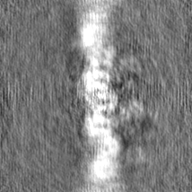
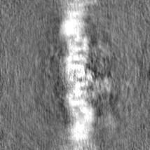
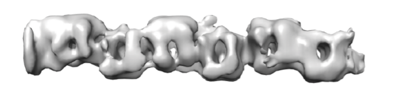 emd_35112.png
 http://ftp.pdbj.org/pub/emdb/structures/EMD-35112
 Z (Sec.)
 Y (Row.)
 X (Col.)


 Sample components
 Processing
 Electron microscopy
 FIELD EMISSION GUN