ジャーナル: Adv Sci (Weinh) / 年: 2023 タイトル: Cryo-Electron Tomography of Toxoplasma gondii Indicates That the Conoid Fiber May Be Derived from Microtubules. 著者: Zhixun Li / Wenjing Du / Jiong Yang / De-Hua Lai / Zhao-Rong Lun / Qiang Guo / 要旨: Toxoplasma gondii (T. gondii) is the causative agent of toxoplasmosis and can infect numerous warm-blooded animals. An improved understanding of the fine structure of this parasite can help elucidate ...Toxoplasma gondii (T. gondii) is the causative agent of toxoplasmosis and can infect numerous warm-blooded animals. An improved understanding of the fine structure of this parasite can help elucidate its replication mechanism. Previous studies have resolved the ultrastructure of the cytoskeleton using purified samples, which eliminates their cellular context. Here the application of cryo-electron tomography to visualize T. gondii tachyzoites in their native state is reported. The fine structure and cellular distribution of the cytoskeleton are resolved and analyzed at nanometer resolution. Additionally, the tachyzoite structural characteristics are annotated during its endodyogeny for the first time. By comparing the structural features in mature tachyzoites and their daughter buds, it is proposed that the conoid fiber of the Apicomplexa originates from microtubules. This work represents the detailed molecular anatomy of T. gondii, particularly during the budding replication stage of tachyzoite, and provides a reference for further studies of this fascinating organism.
 ムービー
ムービー コントローラー


 データを開く
 基本情報
基本情報
 マップデータ
 試料
試料 キーワード
キーワード
 データ登録者
 引用
引用
 構造の表示
構造の表示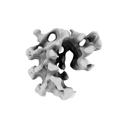
 ダウンロードとリンク
 EMDBマップデータ形式
 emd_35096.png
 http://ftp.pdbj.org/pub/emdb/structures/EMD-35096

 Z
Z Y
Y X
X


 試料の構成要素
 解析
解析 電子顕微鏡法
 FIELD EMISSION GUN
