 Movie
Movie Controller

[English] 日本語
 Yorodumi
- EMDB-35096: The in situ structure of conoid fiber from Toxoplasma gondii tach... -
+ Open data

- Basic information
| Entry |  | |||||||||
|---|---|---|---|---|---|---|---|---|---|---|
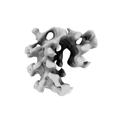
| Title | The in situ structure of conoid fiber from Toxoplasma gondii tachyzoite | |||||||||
 Map data Map data | ||||||||||
 Sample Sample |
| |||||||||
 Keywords Keywords | Toxoplasma gondii / conoid / CELL INVASION | |||||||||
| Biological species |  | |||||||||
| Method | subtomogram averaging / cryo EM / Resolution: 28.0 Å | |||||||||
 Authors Authors | Li Z / Guo Q | |||||||||
| Funding support | 1 items
| |||||||||
 Citation Citation | Journal: Adv Sci (Weinh) / Year: 2023 Title: Cryo-Electron Tomography of Toxoplasma gondii Indicates That the Conoid Fiber May Be Derived from Microtubules. Authors: Zhixun Li / Wenjing Du / Jiong Yang / De-Hua Lai / Zhao-Rong Lun / Qiang Guo /  Abstract: Toxoplasma gondii (T. gondii) is the causative agent of toxoplasmosis and can infect numerous warm-blooded animals. An improved understanding of the fine structure of this parasite can help elucidate ...Toxoplasma gondii (T. gondii) is the causative agent of toxoplasmosis and can infect numerous warm-blooded animals. An improved understanding of the fine structure of this parasite can help elucidate its replication mechanism. Previous studies have resolved the ultrastructure of the cytoskeleton using purified samples, which eliminates their cellular context. Here the application of cryo-electron tomography to visualize T. gondii tachyzoites in their native state is reported. The fine structure and cellular distribution of the cytoskeleton are resolved and analyzed at nanometer resolution. Additionally, the tachyzoite structural characteristics are annotated during its endodyogeny for the first time. By comparing the structural features in mature tachyzoites and their daughter buds, it is proposed that the conoid fiber of the Apicomplexa originates from microtubules. This work represents the detailed molecular anatomy of T. gondii, particularly during the budding replication stage of tachyzoite, and provides a reference for further studies of this fascinating organism. | |||||||||
| History |
|
- Structure visualization
| Supplemental images |
|---|
- Downloads & links
-EMDB archive
| Map data | emd_35096.map.gz | 27.2 MB |  EMDB map data format EMDB map data format | |
|---|---|---|---|---|
| Header (meta data) | emd-35096-v30.xmlemd-35096.xml | 11.4 KB 11.4 KB | Display Display | EMDB header |
| Images |  emd_35096.png emd_35096.png | 43.4 KB | ||
| Others | emd_35096_half_map_1.map.gzemd_35096_half_map_2.map.gz | 24.8 MB 24.9 MB | ||
| Archive directory |  http://ftp.pdbj.org/pub/emdb/structures/EMD-35096ftp://ftp.pdbj.org/pub/emdb/structures/EMD-35096 http://ftp.pdbj.org/pub/emdb/structures/EMD-35096ftp://ftp.pdbj.org/pub/emdb/structures/EMD-35096 | HTTPS FTP |
-Validation report
| Summary document | emd_35096_validation.pdf.gz | 977.4 KB | Display | EMDB validaton report |
|---|---|---|---|---|
| Full document | emd_35096_full_validation.pdf.gz | 977 KB | Display | |
| Data in XML | emd_35096_validation.xml.gz | 11.1 KB | Display | |
| Data in CIF | emd_35096_validation.cif.gz | 13 KB | Display | |
| Arichive directory | https://ftp.pdbj.org/pub/emdb/validation_reports/EMD-35096ftp://ftp.pdbj.org/pub/emdb/validation_reports/EMD-35096 | HTTPS FTP |
-Related structure data


-Links
| EMDB pages | EMDB (EBI/PDBe) / EMDataResource |
|---|
-Map
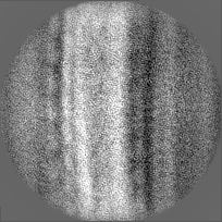
| File | Download / File: emd_35096.map.gz / Format: CCP4 / Size: 32.4 MB / Type: IMAGE STORED AS FLOATING POINT NUMBER (4 BYTES) | ||||||||||||||||||||||||||||||||||||
|---|---|---|---|---|---|---|---|---|---|---|---|---|---|---|---|---|---|---|---|---|---|---|---|---|---|---|---|---|---|---|---|---|---|---|---|---|---|
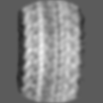
| Projections & slices | Image control
Images are generated by Spider. | ||||||||||||||||||||||||||||||||||||
| Voxel size | X=Y=Z: 3.328 Å | ||||||||||||||||||||||||||||||||||||
| Density |
| ||||||||||||||||||||||||||||||||||||
| Symmetry | Space group: 1 | ||||||||||||||||||||||||||||||||||||
| Details | EMDB XML:
|
 Z (Sec.)
 Y (Row.)
 X (Col.)


-Supplemental data
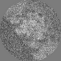
-Half map: #1
| File | emd_35096_half_map_1.map | ||||||||||||
|---|---|---|---|---|---|---|---|---|---|---|---|---|---|
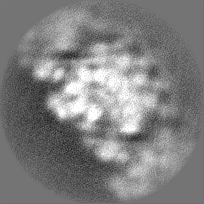
| Projections & Slices |
| ||||||||||||
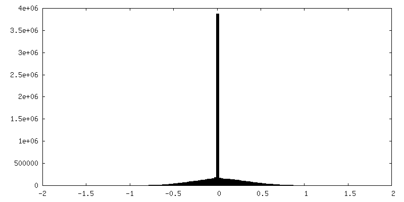
| Density Histograms |


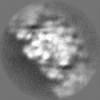
-Half map: #2
| File | emd_35096_half_map_2.map | ||||||||||||
|---|---|---|---|---|---|---|---|---|---|---|---|---|---|
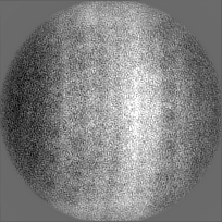
| Projections & Slices |
| ||||||||||||
| Density Histograms |


- Sample components
-Entire : Conoid fiber of mature Toxoplasma gondii tachyzoite
| Entire | Name: Conoid fiber of mature Toxoplasma gondii tachyzoite |
|---|---|
| Components |
|
-Supramolecule #1: Conoid fiber of mature Toxoplasma gondii tachyzoite
| Supramolecule | Name: Conoid fiber of mature Toxoplasma gondii tachyzoite / type: complex / ID: 1 / Parent: 0 |
|---|---|
| Source (natural) | Organism: |
-Experimental details
-Structure determination
| Method | cryo EM |
|---|---|
 Processing Processing | subtomogram averaging |
| Aggregation state | cell |
-Sample preparation
| Buffer | pH: 7 |
|---|---|
| Vitrification | Cryogen name: ETHANE |
- Electron microscopy
| Microscope | FEI TITAN KRIOS |
|---|---|
| Image recording | Film or detector model: GATAN K3 (6k x 4k) / Average electron dose: 3.0 e/Å2 |
| Electron beam | Acceleration voltage: 300 kV / Electron source:  FIELD EMISSION GUN FIELD EMISSION GUN |
| Electron optics | Illumination mode: FLOOD BEAM / Imaging mode: BRIGHT FIELD / Nominal defocus max: 5.0 µm / Nominal defocus min: 3.0 µm |
| Experimental equipment |  Model: Titan Krios / Image courtesy: FEI Company |
-Image processing
| Final reconstruction | Applied symmetry - Point group: C1 (asymmetric) / Resolution.type: BY AUTHOR / Resolution: 28.0 Å / Resolution method: FSC 0.143 CUT-OFF / Number subtomograms used: 1082 |
|---|---|
| Extraction | Number tomograms: 10 / Number images used: 1082 |
| Final angle assignment | Type: MAXIMUM LIKELIHOOD |
