 Movie
Movie Controller


+ Open data

- Basic information
| Entry |  | |||||||||
|---|---|---|---|---|---|---|---|---|---|---|
| Title | Clr4-H3K9 Nucleosome complex | |||||||||
 Map data Map data | ||||||||||
 Sample Sample |
| |||||||||
 Keywords Keywords | H3K9 nucleosome / methyltransferases / Clr4 / PROTEIN BINDING | |||||||||
| Biological species |  | |||||||||
| Method | single particle reconstruction / cryo EM / Resolution: 4.5 Å | |||||||||
 Authors Authors | Akoury E | |||||||||
| Funding support | 1 items
| |||||||||
 Citation Citation | Journal: Sci Rep / Year: 2024 Title: Structural insights into the binding mechanism of Clr4 methyltransferase to H3K9 methylated nucleosome. Authors: Christopher Saab / Joseph Stephan / Elias Akoury /  Abstract: The establishment and maintenance of heterochromatin, a specific chromatin structure essential for genomic stability and regulation, rely on intricate interactions between chromatin-modifying enzymes ...The establishment and maintenance of heterochromatin, a specific chromatin structure essential for genomic stability and regulation, rely on intricate interactions between chromatin-modifying enzymes and nucleosomal histone proteins. However, the precise trigger for these modifications remains unclear, thus highlighting the need for a deeper understanding of how methyltransferases facilitate histone methylation among others. Here, we investigate the molecular mechanisms underlying heterochromatin assembly by studying the interaction between the H3K9 methyltransferase Clr4 and H3K9-methylated nucleosomes. Using a combination of liquid-state nuclear magnetic resonance spectroscopy and cryo-electron microscopy, we elucidate the structural basis of Clr4 binding to H3K9-methylated nucleosomes. Our results reveal that Clr4 engages with nucleosomes through its chromodomain and disordered regions to promote de novo methylation. This study provides crucial insights into the molecular mechanisms governing heterochromatin formation by highlighting the significance of chromatin-modifying enzymes in genome regulation and disease pathology. | |||||||||
| History |
|
- Structure visualization
| Supplemental images |
|---|

- Downloads & links
-EMDB archive
| Map data | emd_35060.map.gz | 1.8 MB |  EMDB map data format EMDB map data format | |
|---|---|---|---|---|
| Header (meta data) | emd-35060-v30.xmlemd-35060.xml | 11.2 KB 11.2 KB | Display Display | EMDB header |
| Images | 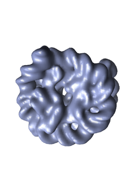 emd_35060.png emd_35060.png | 58.2 KB | ||
| Filedesc metadata | emd-35060.cif.gz | 3.7 KB | ||
| Others | emd_35060_half_map_1.map.gzemd_35060_half_map_2.map.gz | 1.8 MB 1.8 MB | ||
| Archive directory |  http://ftp.pdbj.org/pub/emdb/structures/EMD-35060ftp://ftp.pdbj.org/pub/emdb/structures/EMD-35060 http://ftp.pdbj.org/pub/emdb/structures/EMD-35060ftp://ftp.pdbj.org/pub/emdb/structures/EMD-35060 | HTTPS FTP |
-Validation report
| Summary document | emd_35060_validation.pdf.gz | 460.5 KB | Display | EMDB validaton report |
|---|---|---|---|---|
| Full document | emd_35060_full_validation.pdf.gz | 460 KB | Display | |
| Data in XML | emd_35060_validation.xml.gz | 5.5 KB | Display | |
| Data in CIF | emd_35060_validation.cif.gz | 7 KB | Display | |
| Arichive directory | https://ftp.pdbj.org/pub/emdb/validation_reports/EMD-35060ftp://ftp.pdbj.org/pub/emdb/validation_reports/EMD-35060 | HTTPS FTP |
-Links
| EMDB pages | EMDB (EBI/PDBe) / EMDataResource |
|---|
-Map
| File | Download / File: emd_35060.map.gz / Format: CCP4 / Size: 2 MB / Type: IMAGE STORED AS FLOATING POINT NUMBER (4 BYTES) | ||||||||||||||||||||||||||||||||||||
|---|---|---|---|---|---|---|---|---|---|---|---|---|---|---|---|---|---|---|---|---|---|---|---|---|---|---|---|---|---|---|---|---|---|---|---|---|---|
| Projections & slices | Image control
Images are generated by Spider. | ||||||||||||||||||||||||||||||||||||
| Voxel size | X=Y=Z: 1 Å | ||||||||||||||||||||||||||||||||||||
| Density |
| ||||||||||||||||||||||||||||||||||||
| Symmetry | Space group: 1 | ||||||||||||||||||||||||||||||||||||
| Details | EMDB XML:
|
 Z (Sec.)
 Y (Row.)
 X (Col.)


-Supplemental data
-Half map: #1
| File | emd_35060_half_map_1.map | ||||||||||||
|---|---|---|---|---|---|---|---|---|---|---|---|---|---|
| Projections & Slices |
| ||||||||||||
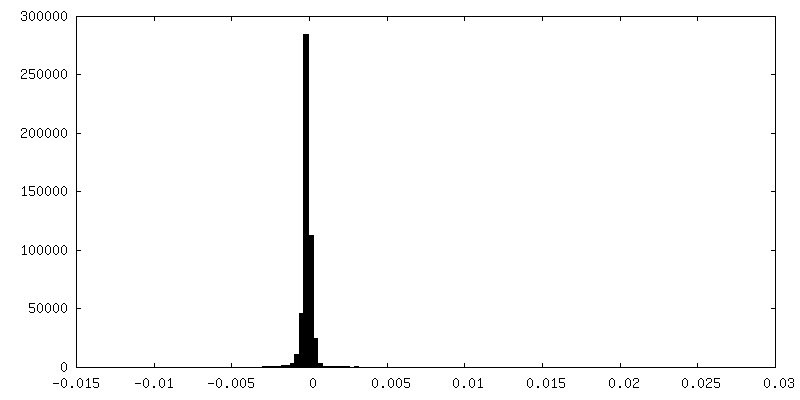
| Density Histograms |


-Half map: #2
| File | emd_35060_half_map_2.map | ||||||||||||
|---|---|---|---|---|---|---|---|---|---|---|---|---|---|
| Projections & Slices |
| ||||||||||||
| Density Histograms |


- Sample components
-Entire : Clr4-H3K9 Nucleosome complex
| Entire | Name: Clr4-H3K9 Nucleosome complex |
|---|---|
| Components |
|
-Supramolecule #1: Clr4-H3K9 Nucleosome complex
| Supramolecule | Name: Clr4-H3K9 Nucleosome complex / type: complex / ID: 1 / Parent: 0 |
|---|---|
| Source (natural) | Organism: |
| Molecular weight | Theoretical: 230 KDa |
-Experimental details
-Structure determination
| Method | cryo EM |
|---|---|
 Processing Processing | single particle reconstruction |
| Aggregation state | particle |
-Sample preparation
| Buffer | pH: 6.8 |
|---|---|
| Grid | Material: COPPER |
| Vitrification | Cryogen name: ETHANE / Instrument: FEI VITROBOT MARK IV |
- Electron microscopy
| Microscope | FEI TITAN KRIOS |
|---|---|
| Image recording | Film or detector model: TVIPS TEMCAM-F816 (8k x 8k) / Average electron dose: 15.0 e/Å2 |
| Electron beam | Acceleration voltage: 200 kV / Electron source:  FIELD EMISSION GUN FIELD EMISSION GUN |
| Electron optics | Illumination mode: SPOT SCAN / Imaging mode: BRIGHT FIELD / Nominal defocus max: 4.0 µm / Nominal defocus min: 1.0 µm |
| Experimental equipment |  Model: Titan Krios / Image courtesy: FEI Company |
-Image processing
| Startup model | Type of model: NONE |
|---|---|
| Final reconstruction | Algorithm: ALGEBRAIC (ARTS) / Resolution.type: BY AUTHOR / Resolution: 4.5 Å / Resolution method: FSC 0.5 CUT-OFF / Number images used: 1200000 |
| Initial angle assignment | Type: ANGULAR RECONSTITUTION |
| Final angle assignment | Type: COMMON LINE |
