 Movie
Movie Controller

[English] 日本語
 Yorodumi
- EMDB-34947: Structure of a GPCR-G protein in complex with a natural peptide a... -
+ Open data

- Basic information
| Entry |  | |||||||||
|---|---|---|---|---|---|---|---|---|---|---|
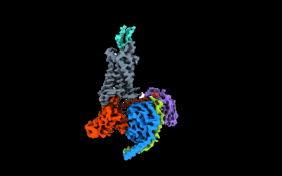
| Title | Structure of a GPCR-G protein in complex with a natural peptide agonist | |||||||||
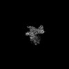
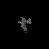
 Map data Map data | Full map corresponding to C5a-C5aR1-Go | |||||||||
 Sample Sample |
| |||||||||
 Keywords Keywords | GPCR / G protein / SIGNALING PROTEIN / SIGNALING PROTEIN-IMMUNE SYSTEM complex | |||||||||
| Function / homology |  Function and homology information Function and homology informationcomplement component C5a binding / cell proliferation in hindbrain / presynapse organization / complement component C5a signaling pathway / response to peptidoglycan / Terminal pathway of complement / Regulation of Complement cascade / membrane attack complex / Peptide ligand-binding receptors / complement component C5a receptor activity ...complement component C5a binding / cell proliferation in hindbrain / presynapse organization / complement component C5a signaling pathway / response to peptidoglycan / Terminal pathway of complement / Regulation of Complement cascade / membrane attack complex / Peptide ligand-binding receptors / complement component C5a receptor activity / G alpha (i) signalling events / Activation of C3 and C5 / complement activation, GZMK pathway / negative regulation of macrophage chemotaxis / other organism cell membrane / mu-type opioid receptor binding / : / corticotropin-releasing hormone receptor 1 binding / complement receptor mediated signaling pathway / complement activation, alternative pathway / positive regulation of neutrophil chemotaxis / chemokine activity / G protein-coupled dopamine receptor signaling pathway / endopeptidase inhibitor activity / positive regulation of macrophage chemotaxis / regulation of heart contraction / parallel fiber to Purkinje cell synapse / amyloid-beta clearance / complement activation, classical pathway / positive regulation of vascular endothelial growth factor production / negative regulation of insulin secretion / postsynaptic modulation of chemical synaptic transmission / positive regulation of chemokine production / neutrophil chemotaxis / Neutrophil degranulation / muscle contraction / positive regulation of epithelial cell proliferation / astrocyte activation / adenylate cyclase-inhibiting serotonin receptor signaling pathway / Regulation of Complement cascade / G protein-coupled serotonin receptor binding / Peptide ligand-binding receptors / locomotory behavior / microglial cell activation / mRNA transcription by RNA polymerase II / cognition / GABA-ergic synapse / G protein-coupled receptor activity / chemotaxis / G-protein beta/gamma-subunit complex binding / positive regulation of angiogenesis / apical part of cell / adenylate cyclase-modulating G protein-coupled receptor signaling pathway / adenylate cyclase-inhibiting G protein-coupled receptor signaling pathway / Olfactory Signaling Pathway / Activation of the phototransduction cascade / G protein-coupled acetylcholine receptor signaling pathway / G beta:gamma signalling through PLC beta / Presynaptic function of Kainate receptors / Thromboxane signalling through TP receptor / Activation of G protein gated Potassium channels / Inhibition of voltage gated Ca2+ channels via Gbeta/gamma subunits / G-protein activation / Glucagon signaling in metabolic regulation / G beta:gamma signalling through CDC42 / Prostacyclin signalling through prostacyclin receptor / Synthesis, secretion, and inactivation of Glucagon-like Peptide-1 (GLP-1) / G beta:gamma signalling through BTK / photoreceptor disc membrane / ADP signalling through P2Y purinoceptor 12 / Glucagon-type ligand receptors / Sensory perception of sweet, bitter, and umami (glutamate) taste / Adrenaline,noradrenaline inhibits insulin secretion / Vasopressin regulates renal water homeostasis via Aquaporins / Glucagon-like Peptide-1 (GLP1) regulates insulin secretion / G alpha (z) signalling events / cellular response to catecholamine stimulus / ADP signalling through P2Y purinoceptor 1 / G beta:gamma signalling through PI3Kgamma / ADORA2B mediated anti-inflammatory cytokines production / adenylate cyclase-activating dopamine receptor signaling pathway / Cooperation of PDCL (PhLP1) and TRiC/CCT in G-protein beta folding / GPER1 signaling / cellular response to prostaglandin E stimulus / heterotrimeric G-protein complex / Inactivation, recovery and regulation of the phototransduction cascade / G alpha (12/13) signalling events / G-protein beta-subunit binding / extracellular vesicle / positive regulation of cytosolic calcium ion concentration / sensory perception of taste / Thrombin signalling through proteinase activated receptors (PARs) / signaling receptor complex adaptor activity / retina development in camera-type eye / fibroblast proliferation / cell body / GTPase binding / presynaptic membrane / G protein activity / cytoplasmic vesicle Similarity search - Function | |||||||||
| Biological species |   Homo sapiens (human) Homo sapiens (human) | |||||||||
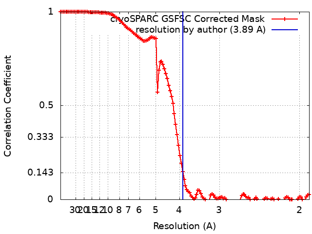
| Method | single particle reconstruction / cryo EM / Resolution: 3.89 Å | |||||||||
 Authors Authors | Saha S / Maharana J / Yadav MK / Sarma P / Chami M / Banerjee R / Shukla AK | |||||||||
| Funding support |  India, 1 items India, 1 items
| |||||||||
 Citation Citation | Journal: Cell / Year: 2023 Title: Molecular basis of anaphylatoxin binding, activation, and signaling bias at complement receptors. Authors: Manish K Yadav / Jagannath Maharana / Ravi Yadav / Shirsha Saha / Parishmita Sarma / Chahat Soni / Vinay Singh / Sayantan Saha / Manisankar Ganguly / Xaria X Li / Samanwita Mohapatra / Sudha ...Authors: Manish K Yadav / Jagannath Maharana / Ravi Yadav / Shirsha Saha / Parishmita Sarma / Chahat Soni / Vinay Singh / Sayantan Saha / Manisankar Ganguly / Xaria X Li / Samanwita Mohapatra / Sudha Mishra / Htet A Khant / Mohamed Chami / Trent M Woodruff / Ramanuj Banerjee / Arun K Shukla / Cornelius Gati /    Abstract: The complement system is a critical part of our innate immune response, and the terminal products of this cascade, anaphylatoxins C3a and C5a, exert their physiological and pathophysiological ...The complement system is a critical part of our innate immune response, and the terminal products of this cascade, anaphylatoxins C3a and C5a, exert their physiological and pathophysiological responses primarily via two GPCRs, C3aR and C5aR1. However, the molecular mechanism of ligand recognition, activation, and signaling bias of these receptors remains mostly elusive. Here, we present nine cryo-EM structures of C3aR and C5aR1 activated by their natural and synthetic agonists, which reveal distinct binding pocket topologies of complement anaphylatoxins and provide key insights into receptor activation and transducer coupling. We also uncover the structural basis of a naturally occurring mechanism to dampen the inflammatory response of C5a via proteolytic cleavage of the terminal arginine and the G-protein signaling bias elicited by a peptide agonist of C3aR identified here. In summary, our study elucidates the innerworkings of the complement anaphylatoxin receptors and should facilitate structure-guided drug discovery to target these receptors in a spectrum of disorders. #1: Journal: Cell(Cambridge,Mass.) / Year: 2023Title: Structure of a GPCR-G protein in complex with a synthetic peptide agonist Authors: Saha S / Maharana J / Yadav MK / Sarma P / Chami M / Banerjee R / Shukla AK | |||||||||
| History |
|
- Structure visualization
| Supplemental images |
|---|
- Downloads & links
-EMDB archive
| Map data | emd_34947.map.gz | 168 MB | EMDB map data format | |
|---|---|---|---|---|
| Header (meta data) | emd-34947-v30.xmlemd-34947.xml | 26.9 KB 26.9 KB | Display Display | EMDB header |
| FSC (resolution estimation) | emd_34947_fsc.xml | 12 KB | Display | FSC data file |
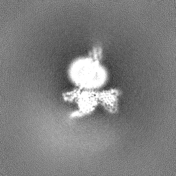
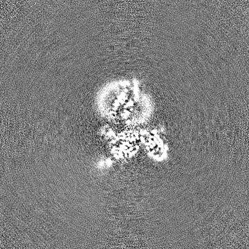
| Images |  emd_34947.png emd_34947.png | 29.4 KB | ||
| Filedesc metadata | emd-34947.cif.gz | 7.2 KB | ||
| Others | emd_34947_half_map_1.map.gzemd_34947_half_map_2.map.gz | 165 MB 165 MB | ||
| Archive directory |  http://ftp.pdbj.org/pub/emdb/structures/EMD-34947ftp://ftp.pdbj.org/pub/emdb/structures/EMD-34947 http://ftp.pdbj.org/pub/emdb/structures/EMD-34947ftp://ftp.pdbj.org/pub/emdb/structures/EMD-34947 | HTTPS FTP |
-Related structure data
| Related structure data |  8hqcMC  8hptC 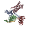 8i95C 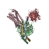 8i97C 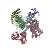 8i9aC 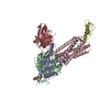 8i9lC 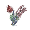 8i9sC  8ia2C 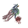 8j6dC 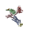 8jzzC M: atomic model generated by this map C: citing same article ( |
|---|---|
| Similar structure data |
-Links
| EMDB pages | EMDB (EBI/PDBe) / EMDataResource |
|---|---|
| Related items in Molecule of the Month |


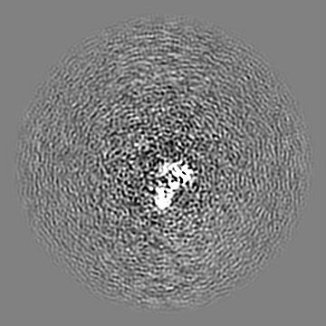
-Map
| File | Download / File: emd_34947.map.gz / Format: CCP4 / Size: 178 MB / Type: IMAGE STORED AS FLOATING POINT NUMBER (4 BYTES) | ||||||||||||||||||||||||||||||||||||
|---|---|---|---|---|---|---|---|---|---|---|---|---|---|---|---|---|---|---|---|---|---|---|---|---|---|---|---|---|---|---|---|---|---|---|---|---|---|
| Annotation | Full map corresponding to C5a-C5aR1-Go | ||||||||||||||||||||||||||||||||||||
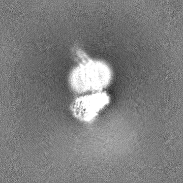
| Projections & slices | Image control
Images are generated by Spider. | ||||||||||||||||||||||||||||||||||||
| Voxel size | X=Y=Z: 0.94756 Å | ||||||||||||||||||||||||||||||||||||
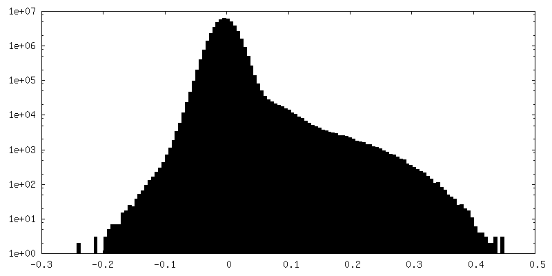
| Density |
| ||||||||||||||||||||||||||||||||||||
| Symmetry | Space group: 1 | ||||||||||||||||||||||||||||||||||||
| Details | EMDB XML:
|
 Z (Sec.)
 Y (Row.)
 X (Col.)


-Supplemental data
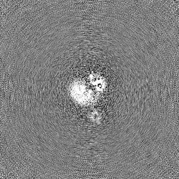
-Half map: Half map B corresponding to C5a-C5aR1-Go
| File | emd_34947_half_map_1.map | ||||||||||||
|---|---|---|---|---|---|---|---|---|---|---|---|---|---|
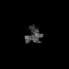
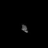
| Annotation | Half map B corresponding to C5a-C5aR1-Go | ||||||||||||
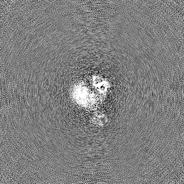
| Projections & Slices |
| ||||||||||||
| Density Histograms |


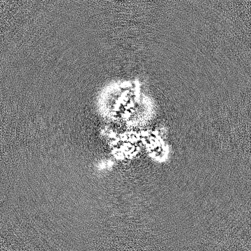

-Half map: Half map A corresponding to C5a-C5aR1-Go
| File | emd_34947_half_map_2.map | ||||||||||||
|---|---|---|---|---|---|---|---|---|---|---|---|---|---|
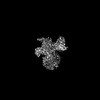
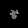
| Annotation | Half map A corresponding to C5a-C5aR1-Go | ||||||||||||
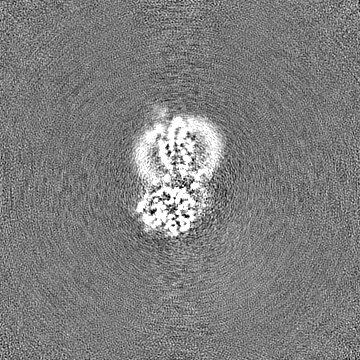
| Projections & Slices |
| ||||||||||||
| Density Histograms |


- Sample components
+Entire : GPCR-G protein in complex with a natural peptide agonist
+Supramolecule #1: GPCR-G protein in complex with a natural peptide agonist
+Supramolecule #2: C5a anaphylatoxin chemotactic receptor 1
+Supramolecule #3: Guanine nucleotide-binding protein G(o) subunit alpha
+Supramolecule #4: Guanine nucleotide-binding protein G(I)/G(S)/G(T) subunit beta-1
+Supramolecule #5: C5a anaphylatoxin
+Supramolecule #6: Guanine nucleotide-binding protein G(I)/G(S)/G(O) subunit gamma-2
+Supramolecule #7: Antibody fragment
+Macromolecule #1: C5a anaphylatoxin chemotactic receptor 1

 Spodoptera frugiperda (fall armyworm)
+Macromolecule #2: Guanine nucleotide-binding protein G(o) subunit alpha

+Macromolecule #3: Guanine nucleotide-binding protein G(I)/G(S)/G(T) subunit beta-1
+Macromolecule #4: C5a anaphylatoxin
+Macromolecule #5: Guanine nucleotide-binding protein G(I)/G(S)/G(O) subunit gamma-2
+Macromolecule #6: Antibody fragment
-Experimental details
-Structure determination
| Method | cryo EM |
|---|---|
 Processing Processing | single particle reconstruction |
| Aggregation state | particle |
-Sample preparation
| Buffer | pH: 7.4 |
|---|---|
| Vitrification | Cryogen name: ETHANE |
- Electron microscopy
| Microscope | FEI TITAN KRIOS |
|---|---|
| Image recording | Film or detector model: GATAN K2 SUMMIT (4k x 4k) / Detector mode: COUNTING / Average electron dose: 42.0 e/Å2 |
| Electron beam | Acceleration voltage: 300 kV / Electron source:  FIELD EMISSION GUN FIELD EMISSION GUN |
| Electron optics | Illumination mode: FLOOD BEAM / Imaging mode: BRIGHT FIELD / Nominal defocus max: 2.5 µm / Nominal defocus min: 0.5 µm |
| Experimental equipment |  Model: Titan Krios / Image courtesy: FEI Company |
+Image processing
-Atomic model buiding 1
| Refinement | Space: REAL / Protocol: FLEXIBLE FIT |
|---|---|
| Output model | PDB-8hqc: |
