 Movie
Movie Controller

[English] 日本語
 Yorodumi
- EMDB-34918: cellodextrin phosphorylase stable variant from Clostridium thermo... -
+ Open data

- Basic information
| Entry |  | |||||||||
|---|---|---|---|---|---|---|---|---|---|---|
| Title | cellodextrin phosphorylase stable variant from Clostridium thermocellum | |||||||||
 Map data Map data | ||||||||||
 Sample Sample |
| |||||||||
 Keywords Keywords | cellulose / phosphorolysis / synthesis / CARBOHYDRATE | |||||||||
| Function / homology |  Function and homology information Function and homology informationglycosyltransferase activity / carbohydrate binding / carbohydrate metabolic process Similarity search - Function | |||||||||
| Biological species |  Acetivibrio thermocellus (bacteria) Acetivibrio thermocellus (bacteria) | |||||||||
| Method | single particle reconstruction / cryo EM / Resolution: 2.28 Å | |||||||||
 Authors Authors | Kuga T / Sunagawa N / Igarashi K | |||||||||
| Funding support |  Japan, 1 items Japan, 1 items
| |||||||||
 Citation Citation | Journal: To Be Published Title: 11 cysteine-to-serine mutations improve the stability of cellodextrin phosphorylase Authors: Kuga T / Sunagawa N / Igarashi K | |||||||||
| History |
|
- Structure visualization
| Supplemental images |
|---|

- Downloads & links
-EMDB archive
| Map data | emd_34918.map.gz | 219.8 MB | EMDB map data format | |
|---|---|---|---|---|
| Header (meta data) | emd-34918-v30.xmlemd-34918.xml | 16.2 KB 16.2 KB | Display Display | EMDB header |
| FSC (resolution estimation) | emd_34918_fsc.xml | 15.8 KB | Display | FSC data file |
| Images | 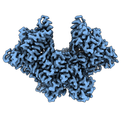 emd_34918.png emd_34918.png | 139.4 KB | ||
| Filedesc metadata | emd-34918.cif.gz | 6.1 KB | ||
| Others | emd_34918_half_map_1.map.gzemd_34918_half_map_2.map.gz | 391.3 MB 391.3 MB | ||
| Archive directory |  http://ftp.pdbj.org/pub/emdb/structures/EMD-34918ftp://ftp.pdbj.org/pub/emdb/structures/EMD-34918 http://ftp.pdbj.org/pub/emdb/structures/EMD-34918ftp://ftp.pdbj.org/pub/emdb/structures/EMD-34918 | HTTPS FTP |
-Validation report
| Summary document | emd_34918_validation.pdf.gz | 733.4 KB | Display | EMDB validaton report |
|---|---|---|---|---|
| Full document | emd_34918_full_validation.pdf.gz | 733 KB | Display | |
| Data in XML | emd_34918_validation.xml.gz | 24.5 KB | Display | |
| Data in CIF | emd_34918_validation.cif.gz | 31.6 KB | Display | |
| Arichive directory | https://ftp.pdbj.org/pub/emdb/validation_reports/EMD-34918ftp://ftp.pdbj.org/pub/emdb/validation_reports/EMD-34918 | HTTPS FTP |
-Related structure data
| Related structure data | 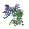 8hnuMC M: atomic model generated by this map C: citing same article ( |
|---|---|
| Similar structure data |
-Links
| EMDB pages | EMDB (EBI/PDBe) / EMDataResource |
|---|---|
| Related items in Molecule of the Month |


-Map
| File | Download / File: emd_34918.map.gz / Format: CCP4 / Size: 421.9 MB / Type: IMAGE STORED AS FLOATING POINT NUMBER (4 BYTES) | ||||||||||||||||||||||||||||||||||||
|---|---|---|---|---|---|---|---|---|---|---|---|---|---|---|---|---|---|---|---|---|---|---|---|---|---|---|---|---|---|---|---|---|---|---|---|---|---|
| Projections & slices | Image control
Images are generated by Spider. | ||||||||||||||||||||||||||||||||||||
| Voxel size | X=Y=Z: 0.83 Å | ||||||||||||||||||||||||||||||||||||
| Density |
| ||||||||||||||||||||||||||||||||||||
| Symmetry | Space group: 1 | ||||||||||||||||||||||||||||||||||||
| Details | EMDB XML:
|
 Z (Sec.)
 Y (Row.)
 X (Col.)


-Supplemental data
-Half map: #2
| File | emd_34918_half_map_1.map | ||||||||||||
|---|---|---|---|---|---|---|---|---|---|---|---|---|---|
| Projections & Slices |
| ||||||||||||
| Density Histograms |


-Half map: #1
| File | emd_34918_half_map_2.map | ||||||||||||
|---|---|---|---|---|---|---|---|---|---|---|---|---|---|
| Projections & Slices |
| ||||||||||||
| Density Histograms |


- Sample components
-Entire : cellodextrin phosphorylase dimer
| Entire | Name: cellodextrin phosphorylase dimer |
|---|---|
| Components |
|
-Supramolecule #1: cellodextrin phosphorylase dimer
| Supramolecule | Name: cellodextrin phosphorylase dimer / type: complex / ID: 1 / Parent: 0 / Macromolecule list: #1 |
|---|---|
| Source (natural) | Organism: Acetivibrio thermocellus (bacteria) / Strain: YM4 |
| Molecular weight | Theoretical: 240 KDa |
-Macromolecule #1: Cellodextrin phosphorylase
| Macromolecule | Name: Cellodextrin phosphorylase / type: protein_or_peptide / ID: 1 / Number of copies: 2 / Enantiomer: LEVO |
|---|---|
| Source (natural) | Organism: Acetivibrio thermocellus (bacteria) / Strain: YM4 |
| Molecular weight | Theoretical: 112.736172 KDa |
| Recombinant expression | Organism: |
| Sequence | String: MGITKVTARN NKITPVELLN QKFGNKINLG NFADAVFTDA AFKNVAGIAN LPMKAPVMQV LMENSIVSKY LKQFVPDRSV SFVEEGQKF YIVLEDGQKI EVPEDVNKAL KATVSDVKHW AGYLTEDGEH VIDLLKPAPG PHFYVNLLIG NRLGFKRTLQ T TPKSVVDR ...String: MGITKVTARN NKITPVELLN QKFGNKINLG NFADAVFTDA AFKNVAGIAN LPMKAPVMQV LMENSIVSKY LKQFVPDRSV SFVEEGQKF YIVLEDGQKI EVPEDVNKAL KATVSDVKHW AGYLTEDGEH VIDLLKPAPG PHFYVNLLIG NRLGFKRTLQ T TPKSVVDR FGRGSFRSHA ATQVLATRFD MRQEENGFPA NRQFYLYEDG KQIFYSALID DNIVEATSKH SSNRTVIKYK TA SNLEITR TIFLVPHKKG FPLATELQRI EIKNASDKAR NLSITYTGMF GTGAVHAIFE DVTYTNVIMQ SAALYNDKGE FIG ITPDYY PEEFKQDTRF VTMIVRNGDE KSFPQSFSTD YNDFVGTGTL EHPAGGSNLN NKLNRKGPGF FALGAPFTVE PGKT VIIDT FTGLSSSKDN ENYSDAVMLR ELDNLLRYFE KSESVEETLN EIINFHENYG KYFQFNTGNK LFDSGFNRNL AFQVL YQTF MSRSFGQTQK GYREIGFREI QDLFASMYYF INIGYQDFVK ELLFEWTANV YKMGYANHNF YWVGKQPGLY SDDSLW LLQ AYYRYIIYTK DTSVLNEEVP VADGNNEKRA VRETLKAIIQ YSASISVGDH GLPLLDLADW NDSLKIDSNS IDGATKE KL YYEQLKKTNG KYGDRFMSDY SESVMNAFLL KLAIDHLAEI ATLDNDTQLA QQMSELSKEV TDRIQKHAWK ENFFARVL I NRYKDGSYTY LGAKGDKLSA DPNIDGVYFL NSFAWSVLSD VATDEQIAIM VDVIKKHLLT PYGLRLVTPA DLNKIANDT ATGHYFFGDR ENGAVFKHAS MMAVAALIKA AKKVKDNELA KEMARIAYFM IDLVLPYKNL ENPFQVAGNP RISTQYINTD TGENIGPLL SGTATWLNLN LISLAGIEYT RDGISFNPIL REEETQLNFT LKAPKSSYKF SITKPVGFAR MESSEYELFV D GQKIDNTV IPMYTDEKEH IVTLKFKLEH HHHHH UniProtKB: Cellodextrin phosphorylase |
-Macromolecule #2: CHLORIDE ION
| Macromolecule | Name: CHLORIDE ION / type: ligand / ID: 2 / Number of copies: 4 / Formula: CL |
|---|---|
| Molecular weight | Theoretical: 35.453 Da |
-Macromolecule #3: water
| Macromolecule | Name: water / type: ligand / ID: 3 / Number of copies: 316 / Formula: HOH |
|---|---|
| Molecular weight | Theoretical: 18.015 Da |
| Chemical component information |  ChemComp-HOH: |
-Experimental details
-Structure determination
| Method | cryo EM |
|---|---|
 Processing Processing | single particle reconstruction |
| Aggregation state | particle |
-Sample preparation
| Concentration | 3.0 mg/mL |
|---|---|
| Buffer | pH: 7.5 |
| Grid | Model: Quantifoil R1.2/1.3 / Material: GOLD / Mesh: 300 / Support film - Material: CARBON / Pretreatment - Type: GLOW DISCHARGE |
| Vitrification | Cryogen name: ETHANE / Chamber humidity: 100 % / Chamber temperature: 279 K / Instrument: FEI VITROBOT MARK IV |
- Electron microscopy
| Microscope | FEI TITAN KRIOS |
|---|---|
| Image recording | Film or detector model: GATAN K3 (6k x 4k) / Average electron dose: 49.0 e/Å2 |
| Electron beam | Acceleration voltage: 300 kV / Electron source:  FIELD EMISSION GUN FIELD EMISSION GUN |
| Electron optics | Illumination mode: FLOOD BEAM / Imaging mode: BRIGHT FIELD / Nominal defocus max: 1.8 µm / Nominal defocus min: 1.0 µm |
| Experimental equipment |  Model: Titan Krios / Image courtesy: FEI Company |

