 Movie
Movie Controller

[English] 日本語
 Yorodumi
- EMDB-3476: Structure of FANCI-FANCD2 hetero-dimer co-expressed with FANCC-FA... -
+ Open data

- Basic information
| Entry | Database: EMDB / ID: EMD-3476 | |||||||||
|---|---|---|---|---|---|---|---|---|---|---|
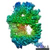
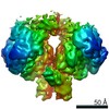
| Title | Structure of FANCI-FANCD2 hetero-dimer co-expressed with FANCC-FANCE-FANCF proteins. | |||||||||
 Map data Map data | 3D Reconstruction by negative stain EM of FANCI-FANCD2 co-expressed with FANCC-FANCE-FANCF. | |||||||||
 Sample Sample |
| |||||||||
| Method | single particle reconstruction / negative staining / Resolution: 18.3 Å | |||||||||
 Authors Authors | Swuec P / Costa A | |||||||||
 Citation Citation | Journal: Cell Rep / Year: 2017 Title: The FA Core Complex Contains a Homo-dimeric Catalytic Module for the Symmetric Mono-ubiquitination of FANCI-FANCD2. Authors: Paolo Swuec / Ludovic Renault / Aaron Borg / Fenil Shah / Vincent J Murphy / Sylvie van Twest / Ambrosius P Snijders / Andrew J Deans / Alessandro Costa /   Abstract: Activation of the main DNA interstrand crosslink repair pathway in higher eukaryotes requires mono-ubiquitination of FANCI and FANCD2 by FANCL, the E3 ligase subunit of the Fanconi anemia core ...Activation of the main DNA interstrand crosslink repair pathway in higher eukaryotes requires mono-ubiquitination of FANCI and FANCD2 by FANCL, the E3 ligase subunit of the Fanconi anemia core complex. FANCI and FANCD2 form a stable complex; however, the molecular basis of their ubiquitination is ill defined. FANCD2 mono-ubiquitination by FANCL is stimulated by the presence of the FANCB and FAAP100 core complex components, through an unknown mechanism. How FANCI mono-ubiquitination is achieved remains unclear. Here, we use structural electron microscopy, combined with crosslink-coupled mass spectrometry, to find that FANCB, FANCL, and FAAP100 form a dimer of trimers, containing two FANCL molecules that are ideally poised to target both FANCI and FANCD2 for mono-ubiquitination. The FANCC-FANCE-FANCF subunits bridge between FANCB-FANCL-FAAP100 and the FANCI-FANCD2 substrate. A transient interaction with FANCC-FANCE-FANCF alters the FANCI-FANCD2 configuration, stabilizing the dimerization interface. Our data provide a model to explain how equivalent mono-ubiquitination of FANCI and FANCD2 occurs. | |||||||||
| History |
|
- Structure visualization
| Movie |
 Movie viewer Movie viewer |
|---|---|
| Structure viewer | EM map: SurfViewMolmilJmol/JSmol |
| Supplemental images |
 UCSF Chimera
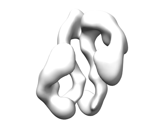
- Downloads & links
-EMDB archive
| Map data | emd_3476.map.gz | 6 MB | EMDB map data format | |
|---|---|---|---|---|
| Header (meta data) | emd-3476-v30.xmlemd-3476.xml | 11.5 KB 11.5 KB | Display Display | EMDB header |
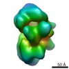
| Images |  emd_3476.png emd_3476.png | 39.9 KB | ||
| Archive directory |  http://ftp.pdbj.org/pub/emdb/structures/EMD-3476ftp://ftp.pdbj.org/pub/emdb/structures/EMD-3476 http://ftp.pdbj.org/pub/emdb/structures/EMD-3476ftp://ftp.pdbj.org/pub/emdb/structures/EMD-3476 | HTTPS FTP |
-Related structure data
| Similar structure data |
|---|
-Links
| EMDB pages | EMDB (EBI/PDBe) / EMDataResource |
|---|
-Map
| File | Download / File: emd_3476.map.gz / Format: CCP4 / Size: 8 MB / Type: IMAGE STORED AS FLOATING POINT NUMBER (4 BYTES) | ||||||||||||||||||||||||||||||||||||||||||||||||||||||||||||
|---|---|---|---|---|---|---|---|---|---|---|---|---|---|---|---|---|---|---|---|---|---|---|---|---|---|---|---|---|---|---|---|---|---|---|---|---|---|---|---|---|---|---|---|---|---|---|---|---|---|---|---|---|---|---|---|---|---|---|---|---|---|
| Annotation | 3D Reconstruction by negative stain EM of FANCI-FANCD2 co-expressed with FANCC-FANCE-FANCF. | ||||||||||||||||||||||||||||||||||||||||||||||||||||||||||||
| Projections & slices | Image control
Images are generated by Spider. | ||||||||||||||||||||||||||||||||||||||||||||||||||||||||||||
| Voxel size | X=Y=Z: 2.7 Å | ||||||||||||||||||||||||||||||||||||||||||||||||||||||||||||
| Density |
| ||||||||||||||||||||||||||||||||||||||||||||||||||||||||||||
| Symmetry | Space group: 1 | ||||||||||||||||||||||||||||||||||||||||||||||||||||||||||||
| Details | EMDB XML:
CCP4 map header:
| ||||||||||||||||||||||||||||||||||||||||||||||||||||||||||||
 Z (Sec.)
 Y (Row.)
 X (Col.)


-Supplemental data
- Sample components
-Entire : Hetero-dimeric complex of FANCI and FANCD2 proteins recombinantly...
| Entire | Name: Hetero-dimeric complex of FANCI and FANCD2 proteins recombinantly co-expressed with the FANCC-FANCE-FANCF ternary complex. |
|---|---|
| Components |
|
-Supramolecule #1: Hetero-dimeric complex of FANCI and FANCD2 proteins recombinantly...
| Supramolecule | Name: Hetero-dimeric complex of FANCI and FANCD2 proteins recombinantly co-expressed with the FANCC-FANCE-FANCF ternary complex. type: complex / ID: 1 / Parent: 0 |
|---|---|
| Recombinant expression | Organism:   Spodoptera frugiperda (fall armyworm) Spodoptera frugiperda (fall armyworm) |
-Experimental details
-Structure determination
| Method | negative staining |
|---|---|
 Processing Processing | single particle reconstruction |
| Aggregation state | particle |
-Sample preparation
| Concentration | 0.005 mg/mL | ||||||||||||
|---|---|---|---|---|---|---|---|---|---|---|---|---|---|
| Buffer | pH: 8 Component:
| ||||||||||||
| Staining | Type: NEGATIVE / Material: Uranyl formate Details: The sample stained with a fresh 2% (wt/vol) uranyl formate solution. | ||||||||||||
| Grid | Model: 3.05mm diameter, square mesh grids. / Material: COPPER / Mesh: 600 / Support film - Material: CARBON / Support film - topology: CONTINUOUS / Support film - Film thickness: 4.0 nm / Pretreatment - Type: GLOW DISCHARGE / Pretreatment - Atmosphere: AIR Details: Prior to sample incubation, the carbon-coated grid was glow-discharged for 30 seconds at 45 mA. |
- Electron microscopy
| Microscope | JEOL 2100 |
|---|---|
| Image recording | Film or detector model: GATAN ULTRASCAN 4000 (4k x 4k) / Number real images: 447 / Average electron dose: 35.0 e/Å2 |
| Electron beam | Acceleration voltage: 120 kV / Electron source: LAB6 |
| Electron optics | Illumination mode: FLOOD BEAM / Imaging mode: BRIGHT FIELD / Cs: 2.0 mm / Nominal defocus max: 2.0 µm / Nominal defocus min: 0.5 µm |
| Sample stage | Specimen holder model: JEOL |
