Movie
Movie Controller

[English] 日本語
 Yorodumi
- EMDB-34559: Immune complex of W313-6D11 IgG binding the RBD of SARS-CoV-1 2P ... -
+ Open data

- Basic information
| Entry |  | |||||||||
|---|---|---|---|---|---|---|---|---|---|---|
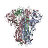
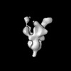
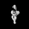
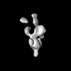
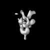
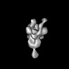
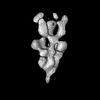
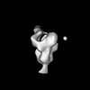
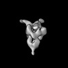
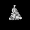
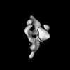
| Title | Immune complex of W313-6D11 IgG binding the RBD of SARS-CoV-1 2P spike protein | |||||||||
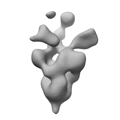
 Map data Map data | Immune complex of W313-6D11 IgG binding the RBD of SARS-CoV-1 2P spike protein | |||||||||
 Sample Sample |
| |||||||||
 Keywords Keywords | Complex / SARS-CoV-1 / antibody / Homo sapiens / RBD / VIRAL PROTEIN | |||||||||
| Biological species |  Homo sapiens (human) / Homo sapiens (human) /  Severe acute respiratory syndrome coronavirus Severe acute respiratory syndrome coronavirus | |||||||||
| Method | single particle reconstruction / negative staining / Resolution: 25.0 Å | |||||||||
 Authors Authors | Zhu JY / Zhou BN | |||||||||
| Funding support |  China, 2 items China, 2 items
| |||||||||
 Citation Citation | Journal: Immunity / Year: 2023 Title: Dissecting the intricacies of human antibody responses to SARS-CoV-1 and SARS-CoV-2 infection. Authors: Ruoke Wang / Yang Han / Rui Zhang / Jiayi Zhu / Xuanyu Nan / Yaping Liu / Ziqing Yang / Bini Zhou / Jinfang Yu / Zichun Lin / Jinqian Li / Peng Chen / Yangjunqi Wang / Yujie Li / Dongsheng ...Authors: Ruoke Wang / Yang Han / Rui Zhang / Jiayi Zhu / Xuanyu Nan / Yaping Liu / Ziqing Yang / Bini Zhou / Jinfang Yu / Zichun Lin / Jinqian Li / Peng Chen / Yangjunqi Wang / Yujie Li / Dongsheng Liu / Xuanling Shi / Xinquan Wang / Qi Zhang / Yuhe R Yang / Taisheng Li / Linqi Zhang / Abstract: The 2003 severe acute respiratory syndrome coronavirus (SARS-CoV-1) causes more severe disease than SARS-CoV-2, which is responsible for COVID-19. However, our understanding of antibody response to ...The 2003 severe acute respiratory syndrome coronavirus (SARS-CoV-1) causes more severe disease than SARS-CoV-2, which is responsible for COVID-19. However, our understanding of antibody response to SARS-CoV-1 infection remains incomplete. Herein, we studied the antibody responses in 25 SARS-CoV-1 convalescent patients. Plasma neutralization was higher and lasted longer in SARS-CoV-1 patients than in severe SARS-CoV-2 patients. Among 77 monoclonal antibodies (mAbs) isolated, 60 targeted the receptor-binding domain (RBD) and formed 7 groups (RBD-1 to RBD-7) based on their distinct binding and structural profiles. Notably, RBD-7 antibodies bound to a unique RBD region interfaced with the N-terminal domain of the neighboring protomer (NTD proximal) and were more prevalent in SARS-CoV-1 patients. Broadly neutralizing antibodies for SARS-CoV-1, SARS-CoV-2, and bat and pangolin coronaviruses were also identified. These results provide further insights into the antibody response to SARS-CoV-1 and inform the design of more effective strategies against diverse human and animal coronaviruses. | |||||||||
| History |
|
- Structure visualization
| Supplemental images |
|---|
- Downloads & links
-EMDB archive
| Map data | emd_34559.map.gz | 20.7 MB |  EMDB map data format EMDB map data format | |
|---|---|---|---|---|
| Header (meta data) | emd-34559-v30.xmlemd-34559.xml | 14 KB 14 KB | Display Display | EMDB header |
| Images |  emd_34559.png emd_34559.png | 38.5 KB | ||
| Filedesc metadata | emd-34559.cif.gz | 4 KB | ||
| Others | emd_34559_half_map_1.map.gzemd_34559_half_map_2.map.gz | 20.7 MB 20.7 MB | ||
| Archive directory |  http://ftp.pdbj.org/pub/emdb/structures/EMD-34559ftp://ftp.pdbj.org/pub/emdb/structures/EMD-34559 http://ftp.pdbj.org/pub/emdb/structures/EMD-34559ftp://ftp.pdbj.org/pub/emdb/structures/EMD-34559 | HTTPS FTP |
-Related structure data
| Related structure data | C: citing same article ( |
|---|


-Links
| EMDB pages | EMDB (EBI/PDBe) / EMDataResource |
|---|
-Map
| File | Download / File: emd_34559.map.gz / Format: CCP4 / Size: 27 MB / Type: IMAGE STORED AS FLOATING POINT NUMBER (4 BYTES) | ||||||||||||||||||||||||||||||||||||
|---|---|---|---|---|---|---|---|---|---|---|---|---|---|---|---|---|---|---|---|---|---|---|---|---|---|---|---|---|---|---|---|---|---|---|---|---|---|
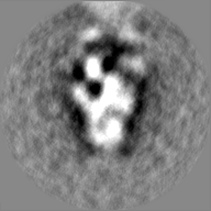
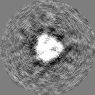
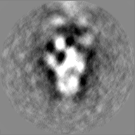
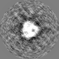
| Annotation | Immune complex of W313-6D11 IgG binding the RBD of SARS-CoV-1 2P spike protein | ||||||||||||||||||||||||||||||||||||
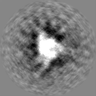
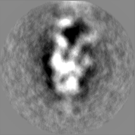
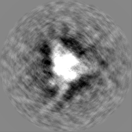
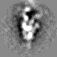
| Projections & slices | Image control
Images are generated by Spider. | ||||||||||||||||||||||||||||||||||||
| Voxel size | X=Y=Z: 2.21 Å | ||||||||||||||||||||||||||||||||||||
| Density |
| ||||||||||||||||||||||||||||||||||||
| Symmetry | Space group: 1 | ||||||||||||||||||||||||||||||||||||
| Details | EMDB XML:
|
 Z (Sec.)
 Y (Row.)
 X (Col.)


-Supplemental data
-Half map: Immune complex of W313-6D11 IgG binding the RBD...
| File | emd_34559_half_map_1.map | ||||||||||||
|---|---|---|---|---|---|---|---|---|---|---|---|---|---|
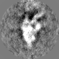
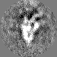
| Annotation | Immune complex of W313-6D11 IgG binding the RBD of SARS-CoV-1 2P spike protein | ||||||||||||
| Projections & Slices |
| ||||||||||||
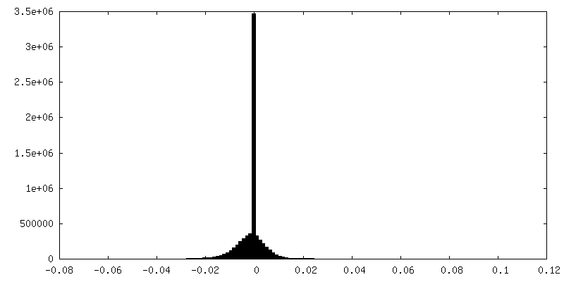
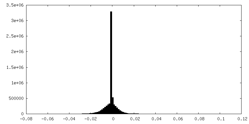
| Density Histograms |
-Half map: Immune complex of W313-6D11 IgG binding the RBD...
| File | emd_34559_half_map_2.map | ||||||||||||
|---|---|---|---|---|---|---|---|---|---|---|---|---|---|
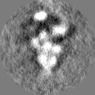
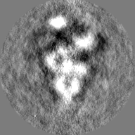
| Annotation | Immune complex of W313-6D11 IgG binding the RBD of SARS-CoV-1 2P spike protein | ||||||||||||
| Projections & Slices |
| ||||||||||||
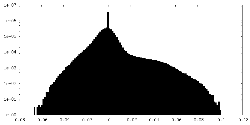
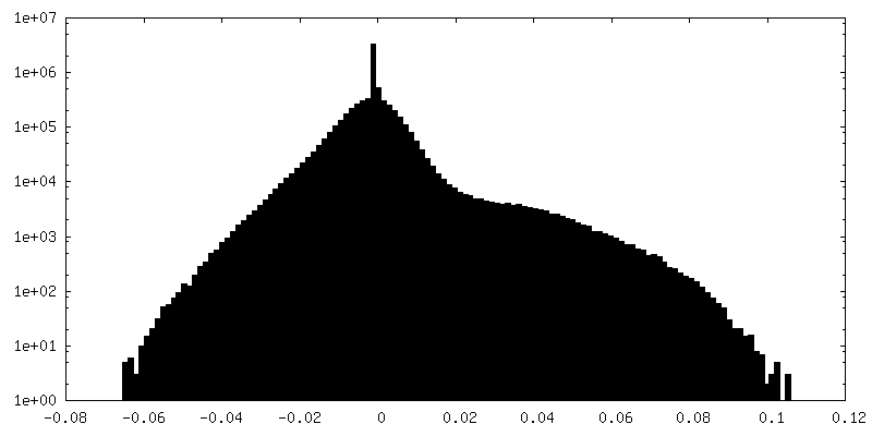
| Density Histograms |
- Sample components
-Entire : SARS-CoV-1 2P in complex with W313-6D11 IgG
| Entire | Name: SARS-CoV-1 2P in complex with W313-6D11 IgG |
|---|---|
| Components |
|
-Supramolecule #1: SARS-CoV-1 2P in complex with W313-6D11 IgG
| Supramolecule | Name: SARS-CoV-1 2P in complex with W313-6D11 IgG / type: complex / ID: 1 / Parent: 0 Details: Immune complex of W313-6D11 IgG binding the RBD of SARS-CoV-1 2P spike protein |
|---|---|
| Source (natural) | Organism: Homo sapiens (human) |
-Supramolecule #2: Severe acute respiratory syndrome coronavirus 1-2P
| Supramolecule | Name: Severe acute respiratory syndrome coronavirus 1-2P / type: complex / ID: 2 / Parent: 1 / Details: SARS-CoV-1 2P |
|---|---|
| Source (natural) | Organism: Severe acute respiratory syndrome coronavirus |
-Supramolecule #3: W313-6D11 IgG
| Supramolecule | Name: W313-6D11 IgG / type: complex / ID: 3 / Parent: 1 / Details: W313-6D11 IgG |
|---|---|
| Source (natural) | Organism: Homo sapiens (human) |
-Experimental details
-Structure determination
| Method | negative staining |
|---|---|
 Processing Processing | single particle reconstruction |
| Aggregation state | particle |
-Sample preparation
| Concentration | 0.015 mg/mL |
|---|---|
| Buffer | pH: 7.4 |
| Staining | Type: NEGATIVE / Material: Uranyl Acetate |
- Electron microscopy
| Microscope | JEOL 2100F |
|---|---|
| Image recording | Film or detector model: OTHER / Average electron dose: 25.0 e/Å2 |
| Electron beam | Acceleration voltage: 200 kV / Electron source:  FIELD EMISSION GUN FIELD EMISSION GUN |
| Electron optics | Illumination mode: FLOOD BEAM / Imaging mode: BRIGHT FIELD / Cs: 2.7 mm / Nominal defocus max: 1.5 µm / Nominal defocus min: 1.5 µm |
-Image processing
| Startup model | Type of model: PDB ENTRY PDB model - PDB ID: |
|---|---|
| Final reconstruction | Resolution.type: BY AUTHOR / Resolution: 25.0 Å / Resolution method: FSC 0.5 CUT-OFF / Number images used: 1366 |
| Initial angle assignment | Type: MAXIMUM LIKELIHOOD |
| Final angle assignment | Type: MAXIMUM LIKELIHOOD |