 ムービー
ムービー コントローラー


+ データを開く

- 基本情報
基本情報
| 登録情報 |  | |||||||||
|---|---|---|---|---|---|---|---|---|---|---|
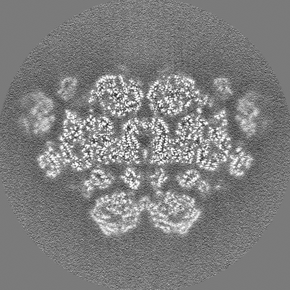
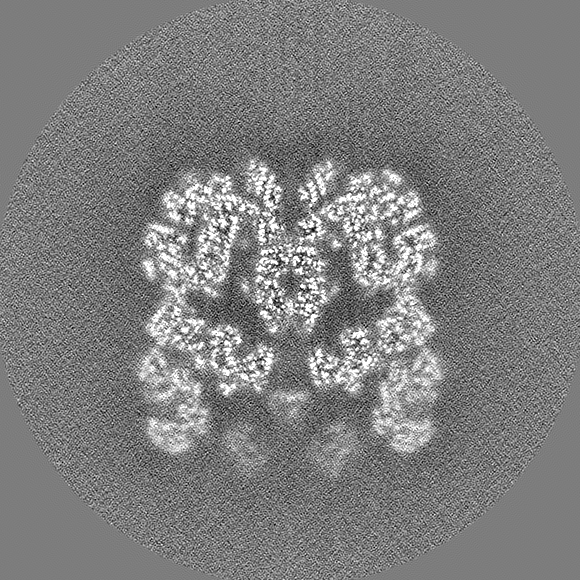
| タイトル | Un-sharpened map of phycobilisome from Porphyridium purpureum under Middle light | |||||||||
 マップデータ マップデータ | Un-sharpened map of phycobilisome under Middle light | |||||||||
 試料 試料 |
| |||||||||
 キーワード キーワード | light-harvesting complex / photosynthesis | |||||||||
| 生物種 |  Porphyridium purpureum (真核生物) Porphyridium purpureum (真核生物) | |||||||||
| 手法 | 単粒子再構成法 / クライオ電子顕微鏡法 / 解像度: 3.6 Å | |||||||||
 データ登録者 データ登録者 | Ma JF / Sui S-F | |||||||||
| 資金援助 |  中国, 2件 中国, 2件
| |||||||||
 引用 引用 | ジャーナル: Commun Biol / 年: 2023 タイトル: The structural basis for light acclimation in phycobilisome light harvesting systems systems in Porphyridium purpureum. 著者: Emma Joy Dodson / Jianfei Ma / Maayan Suissa Szlejf / Naama Maroudas-Sklare / Yossi Paltiel / Noam Adir / Shan Sun / Sen-Fang Sui / Nir Keren /  要旨: Photosynthetic organisms adapt to changing light conditions by manipulating their light harvesting complexes. Biophysical, biochemical, physiological and genetic aspects of these processes are ...Photosynthetic organisms adapt to changing light conditions by manipulating their light harvesting complexes. Biophysical, biochemical, physiological and genetic aspects of these processes are studied extensively. The structural basis for these studies is lacking. In this study we address this gap in knowledge by focusing on phycobilisomes (PBS), which are large structures found in cyanobacteria and red algae. In this study we focus on the phycobilisomes (PBS), which are large structures found in cyanobacteria and red algae. Specifically, we examine red algae (Porphyridium purpureum) grown under a low light intensity (LL) and a medium light intensity (ML). Using cryo-electron microscopy, we resolve the structure of ML-PBS and compare it to the LL-PBS structure. The ML-PBS is 13.6 MDa, while the LL-PBS is larger (14.7 MDa). The LL-PBS structure have a higher number of closely coupled chromophore pairs, potentially the source of the red shifted fluorescence emission from LL-PBS. Interestingly, these differences do not significantly affect fluorescence kinetics parameters. This indicates that PBS systems can maintain similar fluorescence quantum yields despite an increase in LL-PBS chromophore numbers. These findings provide a structural basis to the processes by which photosynthetic organisms adapt to changing light conditions. | |||||||||
| 履歴 |
|
- 構造の表示
構造の表示
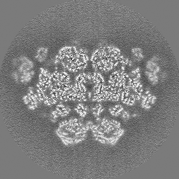
| 添付画像 |
|---|

- ダウンロードとリンク
-EMDBアーカイブ
| マップデータ | emd_34534.map.gz | 597.8 MB |  EMDBマップデータ形式 EMDBマップデータ形式 | |
|---|---|---|---|---|
| ヘッダ (付随情報) | emd-34534-v30.xmlemd-34534.xml | 21.4 KB 21.4 KB | 表示 表示 | EMDBヘッダ |
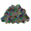
| 画像 | 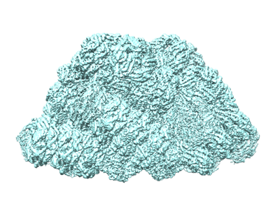 emd_34534.png emd_34534.png | 143 KB | ||
| Filedesc metadata | emd-34534.cif.gz | 4.7 KB | ||
| その他 | emd_34534_half_map_1.map.gzemd_34534_half_map_2.map.gz | 697.6 MB 697.6 MB | ||
| アーカイブディレクトリ |  http://ftp.pdbj.org/pub/emdb/structures/EMD-34534ftp://ftp.pdbj.org/pub/emdb/structures/EMD-34534 http://ftp.pdbj.org/pub/emdb/structures/EMD-34534ftp://ftp.pdbj.org/pub/emdb/structures/EMD-34534 | HTTPS FTP |
-関連構造データ
| 関連構造データ |
|---|

-リンク
| EMDBのページ | EMDB (EBI/PDBe) / EMDataResource |
|---|
-マップ
| ファイル | ダウンロード / ファイル: emd_34534.map.gz / 形式: CCP4 / 大きさ: 744.3 MB / タイプ: IMAGE STORED AS FLOATING POINT NUMBER (4 BYTES) | ||||||||||||||||||||||||||||||||||||
|---|---|---|---|---|---|---|---|---|---|---|---|---|---|---|---|---|---|---|---|---|---|---|---|---|---|---|---|---|---|---|---|---|---|---|---|---|---|
| 注釈 | Un-sharpened map of phycobilisome under Middle light | ||||||||||||||||||||||||||||||||||||
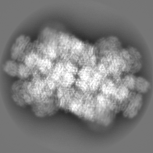
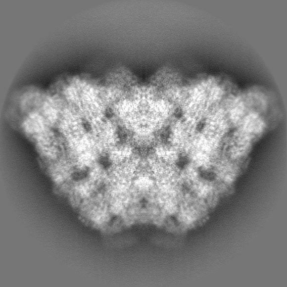
| 投影像・断面図 | 画像のコントロール
画像は Spider により作成 | ||||||||||||||||||||||||||||||||||||
| ボクセルのサイズ | X=Y=Z: 1.066 Å | ||||||||||||||||||||||||||||||||||||
| 密度 |
| ||||||||||||||||||||||||||||||||||||
| 対称性 | 空間群: 1 | ||||||||||||||||||||||||||||||||||||
| 詳細 | EMDB XML:
|
 Z (Sec.)
 Y (Row.)
 X (Col.)


-添付データ
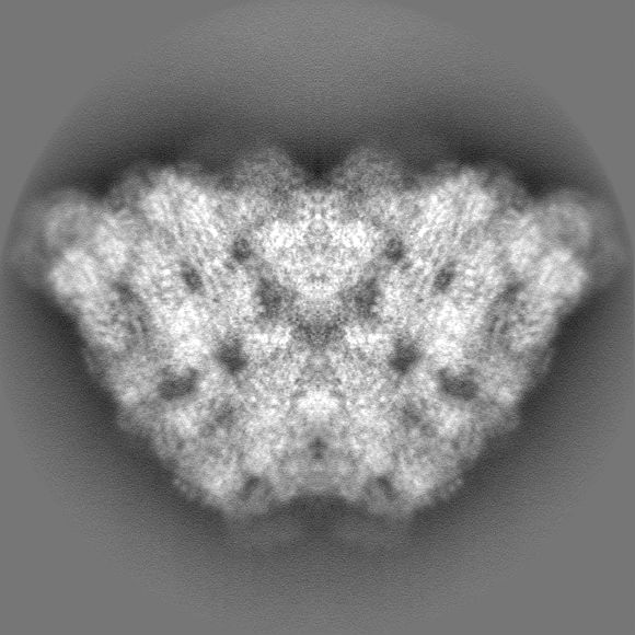
-ハーフマップ: Un-sharpened map of phycobilisome under Middle light half2
| ファイル | emd_34534_half_map_1.map | ||||||||||||
|---|---|---|---|---|---|---|---|---|---|---|---|---|---|
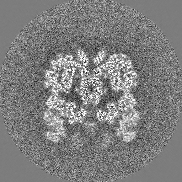
| 注釈 | Un-sharpened map of phycobilisome under Middle light half2 | ||||||||||||
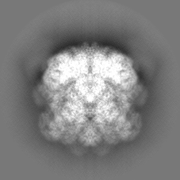
| 投影像・断面図 |
| ||||||||||||
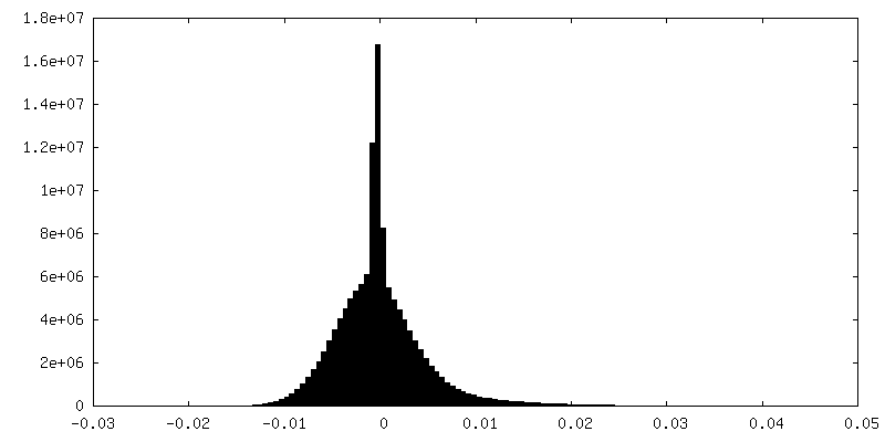
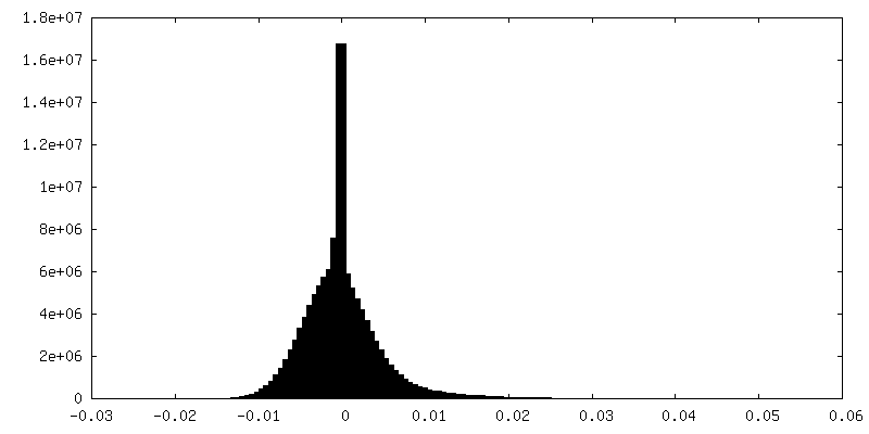
| 密度ヒストグラム |
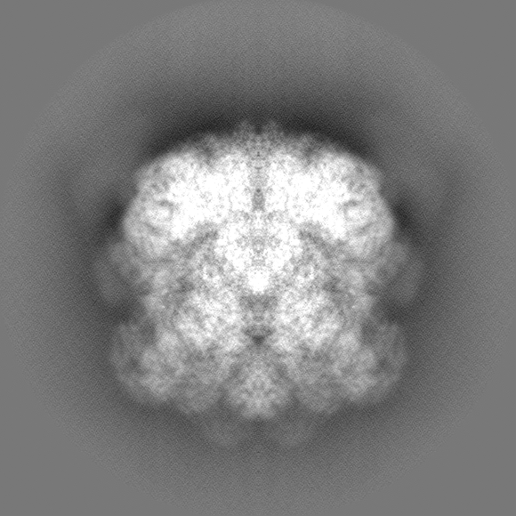

-ハーフマップ: Un-sharpened map of phycobilisome under Middle light half1
| ファイル | emd_34534_half_map_2.map | ||||||||||||
|---|---|---|---|---|---|---|---|---|---|---|---|---|---|
| 注釈 | Un-sharpened map of phycobilisome under Middle light half1 | ||||||||||||
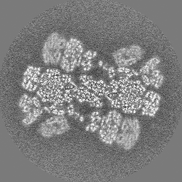
| 投影像・断面図 |
| ||||||||||||
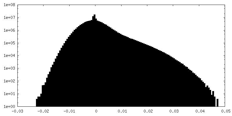
| 密度ヒストグラム |


- 試料の構成要素
-全体 : phycobilisome
| 全体 | 名称: phycobilisome |
|---|---|
| 要素 |
|
-超分子 #1: phycobilisome
| 超分子 | 名称: phycobilisome / タイプ: complex / ID: 1 / 親要素: 0 / 含まれる分子: #1-#28 |
|---|---|
| 由来(天然) | 生物種: Porphyridium purpureum (真核生物) |
| 分子量 | 理論値: 14700 kDa/nm |
-実験情報
-構造解析
| 手法 | クライオ電子顕微鏡法 |
|---|---|
 解析 解析 | 単粒子再構成法 |
| 試料の集合状態 | particle |
-試料調製
| 緩衝液 | pH: 7 構成要素:
| |||||||||
|---|---|---|---|---|---|---|---|---|---|---|
| グリッド | モデル: PELCO Ultrathin Carbon with Lacey Carbon / 支持フィルム - 材質: CARBON / 支持フィルム - トポロジー: LACEY / 支持フィルム - Film thickness: 2 | |||||||||
| 凍結 | 凍結剤: ETHANE / チャンバー内湿度: 100 % / チャンバー内温度: 18 K / 装置: FEI VITROBOT MARK IV |
- 電子顕微鏡法
| 顕微鏡 | FEI TITAN KRIOS |
|---|---|
| 温度 | 最低: 70.0 K / 最高: 70.0 K |
| 特殊光学系 | エネルギーフィルター - 名称: GIF Quantum ER |
| 撮影 | フィルム・検出器のモデル: GATAN K2 SUMMIT (4k x 4k) 検出モード: SUPER-RESOLUTION / デジタル化 - サイズ - 横: 4096 pixel / デジタル化 - サイズ - 縦: 4096 pixel / デジタル化 - 画像ごとのフレーム数: 1-32 / 撮影したグリッド数: 2 / 実像数: 6438 / 平均露光時間: 8.2 sec. / 平均電子線量: 50.0 e/Å2 |
| 電子線 | 加速電圧: 300 kV / 電子線源:  FIELD EMISSION GUN FIELD EMISSION GUN |
| 電子光学系 | C2レンズ絞り径: 100.0 µm 最大 デフォーカス(補正後): 2.3000000000000003 µm 最小 デフォーカス(補正後): 1.3 µm / 倍率(補正後): 130000 / 照射モード: FLOOD BEAM / 撮影モード: BRIGHT FIELD / Cs: 2.7 mm 最大 デフォーカス(公称値): 2.3000000000000003 µm 最小 デフォーカス(公称値): 1.3 µm / 倍率(公称値): 130000 |
| 試料ステージ | 試料ホルダーモデル: FEI TITAN KRIOS AUTOGRID HOLDER ホルダー冷却材: NITROGEN |
| 実験機器 |  モデル: Titan Krios / 画像提供: FEI Company |
+画像解析
-原子モデル構築 1
| 初期モデル | PDB ID: Chain - Source name: PDB / Chain - Initial model type: experimental model |
|---|---|
| 精密化 | 空間: REAL / プロトコル: RIGID BODY FIT / 温度因子: 62.05 / 当てはまり具合の基準: 0,99 |
