 Movie
Movie Controller


+ Open data

- Basic information
| Entry | 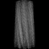 | |||||||||
|---|---|---|---|---|---|---|---|---|---|---|
| Title | Cryo-EM structure of Abeta2 fibril polymorph2 | |||||||||
 Map data Map data | ||||||||||
 Sample Sample |
| |||||||||
 Keywords Keywords | antimicrobial fibrils / amyloid / ANTIMICROBIAL PROTEIN | |||||||||
| Biological species |  Homo sapiens (human) Homo sapiens (human) | |||||||||
| Method | helical reconstruction / cryo EM / Resolution: 2.62 Å | |||||||||
 Authors Authors | Xia WC / Zhang MM / Liu C | |||||||||
| Funding support | 1 items
| |||||||||
 Citation Citation | Journal: Chem Sci / Year: 2023 Title: Engineering of antimicrobial peptide fibrils with feedback degradation of bacterial-secreted enzymes. Authors: Fenghua Wang / Wencheng Xia / Mingming Zhang / Rongrong Wu / Xiaolu Song / Yun Hao / Yonghai Feng / Liwei Zhang / Dan Li / Wenyan Kang / Cong Liu / Lei Liu /  Abstract: Proteins and peptides can assemble into functional amyloid fibrils with distinct architectures. These amyloid fibrils can fulfil various biological functions in living organisms, and then be degraded. ...Proteins and peptides can assemble into functional amyloid fibrils with distinct architectures. These amyloid fibrils can fulfil various biological functions in living organisms, and then be degraded. By incorporating an amyloidogenic segment and enzyme-cleavage segment together, we designed a peptide (enzyme-cleavage amyloid peptides (EAP))-based functional fibril which could be degraded specifically by gelatinase. To gain molecular insights into the assembly and degradation of EAP fibrils, we determined the atomic structure of the EAP fibril using cryo-electron microscopy. The amyloidogenic segment of EAP adopted a β-strand conformation and mediated EAP-fibril formation mainly steric zipper-like interactions. The enzyme-cleavage segment was partially involved in self-assembly, but also exhibited high flexibility in the fibril structure, with accessibility to gelatinase binding and degradation. Moreover, we applied the EAP fibril as a tunable scaffold for developing degradable self-assembled antimicrobial fibrils (SANs) by integrating melittin and EAP together. SANs exhibited superior activity for killing bacteria, and significantly improved the stability and biocompatibility of melittin. SANs were eliminated automatically by the gelatinase secreted from targeted bacteria. Our work provides a new strategy for rational design of functional fibrils with a feedback regulatory loop for optimizing the biocompatibility and biosafety of designed fibrils. Our work may aid further developments of "smart" peptide-based biomaterials for biomedical applications. | |||||||||
| History |
|
- Structure visualization
| Supplemental images |
|---|

- Downloads & links
-EMDB archive
| Map data | emd_34393.map.gz | 38 MB |  EMDB map data format EMDB map data format | |
|---|---|---|---|---|
| Header (meta data) | emd-34393-v30.xmlemd-34393.xml | 13.2 KB 13.2 KB | Display Display | EMDB header |
| FSC (resolution estimation) | emd_34393_fsc.xml | 12.7 KB | Display | FSC data file |
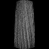
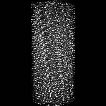
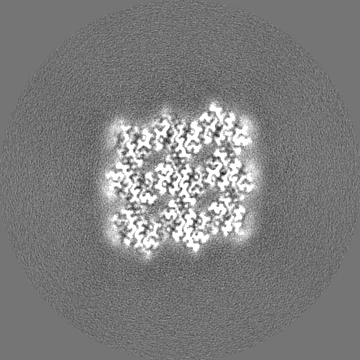
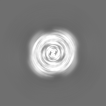
| Images | 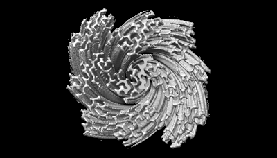 emd_34393.png emd_34393.png | 65.6 KB | ||
| Filedesc metadata | emd-34393.cif.gz | 4.3 KB | ||
| Others | emd_34393_half_map_1.map.gzemd_34393_half_map_2.map.gz | 140.1 MB 140.1 MB | ||
| Archive directory |  http://ftp.pdbj.org/pub/emdb/structures/EMD-34393ftp://ftp.pdbj.org/pub/emdb/structures/EMD-34393 http://ftp.pdbj.org/pub/emdb/structures/EMD-34393ftp://ftp.pdbj.org/pub/emdb/structures/EMD-34393 | HTTPS FTP |
-Related structure data
-Links
| EMDB pages | EMDB (EBI/PDBe) / EMDataResource |
|---|
-Map
| File | Download / File: emd_34393.map.gz / Format: CCP4 / Size: 178 MB / Type: IMAGE STORED AS FLOATING POINT NUMBER (4 BYTES) | ||||||||||||||||||||||||||||||||||||
|---|---|---|---|---|---|---|---|---|---|---|---|---|---|---|---|---|---|---|---|---|---|---|---|---|---|---|---|---|---|---|---|---|---|---|---|---|---|
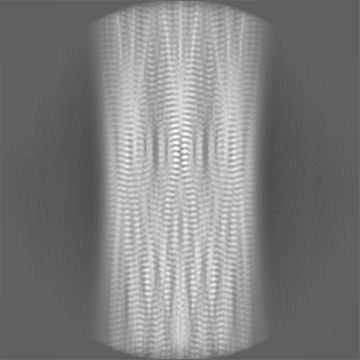
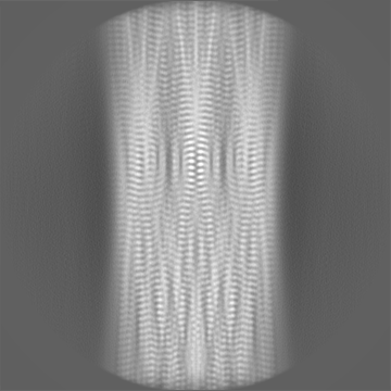
| Projections & slices | Image control
Images are generated by Spider. | ||||||||||||||||||||||||||||||||||||
| Voxel size | X=Y=Z: 0.83 Å | ||||||||||||||||||||||||||||||||||||
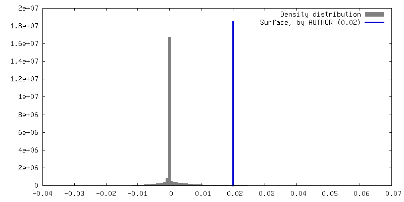
| Density |
| ||||||||||||||||||||||||||||||||||||
| Symmetry | Space group: 1 | ||||||||||||||||||||||||||||||||||||
| Details | EMDB XML:
|
 Z (Sec.)
 Y (Row.)
 X (Col.)


-Supplemental data
-Half map: #1
| File | emd_34393_half_map_1.map | ||||||||||||
|---|---|---|---|---|---|---|---|---|---|---|---|---|---|
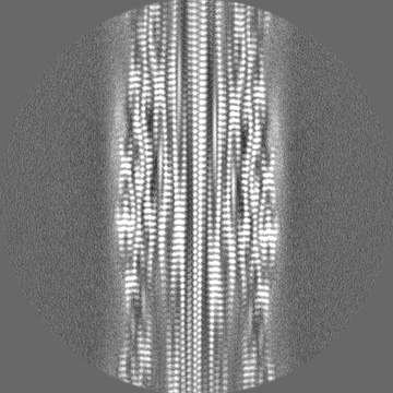
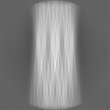
| Projections & Slices |
| ||||||||||||
| Density Histograms |


-Half map: #2
| File | emd_34393_half_map_2.map | ||||||||||||
|---|---|---|---|---|---|---|---|---|---|---|---|---|---|
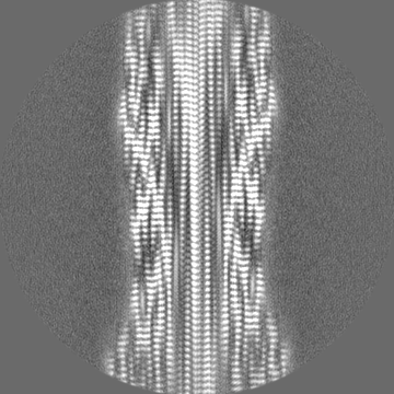
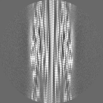
| Projections & Slices |
| ||||||||||||
| Density Histograms |


- Sample components
-Entire : peptide self-assembled antimicrobial fibrils
| Entire | Name: peptide self-assembled antimicrobial fibrils |
|---|---|
| Components |
|
-Supramolecule #1: peptide self-assembled antimicrobial fibrils
| Supramolecule | Name: peptide self-assembled antimicrobial fibrils / type: organelle_or_cellular_component / ID: 1 / Parent: 0 / Macromolecule list: #1 |
|---|---|
| Source (natural) | Organism: Homo sapiens (human) |
-Macromolecule #1: peptide self-assembled antimicrobial fibrils
| Macromolecule | Name: peptide self-assembled antimicrobial fibrils / type: protein_or_peptide / ID: 1 / Number of copies: 34 / Enantiomer: LEVO |
|---|---|
| Source (natural) | Organism: Homo sapiens (human) |
| Molecular weight | Theoretical: 1.083346 KDa |
| Sequence | String: GPLGMLGGVV IA |
-Macromolecule #2: water
| Macromolecule | Name: water / type: ligand / ID: 2 / Number of copies: 24 / Formula: HOH |
|---|---|
| Molecular weight | Theoretical: 18.015 Da |
| Chemical component information |  ChemComp-HOH: |
-Experimental details
-Structure determination
| Method | cryo EM |
|---|---|
 Processing Processing | helical reconstruction |
| Aggregation state | filament |
-Sample preparation
| Buffer | pH: 7 |
|---|---|
| Vitrification | Cryogen name: ETHANE |
- Electron microscopy
| Microscope | FEI TITAN KRIOS |
|---|---|
| Image recording | Film or detector model: GATAN K3 BIOQUANTUM (6k x 4k) / Average electron dose: 55.0 e/Å2 |
| Electron beam | Acceleration voltage: 300 kV / Electron source:  FIELD EMISSION GUN FIELD EMISSION GUN |
| Electron optics | Illumination mode: FLOOD BEAM / Imaging mode: BRIGHT FIELD / Nominal defocus max: 2.2 µm / Nominal defocus min: 1.4000000000000001 µm |
| Experimental equipment |  Model: Titan Krios / Image courtesy: FEI Company |
-Image processing
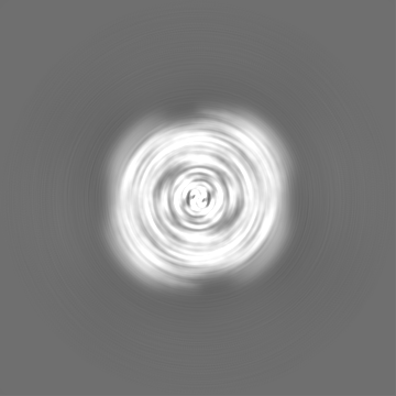
| Final reconstruction | Applied symmetry - Helical parameters - Δz: 2.404 Å Applied symmetry - Helical parameters - Δ&Phi: 179.548 ° Applied symmetry - Helical parameters - Axial symmetry: C1 (asymmetric) Resolution.type: BY AUTHOR / Resolution: 2.62 Å / Resolution method: FSC 0.143 CUT-OFF / Number images used: 53633 |
|---|---|
| Startup model | Type of model: NONE |
| Final angle assignment | Type: NOT APPLICABLE |
| FSC plot (resolution estimation) |  |
