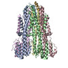
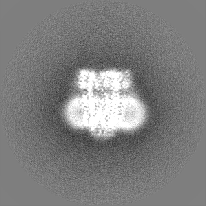
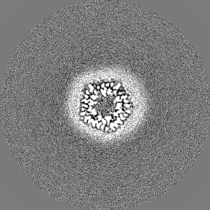
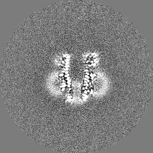
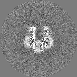
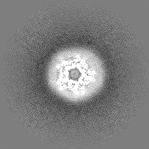
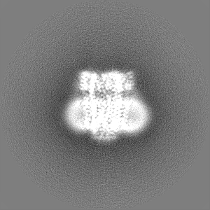
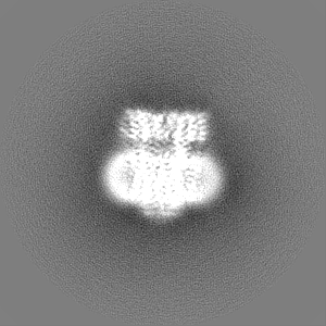
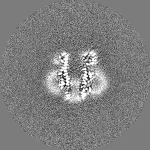
- EMDB-34203: CryoEM structure of pentameric MotA from Aquifex aeolicus -
+
Open data
ID or keywords:
Loading...
-
Basic information
Entry
Database: EMDB / ID: EMD-34203
Title
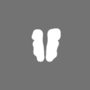
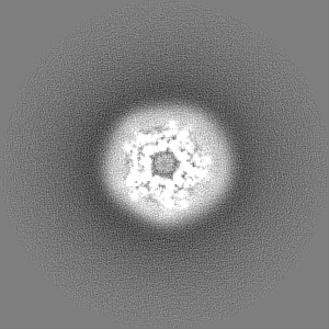
CryoEM structure of pentameric MotA from Aquifex aeolicus
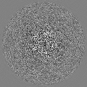
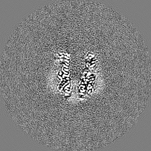
Map data
MotA 5mer postprocessed density map from dataset1
Sample
Complex: Pentameric MotA from Aquifex aeolicus
Protein or peptide: Motility protein A
Keywords
Bacterial flagellum / stator protein / Aquifex aeolicus / single particle Cryo-EM / MotA / MEMBRANE PROTEIN
Function / homology
Function and homology information
bacterial-type flagellum-dependent swarming motility / proton transmembrane transport / chemotaxis / plasma membrane Similarity search - Function
Flagellar motor protein MotA, conserved site / Flagellar motor protein motA family signature. / : / MotA/TolQ/ExbB proton channel / MotA/TolQ/ExbB proton channel family Similarity search - Domain/homology
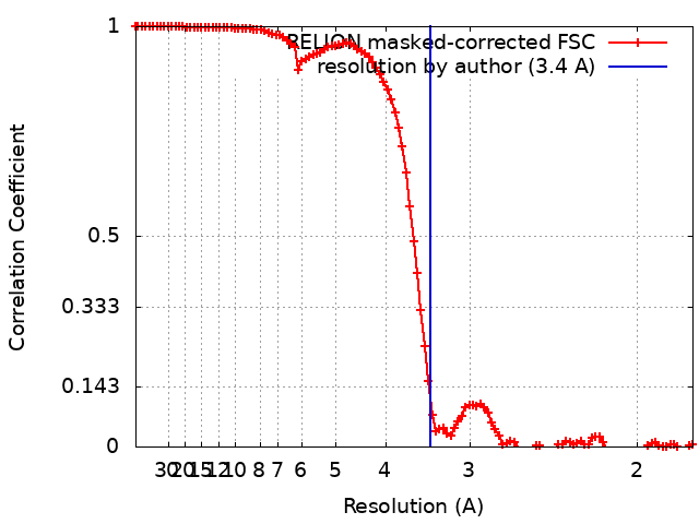
Journal: Biochem Biophys Res Commun / Year: 2022 Title: Structure of MotA, a flagellar stator protein, from hyperthermophile. Authors: Tatsuro Nishikino / Norihiro Takekawa / Duy Phuoc Tran / Jun-Ichi Kishikawa / Mika Hirose / Sakura Onoe / Seiji Kojima / Michio Homma / Akio Kitao / Takayuki Kato / Katsumi Imada / Abstract: Many motile bacteria swim and swarm toward favorable environments using the flagellum, which is rotated by a motor embedded in the inner membrane. The motor is composed of the rotor and the stator, ...Many motile bacteria swim and swarm toward favorable environments using the flagellum, which is rotated by a motor embedded in the inner membrane. The motor is composed of the rotor and the stator, and the motor torque is generated by the change of the interaction between the rotor and the stator induced by the ion flow through the stator. A stator unit consists of two types of membrane proteins termed A and B. Recent cryo-EM studies on the stators from mesophiles revealed that the stator consists of five A and two B subunits, whereas the low-resolution EM analysis showed that purified hyperthermophilic MotA forms a tetramer. To clarify the assembly formation and factors enhancing thermostability of the hyperthermophilic stator, we determined the cryo-EM structure of MotA from Aquifex aeolicus (Aa-MotA), a hyperthermophilic bacterium, at 3.42 Å resolution. Aa-MotA forms a pentamer with pseudo C5 symmetry. A simulated model of the Aa-MotAMotB stator complex resembles the structures of mesophilic stator complexes, suggesting that Aa-MotA can assemble into a pentamer equivalent to the stator complex without MotB. The distribution of hydrophobic residues of MotA pentamers suggests that the extremely hydrophobic nature in the subunit boundary and the transmembrane region is a key factor to stabilize hyperthermophilic Aa-MotA.
Supramolecule #1: Pentameric MotA from Aquifex aeolicus
Supramolecule
Name: Pentameric MotA from Aquifex aeolicus / type: complex / ID: 1 / Parent: 0 / Macromolecule list: all
Source (natural)
Organism: Aquifex aeolicus (bacteria)
Molecular weight
Theoretical: 140 KDa
-
Macromolecule #1: Motility protein A
Macromolecule
Name: Motility protein A / type: protein_or_peptide / ID: 1 Details: Met1 to His6 is translation enhancing element sequence. 6 His residues on C-terminal are purification tag. Number of copies: 5 / Enantiomer: LEVO
In the structure databanks used in Yorodumi, some data are registered as the other names, "COVID-19 virus" and "2019-nCoV". Here are the details of the virus and the list of structure data.
Jan 31, 2019. EMDB accession codes are about to change! (news from PDBe EMDB page)
EMDB accession codes are about to change! (news from PDBe EMDB page)
The allocation of 4 digits for EMDB accession codes will soon come to an end. Whilst these codes will remain in use, new EMDB accession codes will include an additional digit and will expand incrementally as the available range of codes is exhausted. The current 4-digit format prefixed with “EMD-” (i.e. EMD-XXXX) will advance to a 5-digit format (i.e. EMD-XXXXX), and so on. It is currently estimated that the 4-digit codes will be depleted around Spring 2019, at which point the 5-digit format will come into force.
The EM Navigator/Yorodumi systems omit the EMD- prefix.
Related info.:Q: What is EMD? / ID/Accession-code notation in Yorodumi/EM Navigator
Yorodumi is a browser for structure data from EMDB, PDB, SASBDB, etc.
This page is also the successor to EM Navigator detail page, and also detail information page/front-end page for Omokage search.
The word "yorodu" (or yorozu) is an old Japanese word meaning "ten thousand". "mi" (miru) is to see.
Related info.:EMDB / PDB / SASBDB / Comparison of 3 databanks / Yorodumi Search / Aug 31, 2016. New EM Navigator & Yorodumi / Yorodumi Papers / Jmol/JSmol / Function and homology information / Changes in new EM Navigator and Yorodumi
 Movie
Movie Controller


 Open data
 Basic information
 Map data
 Sample
 Keywords
 Function and homology information
 Aquifex aeolicus (bacteria) /
  Authors
 Japan, 7 items
  Citation
 Structure visualization
 Downloads & links
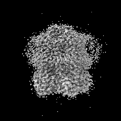 emd_34203.png
 http://ftp.pdbj.org/pub/emdb/structures/EMD-34203
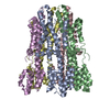
 Z (Sec.)
 Y (Row.)
 X (Col.)


 Sample components
 Processing
 Electron microscopy
 FIELD EMISSION GUN