National Natural Science Foundation of China (NSFC)
91957201
China
National Natural Science Foundation of China (NSFC)
31821091
China
National Natural Science Foundation of China (NSFC)
31870833
China
Citation
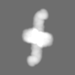
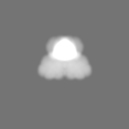
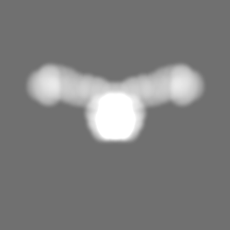
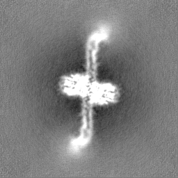
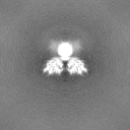
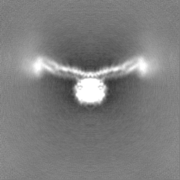
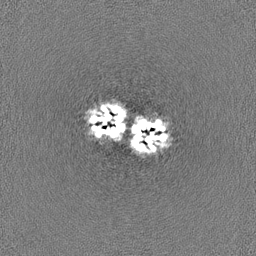
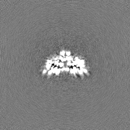
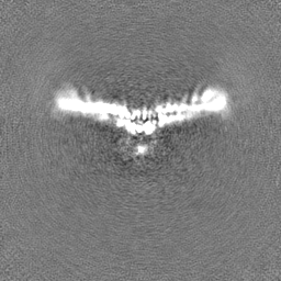
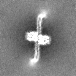
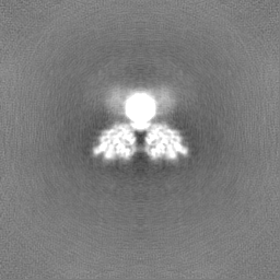
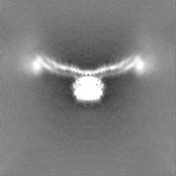
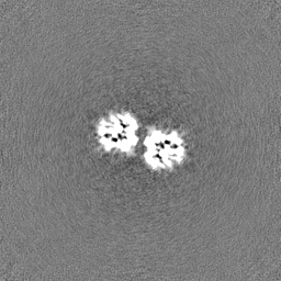
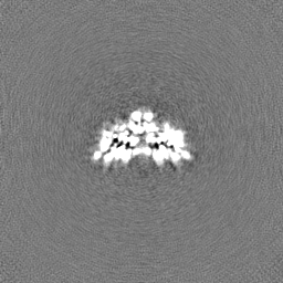
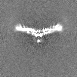
Journal: Biochem J / Year: 2022 Title: Cryo-EM structure of human MG53 homodimer. Authors: Yange Niu / Gengjia Chen / Fengxiang Lv / Rui-Ping Xiao / Xinli Hu / Lei Chen / Abstract: MG53 is a tripartite motif (TRIM) family E3 ligase and plays important biological functions. Here we present the cryo-EM structure of human MG53, showing that MG53 is a homodimer consisting of a ...MG53 is a tripartite motif (TRIM) family E3 ligase and plays important biological functions. Here we present the cryo-EM structure of human MG53, showing that MG53 is a homodimer consisting of a 'body' and two 'wings'. Intermolecular interactions are mainly distributed in the 'body' which is relatively stable, while two 'wings' are more dynamic. The overall architecture of MG53 is distinct from those of TRIM20 and TRIM25, illustrating the broad structural diversity of this protein family.
In the structure databanks used in Yorodumi, some data are registered as the other names, "COVID-19 virus" and "2019-nCoV". Here are the details of the virus and the list of structure data.
Jan 31, 2019. EMDB accession codes are about to change! (news from PDBe EMDB page)
EMDB accession codes are about to change! (news from PDBe EMDB page)
The allocation of 4 digits for EMDB accession codes will soon come to an end. Whilst these codes will remain in use, new EMDB accession codes will include an additional digit and will expand incrementally as the available range of codes is exhausted. The current 4-digit format prefixed with “EMD-” (i.e. EMD-XXXX) will advance to a 5-digit format (i.e. EMD-XXXXX), and so on. It is currently estimated that the 4-digit codes will be depleted around Spring 2019, at which point the 5-digit format will come into force.
The EM Navigator/Yorodumi systems omit the EMD- prefix.
Related info.:Q: What is EMD? / ID/Accession-code notation in Yorodumi/EM Navigator
Yorodumi is a browser for structure data from EMDB, PDB, SASBDB, etc.
This page is also the successor to EM Navigator detail page, and also detail information page/front-end page for Omokage search.
The word "yorodu" (or yorozu) is an old Japanese word meaning "ten thousand". "mi" (miru) is to see.
Related info.:EMDB / PDB / SASBDB / Comparison of 3 databanks / Yorodumi Search / Aug 31, 2016. New EM Navigator & Yorodumi / Yorodumi Papers / Jmol/JSmol / Function and homology information / Changes in new EM Navigator and Yorodumi
 Movie
Movie Controller


 Open data
 Basic information
 Map data
 Sample
 Keywords
 Function and homology information
 Homo sapiens (human)
 Authors
 China, 3 items
  Citation
 Structure visualization
 Downloads & links
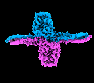 emd_33606.png
 http://ftp.pdbj.org/pub/emdb/structures/EMD-33606


 Z (Sec.)
 Y (Row.)
 X (Col.)


 Sample components
 Processing
 Electron microscopy
 FIELD EMISSION GUN
