Movie
Movie Controller


+ Open data

- Basic information
| Entry |  | |||||||||
|---|---|---|---|---|---|---|---|---|---|---|
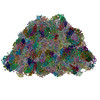
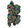
| Title | PSI-LHCs of PBS-PSII-PSI-LHCs from Porphyridium purpureum. | |||||||||
 Map data Map data | ||||||||||
 Sample Sample |
| |||||||||
| Biological species |  Porphyridium purpureum (eukaryote) Porphyridium purpureum (eukaryote) | |||||||||
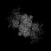
| Method | single particle reconstruction / cryo EM / Resolution: 3.6 Å | |||||||||
 Authors Authors | You X / Zhang X / Cheng J / Xiao YN / Sun S / Sui SF | |||||||||
| Funding support | 1 items
| |||||||||
 Citation Citation | Journal: To Be Published Title: Structure of lateral hexamer of PBS-PSII-PSI-LHCs megacomplex at 6.3 Angstroms resolution. Authors: You X / Zhang X / Cheng J / Xiao YN / Sun S / Sui SF | |||||||||
| History |
|
- Structure visualization
| Supplemental images |
|---|
- Downloads & links
-EMDB archive
| Map data | emd_33561.map.gz | 59.3 MB |  EMDB map data format EMDB map data format | |
|---|---|---|---|---|
| Header (meta data) | emd-33561-v30.xmlemd-33561.xml | 44 KB 44 KB | Display Display | EMDB header |
| Images |  emd_33561.png emd_33561.png | 118.7 KB | ||
| Others | emd_33561_half_map_1.map.gzemd_33561_half_map_2.map.gz | 59.4 MB 59.4 MB | ||
| Archive directory |  http://ftp.pdbj.org/pub/emdb/structures/EMD-33561ftp://ftp.pdbj.org/pub/emdb/structures/EMD-33561 http://ftp.pdbj.org/pub/emdb/structures/EMD-33561ftp://ftp.pdbj.org/pub/emdb/structures/EMD-33561 | HTTPS FTP |
-Related structure data
| Related structure data |  7y1e  8jngC  8jnlC 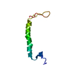 8jnmC 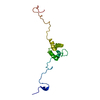 8jnnC M: atomic model generated by this map C: citing same article ( |
|---|


-Links
| EMDB pages | EMDB (EBI/PDBe) / EMDataResource |
|---|
-Map
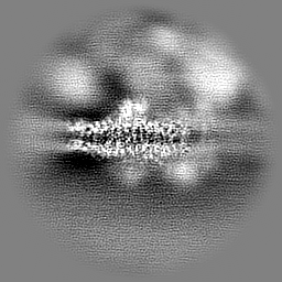
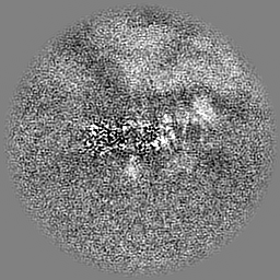
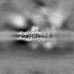
| File | Download / File: emd_33561.map.gz / Format: CCP4 / Size: 64 MB / Type: IMAGE STORED AS FLOATING POINT NUMBER (4 BYTES) | ||||||||||||||||||||||||||||||||||||
|---|---|---|---|---|---|---|---|---|---|---|---|---|---|---|---|---|---|---|---|---|---|---|---|---|---|---|---|---|---|---|---|---|---|---|---|---|---|
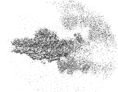
| Projections & slices | Image control
Images are generated by Spider. | ||||||||||||||||||||||||||||||||||||
| Voxel size | X=Y=Z: 1.632 Å | ||||||||||||||||||||||||||||||||||||
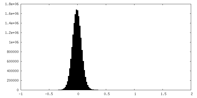
| Density |
| ||||||||||||||||||||||||||||||||||||
| Symmetry | Space group: 1 | ||||||||||||||||||||||||||||||||||||
| Details | EMDB XML:
|
 Z (Sec.)
 Y (Row.)
 X (Col.)


-Supplemental data
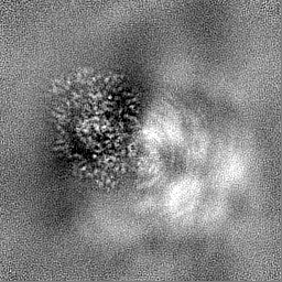
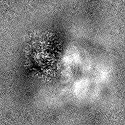
-Half map: #2
| File | emd_33561_half_map_1.map | ||||||||||||
|---|---|---|---|---|---|---|---|---|---|---|---|---|---|
| Projections & Slices |
| ||||||||||||
| Density Histograms |
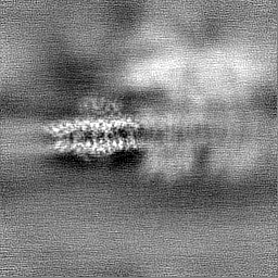


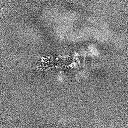
-Half map: #1
| File | emd_33561_half_map_2.map | ||||||||||||
|---|---|---|---|---|---|---|---|---|---|---|---|---|---|
| Projections & Slices |
| ||||||||||||
| Density Histograms |


- Sample components
+Entire : Lateral hexamer of PBS
+Supramolecule #1: Lateral hexamer of PBS
+Macromolecule #1: Chlorophyll a-b binding protein, chloroplastic
+Macromolecule #2: Chlorophyll a-b binding protein of LHCII type III, chloroplastic
+Macromolecule #3: Chlorophyll a-b binding protein, chloroplastic
+Macromolecule #4: Chlorophyll a-b binding protein 1B-21, chloroplastic
+Macromolecule #5: Chlorophyll a-b binding protein 1B-21, chloroplastic
+Macromolecule #6: Chlorophyll a-b binding protein 1B-21, chloroplastic
+Macromolecule #7: Fucoxanthin-chlorophyll a-c binding protein, chloroplastic
+Macromolecule #8: RedCAP
+Macromolecule #9: Photosystem I P700 chlorophyll a apoprotein A1
+Macromolecule #10: Photosystem I P700 chlorophyll a apoprotein A2
+Macromolecule #11: Photosystem I reaction center subunit II
+Macromolecule #12: Photosystem I reaction center subunit IV
+Macromolecule #13: Photosystem I reaction center subunit III
+Macromolecule #14: Photosystem I reaction center subunit IX
+Macromolecule #15: Photosystem I reaction center subunit PsaK
+Macromolecule #16: Photosystem I reaction center subunit XI
+Macromolecule #17: Photosystem I reaction center subunit XII
+Macromolecule #18: Ferredoxin
+Macromolecule #19: Photosystem I subunit O
+Macromolecule #20: PsaR
+Macromolecule #21: LPS1
+Macromolecule #22: Photosystem I iron-sulfur center
+Macromolecule #23: Photosystem I reaction center subunit VIII
+Macromolecule #24: Cytochrome c6
+Macromolecule #25: CHLOROPHYLL A

+Macromolecule #26: beta-D-glucopyranose

+Macromolecule #27: (1R,2S)-4-{(1E,3E,5E,7E,9E,11E,13E,15E,17E)-18-[(4S)-4-hydroxy-2,...

+Macromolecule #28: PHYLLOQUINONE

+Macromolecule #29: 1,2-DIPALMITOYL-PHOSPHATIDYL-GLYCEROLE

+Macromolecule #30: BETA-CAROTENE

+Macromolecule #31: IRON/SULFUR CLUSTER

+Macromolecule #32: DIGALACTOSYL DIACYL GLYCEROL (DGDG)

+Macromolecule #33: (2S)-2,3-dihydroxypropyl octadecanoate
+Macromolecule #34: PROLINE

+Macromolecule #35: FE2/S2 (INORGANIC) CLUSTER

+Macromolecule #36: PROTOPORPHYRIN IX CONTAINING FE

-Experimental details
-Structure determination
| Method | cryo EM |
|---|---|
 Processing Processing | single particle reconstruction |
| Aggregation state | cell |
-Sample preparation
| Buffer | pH: 7 |
|---|---|
| Vitrification | Cryogen name: ETHANE |
- Electron microscopy
| Microscope | FEI TITAN KRIOS |
|---|---|
| Image recording | Film or detector model: GATAN K3 (6k x 4k) / Average electron dose: 35.0 e/Å2 |
| Electron beam | Acceleration voltage: 300 kV / Electron source:  FIELD EMISSION GUN FIELD EMISSION GUN |
| Electron optics | Illumination mode: SPOT SCAN / Imaging mode: BRIGHT FIELD / Nominal defocus max: 6.0 µm / Nominal defocus min: 1.0 µm |
| Experimental equipment |  Model: Titan Krios / Image courtesy: FEI Company |
-Image processing
| Startup model | Type of model: PDB ENTRY PDB model - PDB ID: |
|---|---|
| Final reconstruction | Resolution.type: BY AUTHOR / Resolution: 3.6 Å / Resolution method: FSC 0.143 CUT-OFF / Number images used: 112000 |
| Initial angle assignment | Type: MAXIMUM LIKELIHOOD |
| Final angle assignment | Type: MAXIMUM LIKELIHOOD |